
Abstract
Background and purpose
Previous research has suggested that deficits in emotion recognition are involved in the pathogenesis of persecutory delusion in schizophrenia. Although disruption in auditory and language processing is crucial in the pathophysiology of schizophrenia, the neural basis for the deficits in emotion recognition of auditorily presented language stimuli and its relation to persecutory delusion have not yet been clarified.
Patients and methods
The current functional magnetic resonance imaging study used a dichotic listening task for 15 patients with schizophrenia and 23 healthy controls matched for age, sex, parental socioeconomic background, handedness, dexterous ear, and intelligence quotient. The participants completed a word recognition task on the attended side in which a word with emotionally valenced content (negative/neutral) was presented to one ear and a different neutral word was presented to the other ear. Participants selectively attended to either ear.
Results
The whole brain analysis detected the aberrant neural activity in the right inferior frontal gyrus in the patients with schizophrenia compared to that in the controls (P<0.05, false discovery rate-corrected). Brain activity in the right pars triangularis of the inferior frontal gyrus was significantly reduced when negatively valenced words were presented to the right ear, whereas the activity of the same region was significantly enhanced when these words were presented to the left ear, irrespective of the attended ear, in the participants with schizophrenia compared to the controls. Furthermore, this diminished brain response to auditorily presented negatively valenced words significantly correlated with severe positive symptoms (r=−0.67, P=0.006) and delusional behavior (r=−0.62, P=0.014) in the patients with schizophrenia.
Conclusion
The present results indicate that the significantly impaired brain activity in response to auditorily presented negatively valenced words in the right pars triangularis of the inferior frontal gyrus is associated with the pathogenesis of positive symptoms such as persecutory delusion.
Introduction
Individuals with schizophrenia show deficits in emotion recognition,Citation1 which is considered a key determinant of their functional outcome.Citation2,Citation3 Especially, aberrant recognition of negative emotion is reportedly associated with the pathogenesis of persecutory delusion.Citation4 Deficits have been suggested in the recognition of emotional prosody,Citation5,Citation6 facial expressions,Citation7,Citation8 and emotionally valenced wordsCitation9 in schizophrenia. Deficits in language-related information processing play an important role in the pathophysiology of schizophrenia,Citation10,Citation11 particularly in the formation of positive symptoms such as delusions.Citation12–Citation14 Brain network dysfunction for language processing has been indicated to predict the outcome in youth at clinically high risk for schizophrenia.Citation15 Brain network dysfunction for language processing,Citation15 impaired brain activity for semantic retrieval,Citation16 and impairment in the neural network for semantic tasksCitation17 are reported to be correlated with the severity of positive symptoms. Hence, to examine emotion recognition, visually presented negatively valenced words have often been utilized as stimuli in experiments with patients with schizophrenia.Citation9 However, abnormality in auditory information processing is crucial for the pathophysiology of schizophrenia considering the emphasis on auditory hallucinations in the diagnostic criteriaCitation18,Citation19 and repeatedly reported deficits associated with auditory processing.Citation20,Citation21 Especially, abnormal brain activity during auditory sentence perception,Citation22 auditory working memory,Citation23 and verbal self-monitoringCitation24 has been indicated to correlate with positive symptoms in schizophrenia by utilizing functional magnetic resonance imaging (fMRI). Taken together, investigating the neural basis for the processing of auditorily presented language stimuli would bring novel and significant findings in the pathophysiology of persecutory delusion in schizophrenia.
General deficits in emotional recognition irrespective of specific emotional category, such as negative or positive emotions, have been implicated in patients with schizophrenia,Citation25,Citation26 and a dual model has been proposed to distinguish inattentive from attentive processing of emotional information.Citation27–Citation29 With respect to these issues, our previous study showed that behavioral abnormality was confined to auditorily presented negatively valenced words, but not to positively valenced words in both inattentive and attentive processing, and the abnormality was correlated with delusion.Citation30
Studies investigating language-related information processing have shown dysfunctions in the inferior frontal gyrus (IFG) of participants with schizophrenia using fMRI.Citation31–Citation33 Several studies have suggested that there is heterogeneity in the functionalCitation34 features of the pars opercularis (PO) and pars triangularis (PT) within the IFG. Particularly, participants with schizophrenia had smaller gray matter volume in the PT rather than that in the PO, and the smaller gray matter volume in the right PT was associated with more severe positive symptoms in individuals with schizophrenia.Citation12,Citation35
In this study, utilizing a similar cognitive task to that in our previous study,Citation30 we explored the brain basis of the disturbed processing for auditorily presented negatively valenced words and its relationship with persecutory delusion in patients with schizophrenia for the first time.
Materials and methods
Participants
Forty-six Japanese adults participated in this study. Of these, 23 inpatients and outpatients with schizophrenia were recruited from the Department of Neuropsychiatry, The University of Tokyo Hospital, Japan. The patients were diagnosed with schizophrenia based on the Structured Clinical Interview for DSM-IV Axis I Disorder Clinical Version.Citation19 Psychiatric symptoms were evaluated using the Positive and Negative Syndrome Scale (PANSS)Citation36 immediately before the current fMRI experiment. Delusional behavior scores were calculated from the subscales of the PANSS.Citation35,Citation37 All clinical evaluations were conducted by a psychiatrist (N.I.), fully trained in maintaining reliability and consistency in diagnoses and evaluation of symptom severity, immediately before the fMRI scanning. All patients were categorized as of paranoid subtype. Twenty-three healthy participants were recruited as controls. The intelligence quotient (IQ) was evaluated using the Japanese version of the National Adult Reading Test.Citation38–Citation40 The dexterous ear was decided using four question items as described in our previous study.Citation30 The patient socioeconomic status (SES) and parental SES were assessed using the Hollingshead scale,Citation41 and the handedness was determined using the Edinburgh Inventory.Citation42 The neuroleptic doses were calculated based on chlorpromazine equivalents.Citation43 The controls were screened for neuropsychiatric disorders using the Structured Clinical Interview for DSM-IV Axis I Disorder Non-Patient Edition.Citation18,Citation44
The exclusion criteria for both groups were current or past neurological brain injury with any known cognitive consequences or loss of consciousness for >5 min, history of electroconvulsive therapy, the presence of auditory abnormalities (refer to our previous study),Citation30 and substance abuse or addiction. An additional exclusion criterion for the control group was history of psychiatric disease or family history of Axis I disorders in a first-degree relative. The ethical committee of The University of Tokyo Hospital approved this study (No 397, 1350, and 2226), and this study was conducted according to the principles expressed in the Declaration of Helsinki. After a complete explanation of the study, written informed consent was obtained from every individual.
MRI scanning
A 3-Tesla MRI scanner (GE Signa HDxt, Waukesha, WI, USA) at the University of Tokyo Hospital was used. The anatomical scanning sequence was a three-dimensional Fourier-transform spoiled gradient recalled acquisition (repetition time=6.8 s, slice thickness=1 mm, in-plane resolution=1×1 mm2). A trained neuroradiologist (W.G.) evaluated the MRI scans to confirm the absence of gross abnormality. Gradient-echo echo-planar sequences were used for functional imaging (repetition time=2 s, echo time=30 ms, flip angle=80°, 3×3×3 mm3, 35 slices, ventral-to-dorsal interleaved acquisition). One run lasted 7 min 54 s. One examination consisted of two such runs. The first 10 functional volumes were excluded from analysis for the equilibrium of longitudinal magnetization.
Experimental procedure
We selected two categories of emotional valence (16 negative; word number 100, 160, 182, 201, 224, 236, 257, 322, 375, 397, 435, 460, 726, 782, 817, 851/32 neutral; word number 47, 78, 83, 148, 227, 434, 477, 496, 547, 548, 562, 564, 642, 664, 681, 742, 752, 784, 825, 828,829, 832, 836, 854, 855, 863, 876, 893, 957, 1009, 1020, 1036) from the Affective Norms for English WordsCitation45 and translated them into Japanese nouns consisted of fourmorae (for example, negative; Go-u-to-u, neutral; Na-i-yo-u). Although some words were adjectives in English, we adjusted them to nouns when we translated them into Japanese. For validation, 20 healthy adults (10 male/10 female) who did not participate in the current experiment rated these words in terms of their valence, arousal, and dominance on the same 9-point Likert scale as that used in the Affective Norms for English Words.Citation45 Word frequency was quoted from a Japanese database,Citation46 and the number of orthographic neighbors was calculated from a Japanese thesaurus.Citation47 The current experiment used two types of word lists that significantly differed only in emotional valence (negative/neutral) (ANOVA; P<0.01), but were matched in all other language features such as arousal (P=0.17), dominance (P=0.97), word frequency (P=0.13), and number of orthographic neighbors (P=0.12).Citation45,Citation48 All words were transformed to auditory stimuli pronounced by a synthesized voice in neutral prosody using VoiceText® (HOYA, Tokyo, Japan). We then processed each auditory stimulus to equalize the duration of the utterances into 1,000 ms, spoken in a naturalistic manner using Sound Forge Pro 10® (Sony Creative Software, Middleton, WI, USA). The amplitude and pitch did not significantly differ among the negative and neutral word categories (P=0.86 and P=0.43 respectively). In addition, the mean acoustic energies of the two words, which were presented simultaneously in a dichotic paradigm, were always matched using Mitsyn® (WLH, Belmont, MA, USA) and CoolEdit Pro® (Syntrillium Software Corporation, Scottsdale, AZ, USA), according to previous studies.Citation49,Citation50
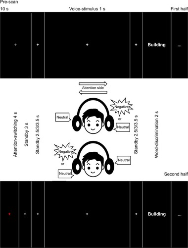
The task design was developed based on that presented in a previous study,Citation49 although that study used emotional prosody in contrast to the current study utilizing the emotional content of words. The participants responded by pressing a button while listening to a pair of words. To manipulate voluntary attention orthogonally to emotional words, we used a dichotic listening paradigm in which stimuli matched for mean acoustic energy and duration were presented simultaneously to both ears (negative–neutral, neutral–neutral, or neutral–negative on the right–left sides, respectively) (see also our previous study).Citation30 The participants were initially instructed to attend either to the right ear or the left ear. After 1 s of speech-stimulus presentation, the participants answered whether the word heard from the attended side was the same as the word presented on the screen by pressing a button within 2 s. Before and after each speech stimulus, a 2.5–3.5-s standby time was provided. After an instruction to switch the attention side in the middle of the session was presented for 4 s, the participants switched their attention to the opposite side and conducted another half session (). Each stimulus condition (negative–neutral, neutral–negative, and neutral–neutral on the right–left sides, respectively) had eight word pairs. Each word pair was used twice, and 48 trials were completed in each session. Two different sessions were completed for each participant. The order of the attention side (right/left ear) and two different sessions was counterbalanced among the participants. For each event, the time taken to answer by pressing the button after the auditory stimuli was presented and the number of correct responses was recorded automatically with E-prime 2.0 (Psychology Software Tools, Pittsburgh, PA, USA).
Figure 1 Experimental paradigm.
Behavioral analysis
For group comparisons of behavioral data in the task, we performed repeated-measures ANOVA using either the reaction time (RT) or the correct rate (CR) as the dependent variable, the groups (healthy control (HC), schizophrenia) as the between-subject factors, and the attention side (right, left) and the stimulus type (negative–neutral, neutral–negative, neutral–neutral) as a within-subject factor. If a significant interaction between the group and any other factor was found, follow-up analyses using ANOVA were performed. The statistical significance level was set at P<0.05. All statistical analyses of behavioral data were conducted using SPSS Statistics, Version 19.0 (IBM Corporation, Armonk, NY, USA).
fMRI analysis
The fMRI data were analyzed using SPM8 (The Wellcome Department of Imaging Neuroscience, London, UK) running MATLAB 2009a (MathWorks Inc., Natick, MA, USA). Functional images were realigned, slice-timing corrected, normalized to the default echo-planar imaging template, smoothed (full width at half maximum=8 mm, Gaussian filtered), and high-pass temporal filtered with a cutoff point of 128 s to remove low-frequency drift from the data. The participants included in the analysis exhibited <3 mm of maximum displacement in the x, y, or z direction and <3° of angular rotation around each axis.Citation51 At the single-subject level, we used a general linear model with three regressors (negative–neutral, neutral–negative, neutral–neutral). The six motion parameters resulting from realignment (x/y/z/pitch/yaw/roll) were also included as regressors to account for residual effects of head motion. We estimated negative–neutral-minus-neutral–neutral activity and neutral–negative-minus-neutral–neutral activity when participants attended to the right ear and when they attended to the left ear, respectively (contrast images of interest), for each participant. We analyzed brain activity only for correctly answered questions. We performed a full factorial model to examine the effects of the following: group (HC, schizophrenia), contrast (negative–neutral-minus-neutral–neutral or neutral–negative-minus-neutral–neutral), attention side (right, left), and group-by-contrast, group-by-attention side, contrast-by-attention side, and group-by-contrast-by-attention side interaction. The statistical threshold was set at P<0.05, false discovery rate-corrected. The whole brain analysis identified brain regions where a significant interaction was found between the group and any other factor. Our task design was developed based on that of a previous study,Citation49 although that study used emotional prosody in contrast to the current study utilizing the emotional content of words. As the previous study used the signal at the peak coordinate for post-hoc tests and figures,Citation49 the blood-oxygen-level-dependent (BOLD) signal of the peak coordinate was extracted. The mean BOLD signal was defined as the “signal intensity” for the contrast of interest. Signal intensity was then used to assess the interaction. As post-hoc analyses, we performed two-sample t-tests between groups. The significance level was set at P<0.05 for the post-hoc analysis.
Correlation between signal intensity and clinical indices
We explored the potential association between clinical symptom severity and signal intensity at the peak coordinate of significant interaction or the main group effect, by using Pearson’s correlation coefficients in the schizophrenia group following the previous studies.Citation37,Citation52 Symptom severity included PANSS scores (positive, negative, and general psychopathology) and delusional behavior scores. Correlations between the BOLD signals and the potential confounding factors (antipsychotic doses, age, age at onset, self/parental SES, IQ, handedness) were also tested. Considering the exploratory nature of the analyses, the statistical threshold was set at P<0.05.
Results
Demographic and clinical characteristics
Five participants with schizophrenia were excluded due to excessive head motion in the fMRI scanning, and three participants with schizophrenia could not complete the fMRI task to the end. Eventually, 15 participants with schizophrenia and 23 controls were included in the statistical analysis. The control participants were matched to the participants with schizophrenia for age, sex, parental SES,Citation41 handedness,Citation42 dexterous ear, and estimated IQ (). In these 38 participants, the extent of head motion was not significantly different between the schizophrenia group and the control group (translation (mean/SD): schizophrenia group 0.37 mm/0.17, HC 0.52 mm/0.32, t(36)=1.6, P=0.11; rotation (mean/SD): schizophrenia group 0.0089°/0.0057, HC 0.011°/0.011, t(36)=0.603, P=0.55).
Table 1 Participant characteristics and symptom scores
Behavioral results
For the CR, the ANOVA showed a significant main effect of group (F[1,36]=11.7, P=0.002), but no significant interaction including group (group-by-attention side: F[1,36]=0.02, P=0.90; group-by-stimulus type: F[2,72]=0.57, P=0.57; group-by-attention side-by-stimulus type: F[2,72]=0.54, P=0.58). For RT, the ANOVA showed no significant main effect of group (F[1,36]=0.7, P=0.40) or interaction including group (group-by-attention side: F[1,36]=0.4, P=0.55; group-by-stimulus type: F[2,72]=3.0, P=0.07; group-by-attention side-by-stimulus type: F[2,72]=0.5, P=0.61). These results indicated that the CR in the schizophrenia group was significantly worse than that in the HCs, but the RTs were not significantly different between the two groups (, ).
Figure 2 Behavioral results.
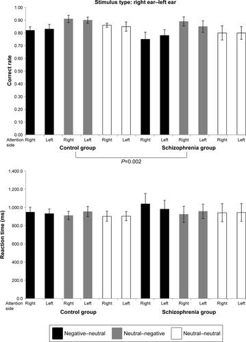
Table 2 Behavioral results
fMRI results
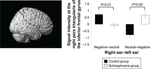
The whole brain analysis showed a significant interaction between group and contrast in the right PT of the IFG ([44 32 4], Montreal Neurological Institute coordinates, k=18, F=27.7, P<0.05, false discovery rate-corrected). The post-hoc analyses revealed that the schizophrenia group showed significantly lower negative–neutral-minus-neutral–neutral activity (P=0.01) but significantly higher neutral–negative-minus-neutral–neutral activity (P<0.01) than the HC group (). No other main effect (group, attention side) or interaction (contrast-by-attention side, group-by-attention side, group-by-contrast-by-attention side) was found.
Figure 3 Functional magnetic resonance imaging results.
Correlations between signal intensity and clinical indices
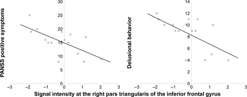
Pearson’s correlation analysis showed that the reduced negative–neutral-minus-neutral–neutral activity in the right PT of the IFG was significantly correlated with the higher PANSS positive symptom score (r=−0.67, P=0.006) and delusional behavior (r=−0.62, P=0.014) in the schizophrenia group (). In contrast, there were no significant correlations between neutral–negative-minus-neutral–neutral activity and clinical symptoms (P>0.31). The activity in each contrast also exhibited no significant correlation with any confounding factors (P>0.18).
Figure 4 Relationships between signal intensity and positive symptoms and delusional behavior.
Abbreviation: PANSS, Positive and Negative Syndrome Scale.
Discussion
To our knowledge, the present study is the first to show that brain activity in the right PT of the IFG was significantly reduced when negatively valenced words were auditorily presented to the right ear, whereas the activity was significantly enhanced when they were presented to the left ear in patients with schizophrenia compared to the control participants irrespective of being inattentively or attentively processed. Furthermore, the decreased brain activity in the right PT when negatively valenced words were auditorily presented to the right ear significantly correlated with severe positive symptoms and delusional behavior in the patients with schizophrenia.
As we predicted in our previous studyCitation30 and based on the available literature,Citation31,Citation32,Citation34 the PT of the IFG was identified as the neural basis of the disturbed processing for the auditorily presented negatively valenced words in schizophrenia. Previous studies have revealed that the PT contributes to semantic processing and the PO to phonological processing in the IFG.Citation53,Citation54 Language processes such as phonology,Citation55 morphology,Citation55 and syntaxCitation56 are dominant in the left hemisphere. Some language functions are mediated by the right hemisphere rather than by the left, such as discourse planning/comprehension; understanding humor, sarcasm, metaphors, and indirect requests; and the generation/comprehension of emotional prosody.Citation10 Regarding the IFG, semantic processing is dominant in the left IFG,Citation34,Citation57 and processing of emotional content is dominant in the right IFG.Citation58,Citation59 Schizophrenia has been previously referred to as “a left hemisphere disorder”.Citation60,Citation61 The higher order language functions mediated by the right hemisphere are essential for accurately understanding someone’s communicative intent, and the deficits displayed by patients with schizophrenia may markedly contribute to their social interaction deficits.Citation10 A previous study showed greater gray matter density reduction in the right IFG in UHR participants who later developed psychosis than in those who did not.Citation62 Right hemisphere network dysfunction for language processing has been proven to be correlated with positive symptoms.Citation16,Citation17
Most of the stimuli presented to the right ear enter contralaterally to the left hemisphere, and few stimuli enter ipsilaterally to the right hemisphere.Citation63 A previous study utilizing magnetoencephalography indicated that emotional context was processed in the right IFG after semantic context being processed in the left IFG regarding language comprehension.Citation64 The individuals with schizophrenia display a bilateral or reversed pattern of lateralization.Citation10,Citation65 The stimuli presented to the right ear entered the left IFG and might have been processed semantically and emotionally in the left IFG, and after that the rate of emotional processing in the right IFG became lower in the patients with schizophrenia than in the HCs. Therefore, brain activity in the right IFG was reduced in the patients with schizophrenia compared to that in the HCs. In the HCs, however, few stimuli entered ipsilaterally for semantic processing in the left IFG from the left ear.Citation63 Hence, emotional processing activity was low in the right IFG after semantic processing. In the schizophrenia group, most stimuli entered from the left ear contralaterally projected to the right IFG, and might be semantically and emotionally processed simultaneously in schizophrenia because of weakness in the lateralization of brain function.Citation10,Citation65 Eventually, brain activity was higher in the schizophrenia group than in the control group when negatively valenced words were presented to the left ear. If semantic and emotional processing occurred simultaneously, this activity corresponded to the early phase of language processing. Hyperactivation in the right hemisphere on early-stage semantic processing has been demonstrated in schizophrenia.Citation66 Thus, this might correspond to pre-attentive orienting bias for threat-related information in schizophrenia.Citation67,Citation68
The IFG has also been implicated in interpersonal interactions through imitation and observation of others’ actions and emotions.Citation69,Citation70 A meta-analysis of fMRI studies showed that the PO reportedly performs “mirror” processing, in that it is activated during both action observation and imitation.Citation70 Conversely, the PT does not exhibit mirror activity; it is activated only during observation.Citation69 The activation of the PT during action observation, not during imitation, is most readily explained by the frontal inhibitory mechanisms involved in suppressing movement execution during observation or motor imagery.Citation71 The cognitive evidence suggests the existence of a pre-attentive orienting bias for threat-related information, but also difficulty in disengaging threatening material from the conscious awareness following it; these cognitive characteristics may be associated with high levels of delusional ideation.Citation67,Citation68 The disruption of frontal inhibitory function in the emotional processing of auditorily presented negatively valenced words in the right PT might cause the difficulty in disengaging threatening material from conscious awareness, and therefore form positive symptoms, especially persecutory delusion.
There are several methodological limitations to our study. First, the present study sample included patients with chronic schizophrenia receiving antipsychotic medications. The effect of medicationCitation72 and chronic illnessCitation73 on our findings cannot be completely excluded, although the neuroleptic dose and duration of illness were not correlated with any neural activities. Second, the present study employing fMRI had poor temporal resolution compared to studies using magne-toencephalography.Citation64,Citation66 In the preceding discussion, changes in brain activity over time were described, but this is only speculation based on past data. Third, we did not execute any field-map corrections. It has been indicated that field-map corrections are not routinely performed in numerous types of fMRI studies,Citation74 and when distortions occur along the anterior–posterior axis and the intrinsic symmetry of the brain is not affected, such benign distortion is corrected by Realign & Unwarp in SPM8. However, EPI images at 3 Tesla can be susceptible to field inhomogeneity that may also lead to artifactual findings, and it has been indicated that field-map correction effectively improves the quality of results of task fMRI with motor tapping and auditory tasks.Citation75 Fourth, it appeared that the neutral word list had more concrete words which have specific shape and entity, and the negative word list had more abstract words which refer to ideas or concepts. This aspect of words had not been controlled in the previous studies.Citation48,Citation76–Citation78 It has been proven that abstract words produce greater activation almost exclusively in the left superior temporal and left inferior frontal cortices in contrast to concrete words.Citation79–Citation81 Furthermore, we recruited another 20 healthy Japanese (10 male/10 female) and asked them to rate the words used in this study for concreteness or abstractness on the same 9-point Likert scale. The ratings of concreteness or abstractness were not significantly different between the neutral words and negative words (mean/SD: negative 3.95/1.55, neutral 3.27/1.95, t(46)=1.23, P=0.23; a low score indicates concrete words). Hence, the results in this study are probably not entirely attributable to differences in the two word lists regarding concreteness and abstractness, but this influence cannot be entirely excluded. Finally, the sample size was relatively small, because the nature of the current study was exploratory. Future studies with patients with first-episode schizophrenia receiving minimal medication and an adequately large sample size, simultaneously employing electroencephalography and fMRI, including field-map scanning, and compiling the word list using only concrete words or abstract words could resolve these limitations.
Conclusion
Overall, taken together with our previous behavioral findings using positive, negative, and neutral auditory word stimuli,Citation30 the present results indicate that the significantly impaired brain activity, especially for auditorily presented negatively valenced words in the right PT of the IFG, relates to the pathogenesis of persecutory delusion in patients with schizophrenia.
Author contributions
N.I. contributed to project management and wrote the manuscript. Y.T., T.N., and Y.A. helped clinical evaluations and contributed to the recruitment of the participants. N.Y. gave technical support for constructing the experimental task. W.G., A.K., and O.A. supervised MRI acquisitions and evaluated all of the acquired images. K.K. and H.Y. coordinated the entire research design and took responsibility for the management of this study. All authors contributed to data analysis, drafting and revising the article, gave final approval of the version to be published, and agree to be accountable for all aspects of the work.
Acknowledgments
The authors would like to thank Chie Shimojyo and Aki Takei for their substantial support in clinical data assessment and management, and Shinsuke Koike, Yoshihiro Satomura, Mariko Tada, and Tatsuya Nagai for their substantial help with recruitment of patients. A portion of this study was also the result of a project entitled “Development of biomarker candidates for social behavior” carried out under the Strategic Research Program for Brain Sciences by the Ministry of Education, Culture, Sports, Science and Technology (MEXT).
Disclosure
This study was supported by grants from the Ministry of Health, Labour, and Welfare (Health and Labour Science Research Grant for Comprehensive Research on Disability Health and Welfare H23-seishin-ippan-002 to H.Y. and H22-seishin-ippan-015 to K.K.) and from the JSPS/MEXT (No 22689034 to H.Y.), and a Grant-in-Aid for Scientific Research on Innovative Areas (Comprehensive Brain Science Network and Ado lescent Mind & Self-Regulation (23118001 and 23118004) to K.K.). The study was also supported by the National Bioscience Database Center (NBDC) of Japan Science and Technology Agency (JST) to K.K. The authors report no other conflicts of interest in this work.
References
- PennDLSannaLJRobertsDLSocial cognition in schizophrenia: an overviewSchizophr Bull200834340841118375928
- PooleJHTobiasFCVinogradovSThe functional relevance of affect recognition errors in schizophreniaJ Inter Neuropsych Soc200066649658
- KeeKSGreenMFMintzJBrekkeJSIs emotion processing a predictor of functional outcome in schizophrenia?Schizophr Bull200329348749714609242
- GreenMJPhillipsMLSocial threat perception and the evolution of paranoiaNeurosci Biobehav Rev200428333334215225975
- HoekertMKahnRSPijnenborgMAlemanAImpaired recognition and expression of emotional prosody in schizophrenia: review and meta-analysisSchizophr Res2007961–313514517766089
- RouxPChristopheAPasserieuxCThe emotional paradox: dissociation between explicit and implicit processing of emotional prosody in schizophreniaNeuropsychologia201048123642364920801135
- LindenSCJacksonMCSubramanianLEmotion-cognition interactions in schizophrenia: implicit and explicit effects of facial expressionNeuropsychologia2010484997100219945472
- van ‘t WoutMAlemanAKesselsRPCahnWde HaanEHKahnRSExploring the nature of facial affect processing deficits in schizophreniaPsychiatry Res2007150322723517313979
- MacleodCMMacdonaldPAInterdimensional interference in the Stroop effect: uncovering the cognitive and neural anatomy of attentionTrends Cogn Sci200041038339111025281
- MitchellRLCrowTJRight hemisphere language functions and schizophrenia: the forgotten hemisphere?Brain2005128Pt 596397815743870
- DelisiLESpeech disorder in schizophrenia: review of the literature and exploration of its relation to the uniquely human capacity for languageSchizophr Bull200127348149611596849
- IwashiroNSugaMTakanoYLocalized gray matter volume reductions in the pars triangularis of the inferior frontal gyrus in individuals at clinical high-risk for psychosis and first episode for schizophreniaSchizophr Res20121371–312413122425035
- FrancisANSeidmanLJJabbarGAAlterations in brain structures underlying language function in young adults at high familial risk for schizophreniaSchizophr Res20121411657122892286
- TakahashiTWoodSJYungARProgressive gray matter reduction of the superior temporal gyrus during transition to psychosisArch Gen Psychiatry200966436637619349306
- SabbFWvan ErpTGHardtMELanguage network dysfunction as a predictor of outcome in youth at clinical high risk for psychosisSchizophr Res20101162–317318319861234
- JamadarSO’NeilKMPearlsonGDImpairment in semantic retrieval is associated with symptoms in schizophrenia but not bipolar disorderBiol Psychiatry201373655556422985694
- WuCHHwangTJChenPJReduced structural integrity and functional lateralization of the dorsal language pathway correlate with hallucinations in schizophrenia: a combined diffusion spectrum imaging and functional magnetic resonance imaging studyPsychiatry Res2014224330331025241043
- American Psychiatric AssociationDiagnostic and statistical manual of mental disorders4th ed.Washington, DCAmerican Psychiatric Press1994
- FirstMBSpitzerRLGibbonMWilliamsJBWStructured clinical interview for DSM-IV axis I disorders: clinical version (SCID-CV)Washington, DCAmerican Psychiatric Press1997
- KasaiKShentonMESalisburyDFProgressive decrease of left superior temporal gyrus gray matter volume in patients with first-episode schizophreniaAm J Psychiatry2003160115616412505815
- FisherMHollandCMerzenichMMVinogradovSUsing neuroplasticity-based auditory training to improve verbal memory in schizophreniaAm J Psychiatry2009166780581119448187
- PlazeMBartrés-FazDMartinotJLLeft superior temporal gyrus activation during sentence perception negatively correlates with auditory hallucination severity in schizophrenia patientsSchizophr Res2006871–310911516828542
- MenonVAnagnosonRTMathalonDHGloverGHPfefferbaumAFunctional neuroanatomy of auditory working memory in schizophrenia: relation to positive and negative symptomsNeuroimage200113343344611170809
- KumariVFannonDFfytcheDHFunctional MRI of verbal self-monitoring in schizophrenia: performance and illness-specific effectsSchizophr Bull201036474075518997158
- BachDRBuxtorfKGrandjeanDStrikWKThe influence of emotion clarity on emotional prosody identification in paranoid schizophreniaPsychol Med200939692793819000339
- LeitmanDIHoptmanMJFoxeJJThe neural substrates of impaired prosodic detection in schizophrenia and its sensorial antecedentsAm J Psychiatry2007164347448217329473
- Gorno-TempiniMLPradelliSSerafiniMExplicit and incidental facial expression processing: an fMRI studyNeuroimage200114246547311467919
- WinstonJSO’DohertyJDolanRJCommon and distinct neural responses during direct and incidental processing of multiple facial emotionsNeuroimage2003201849714527572
- MathersulDPalmerDMGurRCExplicit identification and implicit recognition of facial emotions: II. core domains and relationships with general cognitionJ Clin Exp Neuropsychol200931327829118720178
- IwashiroNYahataNKawamuroYKasaiKYamasueHAberrant interference of auditory negative words on attention in patients with schizophreniaPLoS One2013812e8320124376662
- RaijTTValkonen-KorhonenMHoliMThermanSLehtonenJHariRReality of auditory verbal hallucinationsBrain2009132Pt 112994300119620178
- RaglandJDGurRCValdezJEvent-related fMRI of frontotem-poral activity during word encoding and recognition in schizophreniaAm J Psychiatry200416161004101515169688
- JungWHJangJHShinNYRegional brain atrophy and functional disconnection in Broca’s area in individuals at ultra-high risk for psychosis and schizophreniaPLoS One2012712e5197523251669
- NishitaniNSchürmannMAmuntsKHariRBroca’s region: from action to languagePhysiology200520606915653841
- SugaMYamasueHAbeOReduced gray matter volume of Brodmann’s area 45 is associated with severe psychotic symptoms in patients with schizophreniaEur Arch Psychiatry Clin Neurosci2010260646547320020306
- KaySRFiszbeinAOplerLAThe positive and negative syndrome scale (PANSS) for schizophreniaSchizophr Bull19871322612763616518
- TakanoYAokiYYahataNNeural basis for inferring false beliefs and social emotions in others among individuals with schizophrenia and those at ultra-high risk for psychosisPsychiatry Res Neuroimaging2017259344127960147
- MatsuokaKKimYJapanese Adult Reading Test (JART)TokyoShinkou-Igaku publishers2006
- MatsuokaKUnoMKasaiKKoyamaKKimYEstimation of premorbid IQ in individuals with Alzheimer’s disease using Japanese ideographic script (kanji) compound words: Japanese version of national adult reading testPsychiatry Clin Neurosci200660333233916732750
- UetsukiMMatsuokaKKimYEstimation of premorbid IQ by JART in schizophreniaSeishin Igaku20064811522
- HollingsheadABTwo-factor index of social positionNew Haven, CTYale University Press1957
- OldfieldRCThe assessment and analysis of handedness: the Edinburgh inventoryNeuropsychologia197191971135146491
- AndreasenNCPresslerMNopoulosPMillerDHoBCAntipsy-chotic dose equivalents and dose-years: a standardized method for comparing exposure to different drugsBiol Psychiatry201067325526219897178
- FirstMBSpitzerRLGibbonMWilliamsJBWStructured clinical interview for DSM-IV axis I disorders, non-patient EDNew YorkBiometrics Research Department, New York State Psychiatric Institute1997
- BradleyMMLangPJAffective Norms for English Words (ANEW): stimuli, instruction manual and affective ratings Technical report C-1Gainesville, FLThe Center for Research in Psychophysiology, University of Florida1999
- IkeharaSMiyazakiMShiraiSGoi-Taikei – A Japanese Lexicon CDROMTokyoIwanami Shoten1999
- IkegamiAKanedaHSugisakiKDaijisen. Matsumura aTokyoShogakukan1998
- GaillardRDel CulANaccacheLVinckierFCohenLDehaeneSNonconscious semantic processing of emotional words modulates conscious accessProc Natl Acad Sci U S A2006103197524752916648261
- GrandjeanDSanderDPourtoisGThe voices of wrath: brain responses to angry prosody in meaningless speechNat Neurosci20058214514615665880
- AueTCunyCSanderDGrandjeanDPeripheral responses to attended and unattended angry prosody: a dichotic listening paradigmPsychophysiology201148338539220636295
- LiuYLiangMZhouYDisrupted small-world networks in schizophreniaBrain2008131Pt 494596118299296
- WatanabeTYahataNAbeODiminished medial prefrontal activity behind autistic social judgments of incongruent informationPLoS One201276e3956122745788
- CostafredaSGFuCHLeeLEverittBBrammerMJDavidASA systematic review and quantitative appraisal of fMRI studies of verbal fluency: role of the left inferior frontal gyrusHum Brain Mapp2006271079981016511886
- SaetrevikBSpechtKCognitive conflict and inhibition in primed dichotic listeningBrain Cogn2009711202519403218
- TitoneDLevyDLLexical competition and spoken word identification in schizophreniaSchizophr Res2004681758515037341
- CondrayRSteinhauerSRvan KammenDPKasparekAThe language system in schizophrenia: effects of capacity and linguistic structureSchizophr Bull200228347549012645679
- BookheimerSFunctional MRI of language: new approaches to understanding the cortical organization of semantic processingAnnu Rev Neurosci200225115118812052907
- BuchananTWRetrieval of emotional memoriesPsychol Bull2007133576177917723029
- SassKHabelUKellermannTMathiakKGauggelSKircherTThe influence of positive and negative emotional associations on semantic processing in depression: an fMRI studyHum Brain Mapp201435247148223033120
- OhTMMccarthyRAMckennaPJIs there a schizophasia? A study applying the single case approach to formal thought disorder in schizophreniaNeurocase20028323324412119320
- CrowTJSchizophrenia as the price that Homo sapiens pays for language: a resolution of the central paradox in the origin of the speciesBrain Res Brain Res Rev2000312–311812910719140
- PantelisCVelakoulisDMcgorryPDNeuroanatomical abnormalities before and after onset of psychosis: a cross-sectional and longitudinal MRI comparisonLancet2003361935428128812559861
- NieuwenhuysRAnatomy of the auditory pathways, with emphasis on the brain stemAdv Otorhinolaryngol19843425386393734
- IharaAWeiQMataniALanguage comprehension dependent on emotional context: a magnetoencephalography studyNeurosci Res2012721505822001763
- NatsuboriTHashimotoRYahataNAn fMRI study of visual lexical decision in patients with schizophrenia and clinical high-risk individualsSchizophr Res20141571–321822424893907
- Zeev-WolfMFaustMLevkovitzYHarpazYGoldsteinAMagnetoencephalographic evidence of early right hemisphere overactivation during metaphor comprehension in schizophreniaPsychophysiology201552677078125603893
- ArguedasDGreenMJLangdonRColtheartMSelective attention to threatening faces in delusion-prone individualsCogn Neuropsychiatry200611655757517354088
- BentallRPKaneySContent specific information processing and persecutory delusions: an investigation using the emotional Stroop testBr J Med Psychol198962Pt 43553642597651
- IacoboniMNeurobiology of imitationCurr Opin Neurobiol200919666166519896362
- Molnar-SzakacsIIacoboniMKoskiLMazziottaJCFunctional segregation within pars opercularis of the inferior frontal gyrus: evidence from fMRI studies of imitation and action observationCereb Cortex200515798699415513929
- DeiberMPIbañezVHondaMSadatoNRamanRHallettMCerebral processes related to visuomotor imagery and generation of simple finger movements studied with positron emission tomographyNeuroimage19987273859571132
- VernonACNatesanSModoMKapurSEffect of chronic antipsy-chotic treatment on brain structure: a serial magnetic resonance imaging study with ex vivo and postmortem confirmationBiol Psychiatry2011691093694421195390
- KasaiKShentonMESalisburyDFProgressive decrease of left Heschl gyrus and planum temporale gray matter volume in first-episode schizophrenia: a longitudinal magnetic resonance imaging studyArch Gen Psychiatry200360876677512912760
- TogoHRokickiJYoshinagaKEffects of field-map distortion correction on resting state functional connectivity MRIFront Neurosci20171165629249930
- CusackRBrettMOsswaldKAn evaluation of the use of magnetic field maps to undistort echo-planar imagesNeuroimage200318112714212507450
- RaoNPArasappaRReddyNNVenkatasubramanianGReddyYCEmotional interference in obsessive-compulsive disorder: a neuropsy-chological study using optimized emotional Stroop testPsychiatry Res20101802–39910420546928
- RaoNPJayaramNVishwanathVKalmadySVenkatasubramanianGGangadharBNAntithetical relative emotional interference in bipolar disorder and schizophrenia: an optimized emotional stroop studyBipolar Disorders2011138081
- DemilyCAttalaNFouldrinGThe emotional Stroop task: a comparison between schizophrenic subjects and controlsEur Psychiatry2010252757919541456
- SabsevitzDSMedlerDASeidenbergMBinderJRModulation of the semantic system by word imageabilityNeuroimage200527118820015893940
- NoppeneyUPriceCJRetrieval of abstract semanticsNeuroimage200422116417015110006
- della RosaPACatricalàECaniniMViglioccoGCappaSFThe left inferior frontal gyrus: a neural crossroads between abstract and concrete knowledgeNeuroimage201817544945929655937