

Abstract
Background: Schizophrenia is a neurodevelopmental disorder with high heritability. Widespread cortical thinning has been identified in schizophrenia, suggesting that it is a result of cortical development deficit. However, the findings of other cortical morphological indexes of patients are inconsistent, and the research on their relationship with genetic risk factors for schizophrenia is rare.
Methods: In order to investigate cortical morphology deficits and their disease-related genetic liability in schizophrenia, we analyzed a sample of 33 patients with schizophrenia, 60 biological parents of the patients, as well as 30 young controls for patients and 28 elderly controls for parents with age, sex and education level being well-matched. We calculated vertex-wise measurements of cortical thickness, surface area, local gyrification index, sulcal depth, and their correlation with the clinical and cognitive characteristics.
Results: Widespread cortical thinning of the fronto-temporo-parietal region, sulcal flattening of the insula and gyrification reduction of the frontal cortex were observed in schizophrenia patients. Conjunction analysis revealed that patients with schizophrenia and their parents shared significant cortical thinning of bilateral prefrontal and insula, left lateral occipital and fusiform regions (Monte Carlo correction, P<0.05), as well as a trend-level sulcal depth reduction mainly in bilateral insula and occipital cortex. We observed comprehensive cognitive deficits in patients and similar impairment in the speed of processing of their unaffected parents. Significant associations between lower processing speed and thinning of the frontal cortex and flattening of the parahippocampal gyrus were found in patients and their parents, respectively. However, no significant correlation between abnormal measurements of cortical morphology and clinical characteristics was found.
Conclusion: The results suggest that cortical morphology may be susceptible to a genetic risk of schizophrenia and could underlie the cognitive dysfunction in patients and their unaffected relatives. The abnormalities shared with unaffected parents allow us to better understand the disease-specific genetic effect on cortical development.
Ethics approval and consent to participate
The study was approved by the Medical Ethics Committee of Peking University Sixth Hospital. All available biological parents of the patients were invited to participate in this study, and therefore written consent was given by the patients and their parents, as well as all healthy participants enrolled in this study. This study was conducted in accordance with the Declaration of Helsinki.
Abbreviation list
ROI, regions of interest; ICV, intracranial volume; LGI, local gyrification index; SZ, schizophrenia; PA, unaffected biological parents; HC1, young healthy controls for the patients; HC2, old healthy controls for the unaffected parents; PANSS, Positive and Negative Syndrome Scale.
Data sharing statement
The datasets used and/or analyzed during the current study are available from the corresponding author upon reasonable request.
Acknowledgments
The authors gratefully acknowledge Qiang Zhao from the Radiology Department of Peking University Third Hospital for his assistance in image acquisition. This study was supported by the National Natural Science Foundation of China grant 81370032 and 81771443 presented to H Yan, grant 91432304 presented to D Zhang, and grant 31771076 presented to Y Cui.
Author contributions
All authors contributed to data analysis, drafting and revising the article, gave final approval of the version to be published, and agree to be accountable for all aspects of the work.
Disclosure
The authors report no conflicts of interest in this work.
Supplementary materials
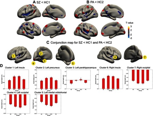
Figure S1 Cortical statistical maps displaying sulcal depth abnormalities in patients with schizophrenia (SZ) compared with young healthy controls for the SZ (HC1) (A), and in unaffected biological parents of patients (PA) compared with old healthy controls for the PA (HC2) (B), and the conjunction map for SZ<HC1 and PA<HC2 (C). (D) Mean ± SEM sulcal depth of the 7 clusters identified in the conjunction analysis. A and B show regions with uncorrected P<0.05. The colour bar indicates T-values.
1. Methods for cognitive assessments and scoring
Several commonly impaired cognitive abilities in schizophrenia were assessed in current study:1) speed of processing assessed with Trail Making Test, Part A (TMT) and Category Fluency Test: Animal Naming (CFT); 2) working memory; Digital Span (DS) forward and backward from the Wechsler Memory Scale-Chinese Revised (WMS-CR); 3) episodic memory: Logical Memory (LM) from the WMS-CR. Except for the TMT, higher rating scores indicated better cognitive abilities. The detailed methods of assessments and scoring were described as follows:
TMT:
Participants were presented some consecutive numbers that are arranged in irregular locations and were asked to draw a line connecting a sequence of 25 numbers in order. Participants need to finish it as quickly as possible while still maintaining accuracy and should not lift the pencil from the paper until they finish this task. We record the completion time. The test can provide information about speed of processing.
CFT:
Participants were asked to produce as many animal words as possible in 1 min. A higher number of correct answers reflects a faster processing speed.
DS:
For Digital Span forward, participants were asked to hear a sequence of numerical digits and tasked to recall the sequence correctly, with increasingly longer sequences being tested in each trial. For Digital Span backward, participants were asked to recall the sequence numbers in reverse order. Two trials for each sequence length are administered. Both trials of an item are administered even if the respondent passes the first trial.
LM:
Participants were presented auditorily two story passages and were asked to recall each story immediately after hearing it using as many of the same words of the original passage as they could remember. There are 50 gists (important story ideas units) in the two story passages. The gist recall was evaluated in each participant. The full score was 25 points (0.5 for each gist), then the raw score was converted to scale score according to converting table in the manual of WMS-CR, which was defined on the basis of the common model of Chinese adults.