

Abstract
Purpose
Infant resting-state networks do not exhibit the same connectivity patterns as those of young children and adults. Current theories of brain development emphasize developmental progression in regional and network specialization. We compared infant and adult functional connectivity, predicting that infants would exhibit less regional specificity and greater internetwork communication compared with adults.
Patients and methods
Functional magnetic resonance imaging at rest was acquired in 12 healthy, term infants and 17 adults. Resting-state networks were extracted, using independent components analysis, and the resulting components were then compared between the adult and infant groups.
Results
Adults exhibited stronger connectivity in the posterior cingulate cortex node of the default mode network, but infants had higher connectivity in medial prefrontal cortex/anterior cingulate cortex than adults. Adult connectivity was typically higher than infant connectivity within structures previously associated with the various networks, whereas infant connectivity was frequently higher outside of these structures. Internetwork communication was significantly higher in infants than in adults.
Conclusion
We interpret these findings as consistent with evidence suggesting that resting-state network development is associated with increasing spatial specificity, possibly reflecting the corresponding functional specialization of regions and their interconnections through experience.
Supplementary materials
Figure S1 Consistency of infant resting-state networks compared with adult template resting-state networks.
Notes: Combined independent components analysis, using both infant and adult subjects, resulted in networks that closely resembled networks from an independent components analysis using the infant subjects only. In both cases, many networks showed spatial distributions matching resting-state network templates.Citation1 (A) V1, (B) V2, (C) auditory, (D) sensorimotor, (E) basal ganglia, (F) precuneus, (G) visuospatial, (H) language, (I) left executive control, (J) right executive control, and (K) anterior salience.
Abbreviations: RSN, resting-state network; ICA, independent components analysis.
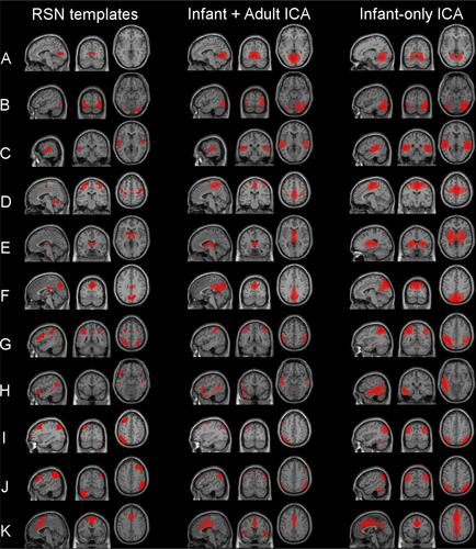
Table S1 Group differences in resting-state networks
References
- ShirerWRRyaliSRykhlevskaiaEMenonVGreiciusMDDecoding subject-driven cognitive states with whole-brain connectivity patternsCereb Cortex201222115816521616982
- Tzourio-MazoyerNLandeauBPapathanassiouDAutomated anatomical labeling of activations in SPM using a macroscopic anatomical parcellation of the MNI MRI single-subject brainNeuroimage200215127328911771995
Acknowledgments
Support for this research came from National Institutes of Health grants R01 MH082820, R01 MH081920, R01 DK089095, and P50 MH086383; the Brain Research Foundation; and the Blowitz-Ridgeway Foundation. The financial sponsors had no role in the design, data collection analysis, data interpretation, or writing of the manuscript or decision to submit the manuscript for publication.
Disclosure
The authors report no conflicts of interest in this work.