
Abstract
Purpose
The aim of this study was to examine the immunolocalization of VEGF-A and CD34, a marker of endothelial cells, in human conjunctival melanoma.
Methods
This study retrospectively analyzed primary conjunctival melanoma patients who underwent surgical resection of the tumor. All excised tissues were fixed with paraformaldehyde and embedded in paraffin, which were then submitted for immunohistochemistry with anti-VEGF and CD34 antibodies.
Results
The study sample comprised 4 female and two male melanoma patients. The age of the patients ranged from 64 to 84 (average age, 73) years. Histopathology of the surgically resected tumor tissues demonstrated accumulation of polygonal atypical malignant cells producing melanin. Cytoplasmic immunoreactivity for VEGF was clearly observed in tumor cells of all six tumors. In contrast, CD34-positive endothelial cells were less marked in the melanoma tissues than in the adjacent noncancerous subconjunctival stroma.
Conclusion
VEGF immunoreactivity was observed in conjunctival melanoma tissues, in which endothelial cells were hardly observed. These results suggest that although VEGF is expressed, conjunctival melanoma is a hypovascular tumor.
Introduction
Conjunctival melanoma is a life-threatening ocular surface tumor with worldwide incidence, although the tumor is rarely found in Japanese population. Conjunctival melanoma can arise from primary acquired melanosis (PAM), nevus, or de novo. Conjunctival melanoma has a 5-year survival rate of over 80%,Citation1 but can develop lymph node and distant metastases such as pulmonary and parotid gland metastases. We recently reported that adjuvant local chemotherapy using topical interferon alpha-2b resulted in preservation of globes in patients with conjunctival melanoma; however, not all patients showed good response to the local therapy.Citation2 Therefore, further additional treatment targets should be developed to save the patients’ vision and life. It has been reported that intratumoral lymphatic vessels were proliferative, which possibly correlated with tumor growth and a worse patients’ prognosis in conjunctival melanoma.Citation3
VEGF-A is a pleiotropic protein, which was historically identified as a tumor angiogenic factor. VEGF can be expressed and secreted from various tumor cells and the stroma, which stimulates endothelial cells and increases the number of microvessels in tumor tissues.Citation4 The increased microvessels are considered to provide oxygen to the tumor tissue and allow the tumor cells to escape from apoptosis. Recent studies have proved that VEGF induced not only tumor angiogenesis and vascular permeability but also tumorigenesis, the latter of which might be independent of angiogenesis.Citation4 It has been reported that cultured conjunctival melanoma cells express VEGF gene and protein;Citation5 however, little is known about the expression of VEGF and the formation of microvessels in human conjunctival melanoma.
The aim of this study is to examine VEGF immunolocalization and microvessel density in human conjunctival melanoma.
Materials and methods
This was a retrospective observational study. We enrolled patients with histology-proven primary conjunctival melanoma who had been treated in the Department of Ophthalmology, Hokkaido University Hospital, from March 2009 to November 2015, based on the medical records. The study sample comprised six patients who underwent local resection of tumor tissues. The tissues were resected with safety margins of 1–2 mm out of the elevated lesions based on no-touch technique.Citation6 Patients with histology-proven conjunctival nevus and PAM were excluded from the study. Each tumor was classified according to TNM classification of the American Joint Committee on Cancer (AJCC), eighth edition. This study was approved by the institutional review board (IRB) of Hokkaido University (IRB number: 015-0062) and was conducted in compliance with the Declaration of Helsinki. The protocol was described in the website of the hospital, and subjects were provided with the opportunity to opt out, and therefore, no new consent was required from the patients.
Histology and immunohistochemistry
The melanoma tissues were fixed with 4% paraformaldehyde immediately after surgical excision in the operating room. After the excised tumor tissues were embedded in paraffin, 5 µm-thick sections were cut. The slides were dewaxed, rehydrated, and rinsed in PBS twice for 10 minutes. Slides were submitted for H&E staining and immunohistochemistry. As a pretreatment for immunohistochemistry, microwave-based antigen retrieval was performed in 10 mM citrate buffer (pH 6.0) after demelanization by incubation with 1 g phosphoric acid in 100 mL of 3% hydrogen peroxide for 4 hours. These slides were immersed in 3% hydrogen peroxide for 10 minutes and then in normal goat serum for 30 minutes. Then, the sections were incubated with anti-VEGF (dilution 1:50, A-20; Santa Cruz Biotechnology Inc., Dallas, TX, USA) and anti-CD34 (dilution 1:50, M7165; Dako Japan Inc) antibodies at 4°C overnight. The secondary antibody reaction was carried out with anti-rabbit antibody (EnVision System HRP-labeled polymer; Dako Japan Inc) for 30 minutes at room temperature. Positive signals were visualized using HistoGreen (LINARIS; catalog number: E109) as a substrate. Slides were examined using a Keyence BZ-9000 (Keyence, Osaka, Japan) microscope. The tumor cells and the adjacent choroidal vessels of the enucleated eyes with choroidal melanomaCitation7 served as a positive control of VEGF and CD34, respectively.
Evaluation of immunohistochemical results
To evaluate VEGF expression, the number of tumor cells with cytoplasmic immunoreactivity was directly counted under light microscope at high magnification (objective ×40) in two or three fields in each section, which was then calculated as positive rate (%) of total tumor cells. Intratumoral microvessel density was calculated according to the previous reports.Citation8,Citation9 Briefly, the number of CD34-immunopositive microvessels in the tumor tissues was directly counted under light microscope (objective ×40). The counting was done in two or three high-power fields, and the numbers were then averaged to determine the microvessel density.
Results
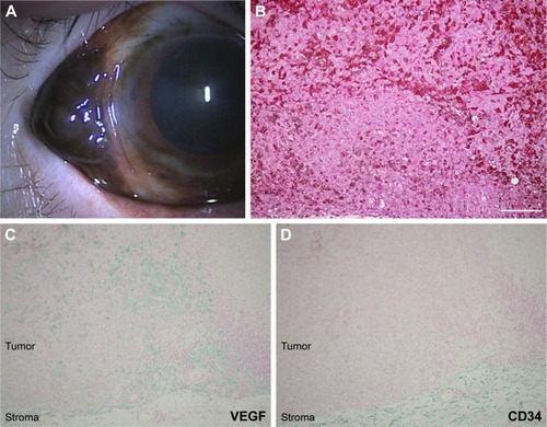
summarizes the clinicopathological features of conjunctival melanoma patients examined in this study. The sample comprised four female and two male melanoma patients. The age of the patients ranged from 65 to 84 (average age, 74) years. Slit lamp examination demonstrated a blackish nodule in the conjunctiva ( and ) in all the patients. Three tumors arose from PAM (), two from nevus (), and one from de novo. Histopathology of the surgically resected tumor tissues demonstrated accumulation of polygonal atypical malignant cells producing melanin ( and ). Cytoplasmic immunoreactivity for VEGF was clearly observed as greenish coloration in a variety of conjunctival melanoma cells ( and ). Four of the six cases showed a high VEGF-immunopositive rate of over 70% (Cases 1–4 in ). In contrast, the other two cases (Cases 5 and 6 in ) revealed a VEGF-immunopositive rate of about 30%, whereas none of the cases showed negative immunoreactivity for VEGF. In contrast, CD34-positive endothelial cells were less marked in the melanoma tissues ( and ). Intratumoral microvessel density was less than one vessel/mm2 in every case examined, while a variety of CD34-positive blood vessels existed in the adjacent noncancerous subconjunctival stroma ().
Table 1 Clinicopathological profile and VEGF-immunopositive rate of patients with conjunctival melanoma
Figure 1 A representative case of conjunctival melanoma in a 65-year-old female.
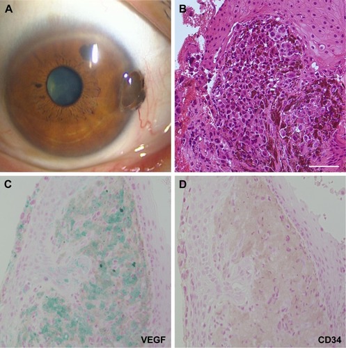
Figure 2 Another representative case of conjunctival melanoma in an 84-year-old female.
Discussion
This study, for the first time, demonstrated VEGF immunoreactivity in tumor cells of human conjunctival melanoma tissues. Our data are also consistent with the laboratory data showing VEGF expression in cultured melanoma cells derived from the conjunctiva.Citation5 However, it still remains controversial how the expressed VEGF pathologically correlates with angiogenesis in melanoma tissues. Simonetti et al showed that intratumoral microvessel density was correlated with increased VEGF expression in oral melanoma tissues.Citation10 On the other hand, Salven et al proved no association between intratumoral microvessel density and VEGF expression in primary and metastatic systemic melanoma tissues.Citation11 This study showed that the number of CD34-positive intratumoral microvessels was quite low in conjunctival melanoma tissues, which was less marked than adjacent normal conjunctiva. These data suggest that conjunctival melanoma is characterized by a hypovascular tumor, although VEGF immunoreactivity was detected in all the tissues examined.
Several authors have shown that VEGF is responsible for the tumor development and metastasis in patients with malignant melanoma.Citation11–Citation13 Recently, Wan et al demonstrated that genifitib, an inhibitor of epidermal growth factor, could suppress cultured melanoma cell proliferation through downregulation of VEGF gene and protein expressions.Citation14 However, it remains unknown whether VEGF-driven angiogenesis affects the tumor growth of melanoma. Although it is well known that VEGF leads to angiogenesis in various solid tumors, VEGF also plays an important role in tumorigenesis, which cannot be associated with pathological neovascularization.Citation4 Therefore, this study suggests that VEGF might be expressed during the tumorigenesis of human conjunctival melanoma, which is independent of tumor angiogenesis.
Recent studies have shown that VEGF is a promising therapeutic target for human diseases. In fact, bevacizumab and ranibizumab, two anti-VEGF agents, have been safely used for patients with neovascular age-related macular degeneration.Citation15,Citation16 Moreover, VEGF may hold a key to a novel therapeutic target for malignant melanoma. VEGF was certainly expressed in cultured uveal melanoma cells, and the cell growth was suppressed under bevacizumab and ranibizumab stimuli.Citation17,Citation18 Taken together, it is likely that anti-VEGF therapy can regulate not only neovascular diseases of the eye but also tumor cell proliferation in uveal melanoma cells. Corrie et al have shown that adjuvant anti-VEGF therapy (bevacizumab) contributed to improvement of disease-free survival in melanoma patients with a high risk of recurrence.Citation19,Citation20 In contrast, although anti-VEGF therapy might not be straightforward in contribution to regulation of primary uveal melanoma,Citation21,Citation22 these results suggest that anti-VEGF therapy might be considered one of the potential therapeutic strategies in patients with conjunctival melanoma.
There are limitations in this study. First, the number of samples examined is very small, since conjunctival melanoma is a rare malignant tumor in Japanese population, because of which, clinicopathological studies on this tumor have not progressed well. Second, although this study indicated that VEGF might play a role in tumorigenesis, VEGF expression could not be examined in premalignant lesions such as PAM and nevus. Further, molecular pathways regarding VEGF signaling are still uncertain. Therefore, further cell biological approaches are needed to examine VEGF receptors and the downstream molecular pathways in conjunctival melanoma as well as the premalignant lesions.
Disclosure
The authors report no conflicts of interest in this work.
References
- ParidaensADMinassianDCMccartneyACHungerfordJLPrognostic factors in primary malignant melanoma of the conjunctiva: a clinicopathological study of 256 casesBr J Ophthalmol19947842522598199108
- KikuchiIKaseSIshijimaKIshidaSLong-term follow-up of conjunctival melanoma treated with topical interferon alpha-2b eye drops as adjunctive therapy following surgical resectionGraefes Arch Clin Exp Ophthalmol2017255112271227628752368
- HeindlLMHofmann-RummeltCAdlerWPrognostic significance of tumor-associated lymphangiogenesis in malignant melanomas of the conjunctivaOphthalmology2011118122351236021835473
- GoelHLMercurioAMVEGF targets the tumour cellNat Rev Cancer2013131287188224263190
- RefaianNSchlerethSLKochKRComparing the hem- and lymphangiogenic profile of conjunctival and uveal melanoma cell linesInvest Ophthalmol Vis Sci20155695691569726313304
- ShieldsJAShieldsCLDe PotterPSurgical management of circumscribed conjunctival melanomasOphthalmic Plast Reconstr Surg19981432082159612814
- XuQZhaoGQZhaoJExpression and significance of factors related to angiogenesis in choroidal melanomaInt J Ophthalmol201141495422553608
- KaseSOsakiMHonjoSExpression of cyclo-oxygenase-2 is correlated with high intratumoral microvessel density and low apoptotic index in human esophageal squamous cell carcinomasVirchows Arch2003442212913512596063
- KinoshitaSKaseSAndoRExpression of vascular endothelial growth factor in human ocular adnexal lymphomaInvest Ophthalmol Vis Sci20145563461346724825110
- SimonettiOLucariniGRubiniCMicrovessel density and VEGF, HIF-1α expression in primary oral melanoma: correlation with prognosisOral Dis201319662062723279259
- SalvenPHeikkiläPJoensuuHEnhanced expression of vascular endothelial growth factor in metastatic melanomaBr J Cancer19977679309349328154
- UgurelSRapplGTilgenWReinholdUIncreased serum concentration of angiogenic factors in malignant melanoma patients correlates with tumor progression and survivalJ Clin Oncol200119257758311208853
- AsciertoPALeonardiEOttaianoANapolitanoMScalaSCastelloGPrognostic value of serum VEGF in melanoma patients: a pilot studyAnticancer Res20042464255425815736481
- WanXZhuYZhangLHouWGefitinib inhibits malignant melanoma cells through the VEGF/AKT signaling pathwayMol Med Rep20181757351735529568946
- MojaLLucenteforteEKwagKHSystemic safety of bevacizumab versus ranibizumab for neovascular age-related macular degenerationCochrane Database Syst Rev20149CD011230
- AbouammohMSharmaSRanibizumab versus bevacizumab for the treatment of neovascular age-related macular degenerationCurr Opin Ophthalmol201122315215821483262
- KochKRRefaianNHosDAutocrine impact of VEGF-A on uveal melanoma cellsInvest Ophthalmol Vis Sci20145542697270424677103
- LiJCuiYWangQThe proliferation of malignant melanoma cells could be inhibited by ranibizumab via antagonizing VEGF through VEGFR1Mol Vis20142064966024868139
- CorriePGMarshallANathanPDAVAST-M InvestigatorsAdjuvant bevacizumab for melanoma patients at high risk of recurrence: survival analysis of the AVAST-M trialAnn Oncol20182981843185230010756
- CorriePGMarshallADunnJAAdjuvant bevacizumab in patients with melanoma at high risk of recurrence (AVAST-M): preplanned interim results from a multicentre, open-label, randomised controlled phase 3 studyLancet Oncol201415662063024745696
- el FilaliMLyLVLuytenGPBevacizumab and intraocular tumors: an intriguing paradoxMol Vis2012182454246723077404
- el FilaliMMissottenGSMaatWRegulation of VEGF-A in uveal melanomaInvest Ophthalmol Vis Sci20105152329233720042655