Abstract
Background
To describe the morphology of a macular hole in the early postoperative period following vitrectomy with primary silicone oil tamponade.
Methods
A case report with optical coherence tomography (OCT) scans prior to surgery, at 20 minutes postoperatively and then at 17 hours postoperatively.
Results
OCT images of a 73-year-old woman with a stage 3 macular hole were obtained. At 20 minutes postoperatively, there was a reduction in intraretinal cysts and a reduction in macular hole size with elevated-open configuration. At 17 hours postoperatively, complete macular hole closure was noted.
Conclusion
OCT Images of a macular hole in the early postoperative period have been successfully obtained. Macular holes can close within 24 hours postoperatively and show morphological changes that may be predictive of closure within 20 minutes postoperatively.
Keywords:
Introduction
Macular hole closure is generally achieved within 3 days following macular hole surgery. If hole closure is not achieved within this period, the hole is unlikely to close without further surgical intervention.Citation1 However, it is unclear how macular holes behave within the first 24 hours postoperatively, with no published reports of findings during this time frame. We present a case where imaging with optical coherence tomography (OCT) was achieved in the early postoperative period and describe the morphological changes following macular hole surgery.
Case report
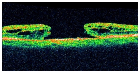
A 73-year-old female of Aboriginal origin from a sparsely populated area of Australia had travelled 4 hours by air to receive specialist assessment and treatment of her visual complaint. She presented with an 8-week history of decreased vision in her left eye. Best corrected visual acuity in the left eye was 20/100 and OCT imaging revealed a stage 3 macular hole ().
Figure 1 Preoperative – stage 3 macular hole.
Following discussion with the patient, and taking into consideration her desire to fly home as soon as possible, the decision was made to undertake macular hole surgery with silicone oil tamponade. Surgery was uncomplicated using a contact lens viewing system and consisted of transconjunctival sutureless 25-gauge vitrectomy with trypan blueassisted internal limiting membrane peel and 1000 centistokes silicone oil tamponade.
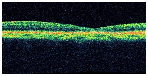
OCT images were taken 20 minutes following surgery. The images showed a persistent but reduced hole size, a decrease in intraretinal cysts, and the absence of subretinal fluid (). The configuration was elevated-open in all sections. Further OCT images were taken at 17 hours and showed hole closure in all image sections with restoration of relatively normal foveal contour ().
Figure 2 20 minutes postoperative – reduction in hole size (elevated-open configuration) with decrease in intraretinal cysts and absence of subretinal fluid.
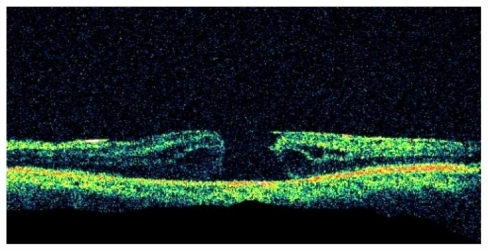
Figure 3 17 hours postoperative – closed hole with restoration of foveal contour.
Discussion
Current treatment of macular holes involves pars plana vitrectomy, intraocular gas tamponade, and face-down posturing. However, our patient preferred to fly home following surgery. Based on this decision and appropriate counseling, the patient was offered an oil tamponade rather than a gas tamponade as the decreased atmospheric pressure during air travel increases the risk of gas expansion, and ultimately increases intraocular pressure. Using primary oil tamponade in macular hole surgery is not a novel technique, and has often been used in those patients who cannot lie face down.Citation2 It also has the advantage of allowing fundus examination and OCT imaging in the early postoperative period with commercially available systems.
Vertical OCT scanning with the patient in a prone position has been used previously to image postoperative macular holes in gas-filled eyes.Citation1 Such a system, however, is not commercially available and to the best of our knowledge, we are not aware of early images being available.
This case displayed rapid macular hole closure. Using OCT, it was possible to observe a reduction in intraretinal cysts and an elevated-open configuration within 20 minutes postoperatively. This reduction in retinal cysts and elevated-open configuration has been proposed as an effective predictor of macular hole closure.Citation3 Complete macular hole closure was achieved at some point between 20 minutes and 17 hours following surgery.
In conclusion, we have successfully imaged a macular hole in the early postoperative period. Our case highlights that macular holes can close well within 24 hours after surgery and show morphological changes which may be predictive of closure within 20 minutes.
Disclosures
The authors report no conflicts of interest in this work. The paper was presented at the Royal Australian and New Zealand College of Ophthalmologists scientif ic conference in Adelaide, Australia in November, 2010.
References
- EckardtCEckertTEckardtUPorkertUGesserCMacular hole surgery with air tamponade and optical coherence tomography-based duration of face-down positioningRetina20082881087109618779715
- GoldbaumMHMcCuenBWHannekenAMBurgessSKChenHHSilicone oil tamponade to seal macular holes without position restrictionsOphthalmology19981051121402147 discussion 2147–21489818619
- HottaKEarly postoperative macular features determined by optical coherence tomography after idiopathic macular hole surgery with silicone oil tamponadeOphthalmic Surg Lasers Imaging200536542643116238045