Abstract
Plateau iris syndrome has been described as persistent angle narrowing or occlusion with intraocular pressure elevation after peripheral iridotomy due to the abnormal plateau iris configuration. Argon laser peripheral iridoplasty (ALPI) is an effective adjunct procedure to treat plateau iris syndrome. Classic theory suggests that the laser causes the contraction of the far peripheral iris stroma, “pulls” the iris away from the angle, and relieves the iris-angle apposition. We report a case of plateau iris syndrome that was successfully treated with ALPI. Spectral domain optical coherence tomography confirmed the angle was open at areas with laser treatment but remained appositionally closed at untreated areas. Further analysis suggested significant cross-sectional thinning of the iris at laser-treated areas in comparison with untreated areas. The findings indicate that APLI opens the angle, not only by contracting the iris stroma, but also by thinning the iris tissue at the crowded angle. This is consistent with the ALPI technique to aim at the iris as far peripheral as possible. This case also suggests that spectral domain optical coherence tomography is a useful adjunct imaging tool to gonioscopy in assessing the angle condition.
Introduction
Plateau iris syndrome is a unique subtype of angle closure glaucoma. It is commonly caused by anteriorly positioned ciliary processes that push the peripheral iris forward, resulting in angle closure. The diagnosis is always based on gonioscopic findings.Citation1,Citation2 Argon laser peripheral iridoplasty (ALPI) has been suggested to be useful in treating this condition by “stretching” the peripheral iris.Citation3,Citation4 High resolution anterior segment spectral domain optical coherence tomography (SD-OCT) is a very useful technology that can detect subtle structural changes in the iris and anterior chamber angles.Citation5 The purpose of this study is to evaluate the mechanism of ALPI in treating plateau iris by anterior segment SD-OCT.
Case report
A 28-year-old Caucasian woman with a history of narrow angles in both eyes, who had bilateral laser peripheral iridotomies one year ago, presented with acute angle closure. Her presenting intraocular pressure was 26 mmHg right eye and 31 mmHg left eye. Gonioscopic exam revealed appositionally closed angles, with steeply elevated far peripheral iris configuration and double hump signs by indentation. High-frequency ultrasound biomicroscopy confirmed the anterior location of the ciliary processes with incidental findings of one iris cyst in each eye. ALPI was performed in both eyes. Postlaser intraocular pressure was reduced to 10 mmHg right eye and 12 mmHg left eye. Anterior segment SD-OCT images were taken at 3, 6, 9, and 12 o’clock of the angle of each eye before and after laser iridoplasty. The anterior segment SD-OCT demonstrated closed angles, even with patent iridotomies. Gonioscopic exam after ALPI revealed angles were open to scleral spur in the areas of iridoplasty but remained appositionally closed in the untreated areas. The anterior segment SD-OCT taken one month after ALPI detected focal iris thinning in the areas which underwent laser treatment (). Iris cross-sectional thickness was 278 μm on average in the iridoplasty-treated areas compared with 315 μm in the closed, untreated areas (). (Student’s t-test, P < 0.001).
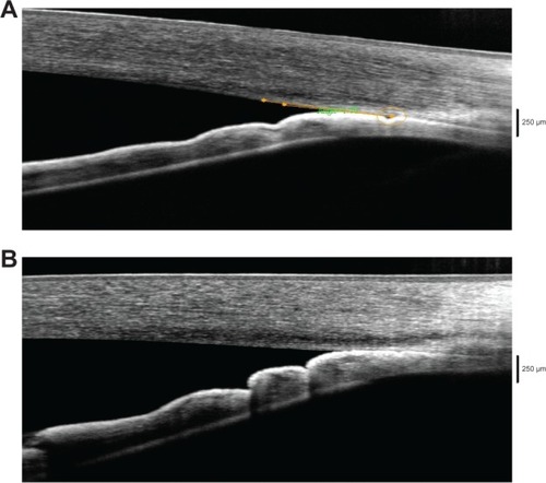
Figure 1 Angles open after laser iridoplasty at treated area.
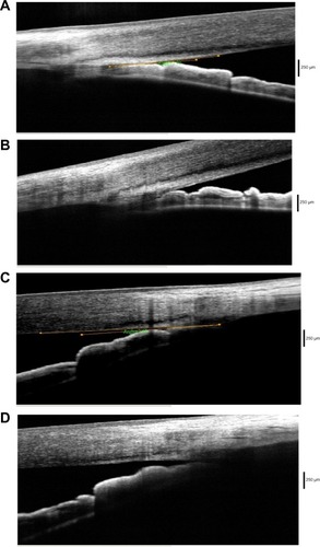
Figure 2 Angle remains closed after laser iridoplasty at untreated area.
Discussion
Anterior segment imaging is a useful adjunct tool to gonioscopy for the evaluation of angles.Citation2,Citation5–Citation8 SD-OCT is a noncontact imaging test that is relatively easy to perform. It provides high resolution images of iris contour and angle configurations when properly performed. Compared to the widely used time-domain OCT, SD-OCT can better reveal details of angle structures such as Schlemn’s canal.Citation5 In our patient, SD-OCT clearly showed the change of angles before and after laser peripheral iridoplasty, which was consistent with gonioscopic findings. It can serve as a convenient adjunct tool in addition to the gonioscopy in the diagnosis and follow-up treatment for plateau iris syndrome. Because both mydriasis and miosis can affect iris shape, volume, cross-sectional thickness and alter the measurements,Citation9–Citation12 it is important to obtain images with the same pupillary dilation status. All our images, before and after ALPI, were taken under the same lighting conditions with pupils undilated. Though plateau iris with ALPI imaging was demonstrated previously with time-domain OCT,Citation13,Citation14 this is the first report that attempts to illustrate the mechanism of ALPI in treating plateau iris syndrome by using SD-OCT.
The traditional theory of ALPI in treating plateau iris is that the laser causes iris tissue shrinking, which stretches peripheral iris tissue away from the anterior chamber angle.Citation3,Citation4 The angle OCT in this patient clearly showed that the “thinning” effect on the peripheral iris tissue also plays an important role to open the angle. This finding can also explain why the ALPI always works best when the laser spots are placed as peripherally as possible.
Conclusion
Plateau iris is a unique subtype of angle closure glaucoma. It is commonly caused by anteriorly positioned ciliary processes that push the peripheral iris forward, resulting in angle closure. Anterior segment SD-OCT demonstrates iris cross-sectional thinning as the mechanism responsible for opening the angle in plateau iris angle closure treated by laser iridoplasty.
Disclosure
The authors report no conflicts of interest in this work.
References
- ChengJBuysYMSpaethGLConfusion with the misuse of plateau iris terminologyJ Glaucoma201322326526623429628
- KiuchiYKanamotoTNakamuraTDouble hump sign in indentation gonioscopy is correlated with presence of plateau iris configuration regardless of patent iridotomyJ Glaucoma200918216116419225356
- RitchRLiebmannJMLaser iridotomy and peripheral iridoplastyRitchRShieldsMBKrupinTThe Glaucomas2nd edSt Louis, MOMosby199615491573
- RitchRThamCCLamDSArgon laser peripheral iridoplasty (ALPI): an updateSurv Ophthalmol200752327928817472803
- DayACGarway-HeathDFBroadwayDCSpectral domain optical coherence tomography imaging of the aqueous outflow structures in normal participants of the EPIC-Norfolk Eye StudyBr J Ophthalmol201397218919523203701
- MandellMAPavlinCJWeisbrodDJSimpsonERAnterior chamber depth in plateau iris syndrome and pupillary block as measured by ultrasound biomicroscopyAm J Ophthalmol2003136590090314597043
- LiuLDeconstructing the mechanisms of angle closure with anterior segment optical coherence tomographyClin Experiment Ophthalmol201139761462221707891
- ShabanaNAquinoMCSeeJQuantitative evaluation of anterior chamber parameters using anterior segment optical coherence tomography in primary angle closure mechanismsClin Experiment Ophthalmol201240879280122594402
- QuigleyHASilverDMFriedmanDSIris cross-sectional area decreases with pupil dilation and its dynamic behavior is a risk factor in angle closureJ Glaucoma200918317317919295366
- AptelFDenisPOptical coherence tomography quantitative analysis of iris volume changes after pharmacologic mydriasisOphthalmology2010117131019923002
- AminiRWhitcombJEAl-QaisiMKThe posterior location of the dilator muscle induces anterior iris bowing during dilation, even in the absence of pupillary blockInvest Ophthalmol Vis Sci20125331188119422281822
- JouzdaniSAminiRBarocasVHContribution of different anatomical and physiologic factors to iris contour and anterior chamber angle changes during pupil dilation: theoretical analysisInvest Ophthalmol Vis Sci20135442977298423482467
- LeungCKChanWMKoCYVisualization of anterior chamber angle dynamics using optical coherence tomographyOphthalmology2005112698098415936438
- Kalev-LandoyMDayACCordeiroMFMigdalCOptical coherence tomography in anterior segment imagingActa Ophthalmol Scand200785442743017355288