
Abstract
Purpose
Here, we present two patients with congenital anterior staphyloma, with mutations in the CYP1B1 gene.
Methods
We reviewed the medical records, including the genetic analysis.
Results
Two unrelated patients presented with congenital anterior staphylomas. Both patients showed mutations in the CYP1B1 gene. The first patient, the product of a consanguineous marriage, showed a homozygous misssense mutation g.3987G>A (p.G61E). The second patient had compound heterozygous misssense mutations [g.4160 G>T (p.A119S) and g.8131 C>G (p.L432V)].
Conclusion
CYP1B1 gene mutation may be associated with congenital anterior staphylomas.
Introduction
Congenital anterior staphyloma is a rare condition characterized by enlarged, opaque, ectatic corneas that protrude through the interpalpebral fissure. This congenital malformation has been described in isolated case reports, but the etiology remains unclear.Citation1 In this paper, we report two cases of congenital staphyloma that were associated with CYP1B1 gene mutations.
Case reports
The history, clinical findings at presentation, the clinical course, and findings after genetic testing are summarized in and illustrated in and .
Figure 1 Patient 1.
Abbreviations: MRI, magnetic resonance imaging; UBM, ultrasound biomicroscopy.
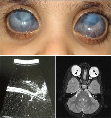
Figure 2 Patient 2.
Abbreviations: MRI, magnetic resonance imaging; UBM, ultrasound biomicroscopy.
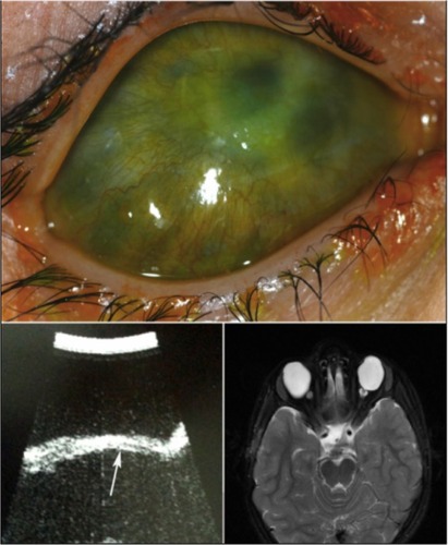
Table 1 Clinical features and course of patients with congenital anterior staphylomas
Discussion
Congenital anterior staphyloma is a rare unilateral or bilateral anomaly and may account for about 11% of congenital corneal opacities.Citation2 The clinical findings include an enlarged, vascularized, ectatic cornea, with thinning and ectasia of the surrounding anterior segment structures. These findings are associated with marked anterior segment anomalies that can include an iris that is adherent to the posterior corneal surface or a partially absent iris. The crystalline lens may be adherent to the posterior corneal surface, similar to Peters anomaly, be subluxed, or show cataractous changes.Citation1,Citation3–Citation5 The degree of staphylomatous changes can be variable. In our patients, the corneal ectasia was not as conspicuous, with the staphylomatous changes being more prominent in the limbal region. Limbal staphylomas can occur due to stretching and thinning of the globe, as a result of high intraocular pressure in severe congenital glaucoma. The clinical course in such patients is progressive, often requiring multiple surgical interventions. In contrast, our patients presented at birth with severe anterior segment changes that remained stable and mild-to-moderate intraocular pressure elevations that appeared controlled with medications. In the differential diagnosis, the pathologies considered included severe primary congenital glaucoma with anterior segment dysgenesis and Peters anomaly.Citation2 There seems to be considerable overlap in the clinical features between these clinical conditions.Citation6 Though the presenting conditions could be considered as a severe form of anterior segment dysgenesis, it was felt that the term was generic since both patients had phenotypic features that were more consistent with those seen in congenital anterior staphylomas. Peters anomaly presents with central corneal opacities with thinning, and with iris adhesions to the posterior surface. Corneal ectasia, staphylomas, and adherence of the crystalline lens have been reported in cases of Peters Plus syndrome with multiple systemic abnormalities.Citation7,Citation8 Both patients in this study showed corneal opacification, ectasia, and vascularization. In addition, our first patient had a dislocated crystalline lenses and the second patient had had congenital aphakia, both of which have been described with Peters anomaly and congenital anterior staphylomas.Citation9,Citation10
Multiple etiological factors have been implicated in the pathogenesis of congenital anterior staphylomas. These include intrauterine infections and specific chromosomal abnormalities with multisystem organ involvement, and an association with amniotic band syndrome has been reported.Citation3,Citation9,Citation11 However, in most cases, the etiology remains undetermined. Both our patients had no family history; however, patient 1 was the product of a consanguineous marriage, alerting us to further look into the association of CYP1B1 mutations with the anterior staphylomas.
We detected a homozygous CYP1B1 mutation g.3987G>A (p.G61E) in patient 1. This mutation is the most common mutation in Saudi Arabian patients with primary congenital glaucoma.Citation12 The mutation was inherited in a homozygous status and was heterozygous in both parents, thus fits well with the fact that both parents were first cousins. A previous study attempting to correlate this mutation with a particular phenotype was unsuccessful.Citation11 This mutation was previously described in Saudi patients with familial juvenile open-angle glaucoma,Citation13 in Peters anomaly,Citation14 and in isolated open-angle glaucoma.Citation15
As for patient 2, he carried compound heterozygous mutations, g.4160 G>T (p.A119S) and g.8131 C>G (p.L432V), which were inherited from his father. Unfortunately, deoxyribonucleic acid (DNA) was not available from the mother, but it is likely that she harbored the other mutation detected in her son. This was consistent with the history that both parents were not related. Both mutations (p.A119S and p.L432V) are less common than the p.G51E, and mutational phenotypes have not been described with the phenotype seen in the two affected patients. Two CYP1B1 mutations detected in our patients (p.G61E and p.A1195) had an established pathogenicity.Citation12 As for the L432V, the literature is inconclusive about its pathogenic role as this mutation has been detected in normal Turkish and Japanese populations, in a heterozygous status with no other mutation in the CYP1B1 gene.Citation16 It is plausible to suggest that this mutation, if inherited in a heterozygous status with no other CYP1B1 mutation, may not cause the development of congenital glaucoma. However, if this mutation is inherited in a homozygous status or in a heterozygous status with another CYP1B1 mutation, then it may be capable of causing the disease.
Homozygous/compound heterozygous mutations in the CYP1B1 gene are typically associated with primary congenital glaucoma.Citation17 Several reports have identified CYP1B1 mutations in patients with other phenotypes, such as isolated Peters or Axenfeld-Rieger anomaly. These phenotypes are typically associated with glaucoma.Citation14,Citation18 Homozygous or compound heterozygous CYP1B1 mutations were identified in eight probands with mild ectropion uvea, partial aniridia, and congenital glaucoma,Citation19 and now this report further expands the ocular phenotype associated with CYP1B1 mutations.
Disclosure
The authors report no conflicts of interest in this work.
References
- LunardelliPMatayoshiSCongenital Anterior StaphylomaJ Pediatr Ophthalmol Strabismus Epub6252009
- ShigeyasuCYamadaMMizunoYYokoiTNishinaSAzumaNClinical features of anterior segment dysgenesis associated with congenital corneal opacitiesCornea201231329329822157569
- StuartJACongenital anterior staphylomaSurv Ophthalmol19635439339514057429
- MillerMMButrusSHidayatAWeiLLPontigoMCorneoscleral transplantation in congenital corneal staphyloma and Peters’ anomalyOphthalmic Genet2003241596312660867
- MullaneyPBRiscoJMHeinzGWCongenital corneal staphylomaArch Ophthalmol19951139120612077661758
- VerschootenRFoetsBDe RavelTVan GinderdeurenRLombaertsRCasteelsIClinical spectrum of congenital corneal staphyloma: a case reportBull Soc Belge Ophtalmol201131871022003758
- ShimizuRSaitoRHoshinoKSevere Peters Plus syndrome-like phenotype with anterior eye staphyloma and hypoplastic left heart syndrome: proposal of a new syndromeCongenit Anom (Kyoto)201050319719920584037
- EberweinPReinhardTAgostiniHPoloschekCMGuthoffRAuw-HaedrichCIntensive intracorneal keloid formation in a case of Peters plus syndrome and in Peters anomaly with maximum manifestationOphthalmologe20101072178181 German19756642
- SchrammCRohrbachJMReinertSAmniotic bands as a cause of congenital anterior staphylomaGraefes Arch Clin Exp Ophthalmol2013251395996523150045
- TrabucchiGPiantanidaABandelloFFreschiMNucciPBrancatoRCongenital aphakia in Peters’ anomaly syndrome. A case reportActa Ophthalmol Scand19977555955979469564
- BessantDAAnwarKKhaliqSPhenotype of autosomal recessive congenital microphthalmia mapping to chromosome 14q32Br J Ophthalmol199983891992210413693
- Abu-AmeroKKOsmanEAMousaAScreening of CYP1B1 and LTBP2 genes in Saudi families with primary congenital glaucoma: genotype-phenotype correlationMol Vis2011172911291922128238
- KhanAOAl-AbdiLMohamedJYAldahmeshMAAlkurayaFSFamilial juvenile glaucoma with underlying homozygous p.G61E CYP1B1 mutationsJ AAPOS201115219819921596299
- EdwardDAl RajhiALewisRACurrySWangZBejjaniBMolecular basis of Peters anomaly in Saudi ArabiaOphthalmic Genet200425425727015621878
- López-GarridoMPSánchez-SánchezFLópez-MartínezFHeterozygous CYP1B1 gene mutations in Spanish patients with primary open-angle glaucomaMol Vis20061274875516862072
- VasiliouVGonzalezFJRole of CYP1B1 in glaucomaAnnu Rev Pharmacol Toxicol20084833335817914928
- StoilovIAkarsuANSarfaraziMIdentification of three different truncating mutations in cytochrome P4501B1 (CYP1B1) as the principal cause of primary congenital glaucoma (Buphthalmos) in families linked to the GLC3A locus on chromosome 2p21Hum Mol Genet1997646416479097971
- TanwarMDadaTDadaRAxenfeld-Rieger Syndrome Associated with Congenital Glaucoma and Cytochrome P4501B1 Gene MutationsCase Rep Med20102010
- KhanAOAldahmeshMAAl-AbdiLMolecular characterization of newborn glaucoma including a distinct aniridic phenotypeOphthalmic Genet201132313814221306220