
Abstract
Glaucoma drainage device implantation is efficacious for the treatment of pediatric glaucoma patients when multiple angle surgeries fail. However, tube touching of the corneal endothelium is one of the major postoperative complications to deal with. A 15-month-old male patient with Wilms’ tumor, aniridia, genitourinary anomalies, and mental retardation (WAGR) syndrome was diagnosed with bilateral developmental glaucoma. He underwent Baerveldt glaucoma implant (BGI) surgeries in both eyes after multiple failed trabeculotomies. The tube in his right eye was touching the cornea 15 months after BGI surgery. To avoid corneal endothelium damage, BGI tube repositioning with scleral fixation was performed without serious complications. The bilateral BGI surgeries achieved successful intraocular pressure reduction for over 2 years and tube repositioning with scleral fixation of BGI tube was successful for BGI tube malposition. Although careful attention to intraocular pressure and tube malposition is essential after glaucoma drainage device implantation, especially in pediatric cases, BGI surgery is effective in the management of developmental glaucoma following unsuccessful angle surgeries.
Introduction
In eyes with developmental glaucoma, surgical therapies such as trabeculotomy or goniotomy are usually essential to control intraocular pressure (IOP). Although these angle surgeries have high success rates in primary developmental glaucoma,Citation1,Citation2 secondary developmental glaucoma, with associated ocular/systemic anomalies, including aniridia, has a poor prognosis, probably because of maldevelopment of the angle structures and Schlemm’s canal.Citation1,Citation2 Wilms’ tumor, aniridia, genitourinary anomalies, and mental retardation (WAGR) syndrome is caused by heterozygous contiguous gene deletions that involve at least PAX6 and Wt1, which has a risk of a developmental glaucoma.Citation3
Even when the first trabeculotomy is unsuccessful, a subsequent trabeculotomy may be effective in lowering IOP.Citation1 However, when multiple angle surgeries fail, further surgical procedures such as trabeculectomy with mitomycin-C, cyclodestructive surgery, and glaucoma drainage device (GDD) implantation are needed.Citation2,Citation4 Trabeculectomy in younger children has many problems: a rapid healing response, postoperative bleb management, and a high rate of bleb-associated endophthalmitis.Citation2 Cyclodestructive procedures require multiple retreatments and have a possible risk for a serious complication, phthisis bulbi. It has been reported that the success rate is significantly higher after Ahmed glaucoma valve or Baerveldt glaucoma implant (BGI) (87%) versus trabeculectomy with mitomycin-C (36%) in children.Citation4 GDD implantation, such as BGI, is suggested as the most predictable and possibly the safest procedure after failed angle surgery.
Here, we report a case of WAGR syndrome in association with developmental glaucoma requiring bilateral BGIs and subsequent tube repositioning. This is the first report of tube repositioning with scleral fixation of BGI tube in a pediatric case.
Case report
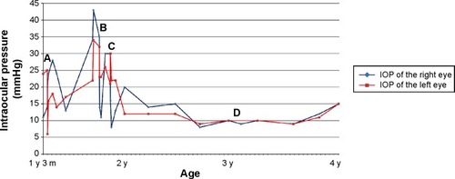
A 15-month-old male patient was referred to Kyoto University Hospital. He was diagnosed with bilateral (OU) aniridia and developmental glaucoma in his right eye (OD) at 1 month of age and subsequently with WAGR syndrome. He had undergone three OD trabeculotomies before 1 year of age. At the initial visit to our hospital, he was using three types of glaucoma eyedrops; his IOP was 11 mmHg OD and 24 mmHg in the left eye (OS). Fundus examination showed macular hypoplasia OU; a tilted disc with 0.9 cup to disc ratio OD; and an oval disc with 0.6 cup to disc ratio OS. and show pre- and postoperative photographs and IOP throughout the follow-up period, respectively. Briefly, after three failed OD and two failed OS trabeculotomies, BGI surgeries were performed in both eyes.
Figure 1 Pre- and postoperative photographs.
Abbreviation: BGI, Baerveldt glaucoma implant.
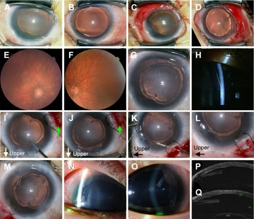
Figure 2 The intraocular pressure (IOP) time course.
Abbreviations: BGI, Baerveldt glaucoma implant; y, year(s); m, month(s).
A BGI103-250 implant was placed in the superior temporal quadrant with a fornix based conjunctival flap (). The plate was secured with 9-0 nylon sutures 9 mm posterior to the limbus. The tube was ligated with 7-0 Vicryl and was slit to manage the transient hypertensive phase. The tube was placed at a level intermediate between the cornea and the lens, because both tube-cornea and tube-lens touch should be prevented. Preserved donor sclera was used to cover the tube.
The IOPs (OU) became stable 2–3 months after the surgeries () and the optic disc findings improved (). The tube positions OU were at the level intermediate between the cornea and the lens until 1 year postoperative. The OD tube was touching the cornea 15 months after BGI surgery (). To avoid corneal endothelium damage, BGI tube repositioning with scleral fixation was performed as previously described.Citation5 Briefly, a conjunctival incision and triangular-shaped scleral flap were made at the inferior temporal area (, green arrows). The end of the tube was pulled out through the corneal side port punctured immediately above the tube using capsulorhexis forceps (). A double-armed 10-0 Prolene straight needle bent using two needle-holders penetrated through the tube end and pulled under the scleral flap using the 27-gauge leading needle (). The second needle with a 10-0 Prolene was also pulled under the scleral flap and the tube end was extended parallel to the corneal surface (). The scleral flap was sutured with a 10-0 nylon suture and the incised conjunctiva was closed with 8-0 Vicryl sutures. After the procedure, the patient maintained good tube positioning OD and good IOP without serious complications (). The tube OS has not shown tube touching of the corneal endothelium without tube repositioning so far ().
Discussion
Tube positioning in the anterior chamber is especially important in pediatric cases due to the growth of the eyeball. Because tube retraction is a possible postoperative complication in pediatric GDD surgeries, we left a longer tube in the anterior chamber than in adults (). Tube touching of the corneal endothelium is another postoperative complication. Beck et alCitation4 reported that tube repositioning was needed in 34.8% of 46 GDD-implanted pediatric eyes. Cutting the tube end or repositioning through a new scleral pathway is a possible choice. However, cutting the tube would be palliative, because it would not change the tube insertion angle. Reopening the conjunctiva and sclera for removal and tube reinsertion through a new pathway might cause insufficient wound closure and disturb the function of the BGI. Our choice was a novel technique of repositioning the tube with scleral fixation, which Ma et alCitation5 reported in adult patients.
The bilateral BGI surgeries achieved successful IOP reduction for over 2 years. This is the first report of tube repositioning with scleral fixation of BGI tube in a pediatric case. This procedure was good for BGI tube malposition even in a pediatric case, at least for a short period. Although careful attention to IOP and tube malposition is essential after GDD implantation, especially in pediatric cases, BGI surgery is effective in the management of developmental glaucoma following unsuccessful angle surgeries.
Disclosure
The authors report no conflicts of interest in this work.
References
- IkedaHIshigookaHMutoTTaniharaHNagataMLong-term outcome of trabeculotomy for the treatment of developmental glaucomaArch Ophthalmol200412281122112815302651
- OuYCaprioliJSurgical management of pediatric glaucomaDev Ophthalmol20125015717222517182
- HanJCLiuQRJonesMBrain-derived neurotrophic factor and obesity in the WAGR syndromeN Engl J Med2008359991892718753648
- BeckADFreedmanSKammerJJinJAqueous shunt devices compared with trabeculectomy with Mitomycin-C for children in the first two years of lifeAm J Ophthalmol20031366994100014644208
- MaKTKimJHSeongGJJangDSKimCYScleral fixation of Ahmed glaucoma valve tube tip for adjustment of cornea-touching malpositionEye (Lond)2014281232524097119