
Abstract
Background
MicroRNA (miRNA) expression is correlated with tumor histology, differentiation, invasiveness and treatment outcome. We aimed to identify miRNAs whose differential expression might enable early diagnosis of lung adenocarcinoma in patients presenting with ground-glass nodules (GGNs).
Methods
To identify potential miRNAs of interest, we analyzed the miRNA expression profile of tumor and adjacent non-para-tumor tissue in three participants by next-generation sequencing (NGS). We then assessed the expression levels of the miRNAs of interest in 73 lung adenocarcinomas presenting with GGNs with matched adjacent non-tumor tissue by quantitative real-time polymerase chain reaction (qRT-PCR). We also detected the miRNA panel in 66 lung benign diseases and 66 lung adenocarcinomas presenting with GGN lesion tissues by qRT-PCR. Target genes of our selected miRNA panel were predicted using Miranda with default parameters.
Results
Twenty-three miRNAs showed differential expression between tumor and adjacent non-tumor tissue by NGS. Five miRNAs exhibited higher expression in tumor tissue compared to adjacent non-tumor tissue (P<0.05); 18 miRNAs demonstrated lower expression in tumor tissue versus adjacent non-tumor tissue (P<0.05). When qRT-PCR was performed for the 23 miRNAs identified by NGS in the pilot stage, seven were found to have statistically significant expression in tumor versus adjacent non-tumor tissue (P<0.05). The sensitivity and specificity of seven-miRNA panel were 86.4% and 60.6%, respectively.
Conclusion
The predicted targets of our miRNAs of interest are frequently associated with cancer signaling pathways. We developed a miRNA panel that could potentially predict the presence of lung adenocarcinoma in patients presenting with GGNs.
Introduction
Lung cancer is one of the leading causes of cancer deaths worldwide. Adenocarcinoma represents over 40% of diagnosed lung cancer cases.Citation1,Citation2 At least in part due to the lack of an effective method for early diagnosis, lung adenocarcinoma is usually locally advanced or metastatic when discovered, and the prognosis is poor (5-year survival rate of 15%).Citation2 Therefore, an effective, dependable, non-invasive tool for the early diagnosis of lung cancer would go very far to improve the prognosis and reduce the mortality of patients.
On computed tomography (CT) scan images, pulmonary ground-glass nodules (GGNs) appear as hazy lesions overlying bronchial structures or pulmonary vessels.Citation3 The presence of GGNs, which are being detected with increasing frequency, is strongly suggestive of lung cancer, particularly lung adenocarcinoma. However, GGNs can also represent an entirely benign process.Citation4 The clinical management of pulmonary GGNs remains controversial and complex. There is a crucial need for better biomarkers to identify which patients presenting with GGNs are at high risk for harboring lung adenocarcinoma and therefore require aggressive and speedy intervention to increase the likelihood of successful treatment.
MicroRNAs (miRNAs) are a category of small non-coding RNAs between 18 and 24 nucleotides in lengthCitation5 that play a role in various pathologic processes including oncogenesis and tumor metastasis. Abnormal miRNA expression and function have been found in multiple human cancers. MiRNAs have the potential both as biomarkers and new drug targets for cancer patients.Citation6–Citation8 For example, differential expression of the precursors of miRNA-155, mature miRNA-15 and miRNA-16 was detected in malignant lymphomas.Citation7 Furthermore, Yanaihara et alCitation10 found 43 aberrantly expressed miRNAs in a comparison of 104 paired primary lung cancer and non-tumor tissues. In the lung cancer samples, expression levels of 28 miRNAs were lower and 15 miRNAs were higher, demonstrating that the comprehensive analysis of miRNA expression can identify miRNAs associated with cancer.
Next-generation sequencing (NGS) is an effective tool for miRNA analysis that quantifies miRNA levels by detecting the exact nucleotide sequence. MiRNA sequencing data for lung cancer has been reported in several studies.Citation6–Citation9 However, this is the first study to report the use of NGS to systematically characterize miRNAs associated with the presence of adenocarcinoma in pulmonary GGNs, which could facilitate earlier diagnosis and more successful treatment of patients with lung cancer.
In this study, we investigated aberrations of miRNAs in resected GGNs to design a miRNA panel that predicts lung adenocarcinoma in patients presenting with GGNs.
Materials and methods
Patients and clinical specimens
Patients enrolled in the study were diagnosed with lung adenocarcinoma and had one or more intrapulmonary pure or partially solid GGNs. In total, 76 pairs of tumor samples from adenocarcinomas located in GGNs and adjacent non-tumor samples were surgically resected from the enrolled patients at Shanghai Pulmonary Hospital between May 2013 and June 2015. A total of 66 lung benign diseases and 66 lung adenocarcinomas presenting with GGNs lesion tissues were surgically resected from the enrolled patients at Shanghai Pulmonary Hospital between September 2014 and June 2015. Immediately following resection, fresh tissue samples were flash frozen with dry ice and then stored at −80°C. All clinical investigations were performed in accordance with the tenets of the Declaration of Helsinki. All participants provided written informed consent to participate prior to inclusion in the study. The protocol was approved by the Shanghai Pulmonary Hospital, Tongji University (issue number 13-786, ethical number 13-768). All participants were competent to provide their consent.
Next-generation sequencing
MiRNA expression in both tumor and adjacent non-tumor tissue was evaluated by NGS in three patients to identify whether it is higher or lower in tumor. Extraction of total RNA was performed by TRIzol solution (Thermo Fisher Scientific), and the RNA quality was measured by Agilent BioAnalyzer2100. To achieve optimal tissue miRNA profiles, we carried out high-throughput NGS. The adaptor sequence was cut by the primer. After the quality inspection and the length screening, the basic sequencing fragments were selected. Subsequently, miRNAs were reverse transcribed and sequenced using a single-ended 50 bp sequencing model on the platform.
To identify miRNAs differentially expressed between lung cancer tissues and normal controls, we applied transcripts per million (TPM) to normalize the expression of miRNA in two groups. Then we calculated fold change (FC) and P-value via double sample t-test, and corrected P-value into false discovery rate (FDR). FDR ≤0.05 and |log2FC| ≥2 were set as the cutoffs to screen out differentially expressed miRNAs.
Validation of the NGS by qRT-PCR
The subset of miRNAs found to be differentially expressed in tumor versus non-tumor analysis by NGS was assayed by quantitative real-time polymerase chain reaction (qRT-PCR) in 73 lung adenocarcinoma patients who presented with GGNs. Total RNA in tumor and adjacent non-tumor tissue was extracted with an RNA Extraction Kit (SLNco, Cinoasia, Shanghai, People’s Republic of China), and cDNA was synthesized by Prime Script RT reagent Kit (TaKaRa Biotechnology, Kusatsu, Japan). Primers for the U6 gene and 23 miRNAs were obtained from Cinoasia (miR-Quant), validated with PRIMER 5.0 (ABI, Foster City, CA, USA) and produced by Generay (Shanghai, People’s Republic of China).
The cDNA was reverse transcribed using the ReverTra Ace® qRT-PCR RT Kit (FSQ-101; Toyobo, Osaka, Japan) according to the manufacturer’s protocol (incubation for 15 minutes at 37°C and then 5 seconds at 85°C). qRT-PCR was performed on a Real time Thermo Cycler (FTC3000; Funglyn, Ontario, Canada) with SYBR Green Real-time qRT-PCR Master Mix (QPK-201; Toyobo) as follows: 5 minutes at 95°C, followed by 45 cycles of 15 seconds at 95°C and 1 minute at 60°C. The specificity of qRT-PCR product was assessed by melting-curve analysis. Relative Ct values were normalized using the U6 Ct value. Data were analyzed with the 2−ΔΔCt formula, where ΔCt = (CTmiRNA − CTU6). Each reaction was performed in triplicates.
Validation of miRNA panel by qRT-PCR
We chose miRNA with significant difference and the same trend for further detection. We also detected the miRNA panel in 66 lung benign diseases and 66 lung adenocarcinomas presenting with GGN lesion tissue by qRT-PCR. Total RNA was extracted with an RNA Extraction Kit (SLNco, Cinoasia), and cDNA was synthesized by Prime Script RT reagent Kit (TaKaRa Biotechnology). Primers for the U6 gene and seven miRNAs were obtained from Cinoasia (miR-Quant), validated with PRIMER 5.0 (ABI) and produced by Generay.
The cDNA was reverse transcribed using the ReverTra Ace® qRT-PCR RT Kit (FSQ-101; Toyobo) kit according to the manufacturer’s protocol (incubation for 15 minutes at 37°C and then 5 seconds at 85°C). qRT-PCR was performed on a Real time Thermo Cycler (FTC3000; Funglyn) with SYBR Green Real-time qRT-PCR Master Mix (QPK-201; Toyobo) as follows: 5 minutes at 95°C, followed by 45 cycles of 15 seconds at 95°C and 1 minute at 60°C. The specificity of qRT-PCR product was assessed by melting-curve analysis. Relative Ct values were normalized using the U6 Ct value. Data were analyzed with the 2−ΔΔCt formula, where ΔCt = (CTmiRNA − CTU6). Each reaction was performed in triplicate.
In silico analysis
The sequencing adapters were clipped from the 3′ end of reads, and short reads of less than 18 bp were filtered. Low quality reads were filtered if the average base quality score was less than 20. The clean reads were aligned to the hg19 reference human genome using Bowtie2, which does not allow any mismatches in the 15 bp seed regions and discards reads mapped to 10 or more genomic regions. Mapped reads were annotated using BED Tools with miRBase database V21. The read counts were transformed to tags per million miRNA alignments (TPM) for normalization, and then the TPM values were log transformed.
Analysis of the differential expression of miRNAs detected by NGS was performed by calculating the FCs of miRNA expression and t-test P-value between three tumor and three normal samples. We defined significantly different expression levels for a miRNA as a FC ≥1.5 and a P-value <0.05. We used TarBase v7.0 to identify target genes of miRNAs with significantly different tumor expression. The TarBase database collects published experimentally validated miRNA/gene interactions. Pathway analysis was performed with the genes that we found to be targeted by differentially expressed miRNAs.
Statistical analysis
Results were presented as mean ± standard error of the mean. Graph Pad Prism 5.0 (GraphPad Software, San Diego, CA, USA) was used for statistical analysis. All P-values were two sided, and P-values <0.05 were considered significant.
Results
Patient characteristics
Three patients analyzed by NGS were all 55 years old, females and non-smokers. Participants’ characteristics, which were analyzed by qRT-PCR, are presented in . The median age was 55 years (range: 24–82 years). The characteristics of the 73 patients were as follows: 54 were female, 55 were never smokers, 78.1% had mixed GGN (median GGN size on CT was 12 mm [range, 4–30 mm]), 54 exhibited one lesion, 17 had two GGNs, and the remaining two patients had three GGNs. Participants’ characteristics with 66 lung benign diseases and 66 lung adenocarcinomas presenting with GGNs are presented in .
Table 1 Characteristics of 73 patients
Table 2 Participants’ characteristics with 66 lung benign diseases and 66 lung adenocarcinomas presenting with GGNs
Next-generation sequencing
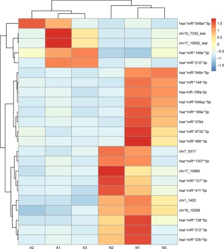
We analyzed expression of 2,231 miRNAs in tumor and paired control tissues in three patients, as shown in . Of these, five miRNAs (hsa−miR−548ar−5p, chr10_7330_star, chr17_10932_star, hsa−miR−148a−3p and hsa−miR−210−3p) exhibited higher expression in the adenocarcinoma samples than that in the paired control non-tumor tissues (P<0.05), and 18 miRNAs (hsa−miR−548x−5p, hsa−miR−144−3p, hsa-miR-106-a-5p, hsa−miR−548ay−5p, hsa−miR−199a−3p, hsa−miR−378d, hsa−miR−4732−3p, hsa−miR−486−3p, chr7_5517, hsa−miR−1307−5p, chr17_10880, hsa−miR−127−3p, hsa−miR−411−5p, chr1_1402, chr16_10269, hsa−miR−138−5p, hsa−miR−212−3p and hsa−miR−33b−5p) demonstrated lower expression in adenocarcinoma samples than that in the paired control non-tumor tissues.
Figure 1 Heat map of expression data of the 23 miRNAs.
Abbreviation: miRNAs, microRNAs.
Validation of NGS results by qRT-PCR
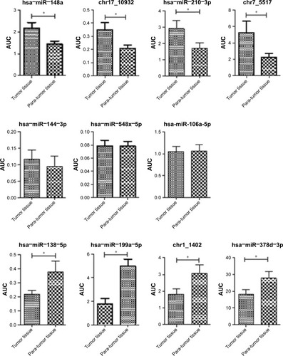
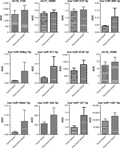
In a validation study using qRT-PCR of 73 tumor and matched non-tumor samples, seven miRNAs (hsa−miR−199a−3p, chr17_10932, hsa−miR−148a−3p, hsa−miR−210−3p, chr1_1402, hsa−miR−378d and hsa−miR−138−5p) were confirmed to have significantly different expression (). chr17_10932, hsa−miR−148a−3p and hsa−miR−210−3p exhibited higher expression in the tumor tissue than in adjacent non-tumor tissue (P<0.05). By contrast, hsa−miR−199a−3p, hsa−miR−378d, chr1_1402 and hsa−miR−138−5p demonstrated lower expression in tumor tissue than that in the adjacent non-tumor tissue (P<0.05; ).
Figure 2 AUC of 23 different microRNAs in tumor and non-tumor tissue by qRT-PCR.
Abbreviations: AUC, area under the curve; miRNAs, microRNAs; qRT-PCR, quantitative real-time polymerase chain reaction.
Validation of miRNA panel by qRT-PCR
We also detected the miRNA panel in 66 lung benign diseases and 66 lung adenocarcinomas presenting with GGN lesion tissue by qRT-PCR. We used more than 75% of the 2−ΔΔCt value in lung adenocarcinoma patients as the cutoff of chr17_10932, hsa−miR−148a−3p and hsa−miR−210−3p. We used less than 75% of the 2−ΔΔCt value in lung adenocarcinoma patients as the cutoff of hsa−miR−199a−3p, hsa−miR−378d, chr1_1402 and hsa−miR−138−5p. The sensitivity and specificity of miRNA panel were 86.4% and 60.6%, respectively.
In silico analysis of potential targets of the seven validated differentially expressed miRNAs
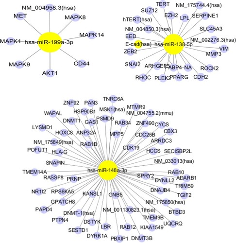
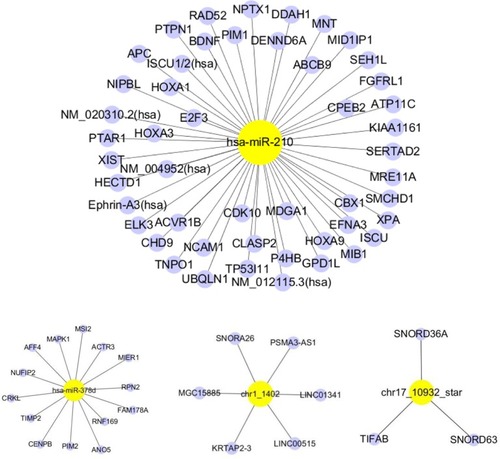
To consider the potential biological functions of the seven validated miRNAs, we used TarBase to predict the targeted mRNAs. We used the miRand algorithm to predict the targets of two novel miRNAs (chr1_1402 and chr17_10932). depicts the mRNAs predicted to be targeted by our miRNAs of interest. Has-miR-138-5p, has-miR-148a-3p and has-miR-210 were predicted to target the largest number of mRNAs. Pathway enrichment analysis was performed for each predicted target mRNA. The enriched pathways are listed in , ordered according to the spell out FDR here adjusted fisher exact test P-value. Most of the enriched pathways were associated with cancer (pancreatic, colorectal, bladder and chronic myeloid leukemia) ().
Figure 3 miRNA target genes.
Abbreviation: miRNA, microRNA.
Discussion
Worldwide, lung cancer is the leading cause of cancer death. Lung cancer’s deadliness is due in large part to the fact that only 20% of lung cancer patients are diagnosed at an early stage.Citation2 Effective biomarkers could permit earlier diagnosis, significantly improving the patient’s prognosis. MiRNAs are involved in a myriad of important biological processes including development, differentiation, apoptosis and proliferation.Citation9 MiRNAs can act as tumor suppressors or oncogenes.Citation9 Abnormal expression of miRNAs has been found to be an effective biomarker for both solid and hematopoietic tumors.Citation6–Citation8,Citation10–Citation12 For example, one previous study suggested that miR-125a-5p, miR-145 and miR-146a may be useful biomarkers for the clinical diagnosis of non-small-lung cancer (NSCLC). However, the data, especially regarding GGNs (a hazy opacity in the lung with the preservation of bronchial and vascular marginsCitation3), remain limited. GGNs can indicate both malignant and benign lesions, including lung adenocarcinoma, focal interstitial fibrosis, hemorrhage or infection.Citation3 The difficulty associated with diagnosis involving GGN increases the need for and value of molecular markers to identify disease.
Several miRNA studies have been reported on various cancer types, including chronic lymphocytic leukemia,Citation13 breast,Citation14 glioblastoma,Citation15 thyroid papillary carcinoma,Citation16 hepatocellular carcinoma,Citation17 lungCitation10,Citation18 and colon.Citation19,Citation20 The downregulation of miR-143 and miR-145 was reported in colorectal neoplasia.Citation20 Garzon et alCitation21 demonstrated that significant differences in miRNA levels can differentiate the stages of megakaryocytopoiesis. Volinia et alCitation8 found distinct miRNA profiles in cancer cells compared with normal cells. In our study, a miRNA panel (hsa−miR−199a−3p, chr17_10932, hsa−miR−148a−3p, hsa−miR−210−3p, chr1_1402, hsa−miR−378d and hsa−miR−138−5p) shows potential as a tool for the early diagnosis for lung adenocarcinoma presenting with GGNs.
In a lung cancer study, Yanaihara et alCitation10 found 43 miRNAs that were expressed at significantly different levels in lung cancer versus matched normal lung tissue. Also, serum concentration of miR-1254 and miR-574-5p could serve as an early diagnostic marker of lung cancer.Citation22 Serum levels of miR-146b, miR-221, let-7a, miR-155, miR-17-5p, miR-27a and miR-106a were significantly decreased in NSCLC, while miR-29c levels were significantly increased.Citation23 Shen et alCitation24 were able to distinguish lung cancer patients from controls by detecting differences in miR-21, miR-126, miR-210 and miR-486-5p in plasma. It has also been demonstrated that the panel of miR-155, miR-197 and miR-182 in plasma can diagnose lung cancer patients.Citation25 Another independent study found seven upregulated and eight downregulated miRNAs in lung cancer patients.Citation26 A meta-analysis using the vote-counting strategy showed miR-210 to be expressed significantly differently in tumor samples in nine studies, and miR-21 to be differentially expressed in seven studies.Citation27 A subsequent study of 250 smokers with CT-detected solitary pulmonary nodules was able to use plasma levels of miR-21, miR-210 and miR-486-5p to discriminate malignant from benign disease with 76% sensitivity and 85% specificity,Citation28 indicating that miRNA biomarkers could potentially supplement and enhance the efficacy of CT screening. This collective body of evidence strongly suggests that miRNAs can be diagnostic and prognostic markers of lung cancer.Citation22–Citation29 However, we did not find data in the literature about miRNA expression profiles in lung adenocarcinoma presenting with GGNs, which are generally present in early-stage disease.
In order to address this lack of data, we employed NGS to identify all the miRNAs that were expressed in lung adenocarcinoma presenting with GGNs in three patients. Our deep sequencing analysis data identified 23 significantly differentially expressed miRNAs in adenocarcinoma tissue with GGNs compared with paired adjacent non-tumor tissue. Among the miRNAs of interest identified by NGS, expressions of hsa−miR−199a−3p, chr17_10932, hsa−miR−148a−3p, hsa−miR−210−3p, chr1_1402, hsa−miR−378d and hsa−miR−138−5p were validated to be statistically different by qRT-PCR in a larger independent group of 73 paired cancer/normal samples. We also detected the miRNA panel in 66 lung benign diseases and 66 lung adenocarcinomas presenting with GGN lesion tissue. The sensitivity and specificity of miRNA panel were 86.4% and 60.6%, respectively, while CEA, NSE and CYFRA21-1 showed sensitivities of 5%, 4% and 17% in stage I and 47%, 21% and 56% in stage IV, respectively.
MiR-210 has consistently been found to be induced by hypoxia and implicated in cancer progression.Citation30–Citation32 Consistent with our data, it is upregulated in multiple tumors and associated with poor patient prognosis. Ding et alCitation31 high-lighted the importance of miR-210 in ovarian cancer progression by demonstrating that it promoted cancer cell mobility by modulating epithelial–mesenchymal transition. Furthermore, one study recently indicated that miR-210 could be considered as a poor prognostic biomarker for lung adenocarcinoma.Citation32 Thus, previous studies support our findings that miR-210 could be a biomarker for the early diagnosis of lung adenocarcinoma.
Altered miR-378 expression has been shown to contribute to human cancers, including breast, prostate, gastric and hepatocellular carcinoma.Citation33,Citation34 Peng et alCitation33 found that miRNA-378 is a biomarker for early detection and diagnosis of colorectal cancer. Decreased expression of miR-378 has been reported in prostate cancer tissue; its overexpression results in inhibition of cancer cell migration and invasion and promotes cell apoptosis.Citation34 These other findings of altered miR-378 in cancer corroborates our finding that miR-378 could be helpful in the diagnosis of early lung adenocarcinoma presenting with GGNs.
Our finding that miR-138 is downregulated in adenocarcinoma presenting with GGNs concurs with mounting evidence presented in other studies that miR-138 is dysregulated in numerous human cancers including lung and may play an important role in tumorigenesis.Citation35,Citation36
Consistent with our findings for lower miR-199a-3p expression in tumor samples, other studies have noted its altered expression in cancer. Kinose et alCitation37 reported that miR-199a-3p dramatically suppressed the progression of ovarian cancer by downregulating expression of c-Met. Wang et alCitation38 found that MiR-199a inhibited proliferation and migration by regulating CD44-ezrin signaling in cutaneous cancer.
We have found that miR-148a expression level was significantly elevated in lung adenocarcinoma patients presenting with GGNs. Several studies have revealed differential expression of miR-148a in tumor versus non-tumor tissue in liver and colorectal cancer.Citation39,Citation40 Li et alCitation41 found that the expression levels of circulating miR-148 family miRNAs might serve as biomarkers for distinguishing NSCLC from benign pulmonary diseases.
Finally, pathway enrichment analysis for the predicted target genes of our validated differentially expressed miRNA results show strong evidence that these miRNAs were linked to cancer pathways. Similar targeting patterns and pathways have been observed in many cancer types including colorectal, pancreatic, bladder and chronic myeloid leukemia, which supports our findings. Others including Kim et alCitation42 have identified some of the same miRNA targets such as the MAPK family as well.
Our work contributes novel data to the literature of miRNAs in cancer because we detected miRNAs via NGS, enabling us to better identify comprehensive potential diagnostic markers. Moreover, we analyzed lung adenocarcinoma patients presenting with GGNs, which are known to be correlated to the early stages of lung adenocarcinoma. In addition, the differential miRNA expression identified in our study was validated in both tumor and matched adjacent non-tumor tissue. We also used TarBase v7.0 to identify target genes of significantly differentially expressed miRNAs.
There are several limitations in our study. Circulating miRNAs in the blood should be analyzed for their potential use as the markers for minimally invasive lung cancer diagnostics, prediction of treatment efficacy and disease prognosis. It has been reported that large numbers of miRNAs are released into peripheral blood when cell damage occurs.Citation43–Citation46 In upcoming research, we plan to compare miRNA levels in the blood of adenocarcinoma patients to healthy individuals with the goal of identifying and verifying a miRNA signature to detect the cancer non-invasively.
This study was aimed to identify characteristic, statistically significantly different levels of miRNA expression in lung adenocarcinoma presenting with GGNs compared to non-cancerous tissue. We found seven miRNAs whose differential expression could potentially illuminate or even initiate lung adenocarcinoma presenting with GGNs, though additional research would be necessary to draw any definitive conclusions. However, this miRNA panel has more immediate potential as a tool for early diagnosis of lung cancer.
Data sharing statement
Our NGS data were uploaded to the website Zenodo: https://zenodo.org/record/50498/export/xm#.Wblpgvl71gk.
Acknowledgments
This study was supported in part by a grant from Shanghai Pujiang Program (17PJD036); 3-year action plan for promoting major disease clinical skills, Shanghai Shenkang Hospital Development Center (16CR1001A); the fundamental research funds for the central universities.
The abstract of this paper was presented at the World Conference on Lung Cancer (WCLC) as an oral presentation with interim findings. The oral abstract was published in “Oral Abstracts” on the website of the International Association for the Study of Lung Cancer (IASLC).
Disclosure
The authors report no conflicts of interest in this work.
References
- FerlayJShinHRBrayFFormanDMathersCParkinDMEstimates of worldwide burden of cancer in 2008: GLOBOCAN 2008Int J Cancer2010127122893291721351269
- TorreLABrayFSiegelRLFerlayJLortet-TieulentJJemalAGlobal cancer statistics, 2012CA Cancer J Clin20156528710825651787
- HansellDMBankierAAMacMahonHMcLoudTCMüllerNLRemyJFleischner Society: glossary of terms for thoracic imagingRadiology2008246369772218195376
- MatsugumaHMoriKNakaharaRCharacteristics of subsolid pulmonary nodules showing growth during follow-up with CT scanningChest2013143243644322814723
- BartelDPMicroRNAs: genomics, biogenesis, mechanism, and functionCell2004116228129714744438
- GarzonRFabbriMCimminoACalinGACroceCMMicroRNA expression and function in cancerTrends Mol Med2006121258058717071139
- CalinGACroceCMMicroRNA signatures in human cancersNat Rev Cancer200661185786617060945
- VoliniaSCalinGALiuCGA microRNA expression signature of human solid tumors defines cancer gene targetsProc Natl Acad Sci U S A200610372257226116461460
- ChenCZMicroRNAs as oncogenes and tumor suppressorsN Engl J Med2005353171768177116251533
- YanaiharaNCaplenNBowmanEUnique microRNA molecular profiles in lung cancer diagnosis and prognosisCancer Cell20069318919816530703
- LuJGetzGMiskaEAMicroRNA expression profiles classify human cancersNature2005435704383483815944708
- RikkeBAWynesMWRozeboomLMBarónAEHirschFRIndependent validation test of the vote-counting strategy used to rank biomarkers from published studiesBiomark Med20159875176126223535
- CalinGALiuCGSevignaniCMicroRNA profiling reveals distinct signatures in B cell chronic lymphocytic leukemiasProc Natl Acad Sci U S A200410132117551176015284443
- IorioMVFerracinMLiuCGMicroRNA gene expression deregulation in human breast cancerCancer Res200565167065707016103053
- CiafrèSAGalardiSMangiolaAExtensive modulation of a set of microRNAs in primary glioblastomaBiochem Biophys Res Commun200533441351135816039986
- HeHJazdzewskiKLiWThe role of microRNA genes in papillary thyroid carcinomaProc Natl Acad Sci U S A200510252190751908016365291
- MurakamiYYasudaTSaigoKComprehensive analysis of microRNA expression patterns in hepatocellular carcinoma and non-tumorous tissuesOncogene200625172537254516331254
- JohnsonSMGrosshansHShingaraJRAS is regulated by the let-7 microRNA familyCell2005120563564715766527
- CumminsJMHeYLearyRJThe colorectal microRNAomeProc Natl Acad Sci U S A2006103103687369216505370
- MichaelMZO’ConnorSMvan Holst PellekaanNGYoungGPJamesRJReduced accumulation of specific microRNAs in colorectal neoplasiaMol Cancer Res200311288289114573789
- GarzonRPichiorriFPalumboTMicroRNA fingerprints during human megakaryocytopoiesisProc Natl Acad Sci U S A2006103135078508316549775
- FossKMSimaCUgoliniDNeriMAllenKEWeissGJmiR-1254 and miR-574-5p: serum-based microRNA biomarkers for early-stage non-small cell lung cancerJ Thorac Oncol20116348248821258252
- HeegaardNHSchetterAJWelshJAYonedaMBowmanEDHarrisCCCirculating micro-RNA expression profiles in early stage nonsmall cell lung cancerInt J Cancer201213061378138621544802
- ShenJToddNWZhangHPlasma microRNAs as potential biomarkers for non-small-cell lung cancerLab Invest201191457958721116241
- ZhengDHaddadinSWangYPlasma microRNAs as novel biomarkers for early detection of lung cancerInt J Clin Exp Pathol20114657558621904633
- VõsaUVooderTKoldeRViloJMetspaluAAnniloTMeta-analysis of microRNA expression in lung cancerInt J Cancer2013132122884289323225545
- GuanPYinZLiXWuWZhouBMeta-analysis of human lung cancer microRNA expression profiling studies comparing cancer tissues with normal tissuesJ Exp Clin Cancer Res2012315422672859
- ShenJLiuZToddNWDiagnosis of lung cancer in individuals with solitary pulmonary nodules by plasma microRNA biomarkersBMC Cancer20111137421864403
- BoeriMSestiniSFortunatoORecent advances of microRNA-based molecular diagnostics to reduce false-positive lung cancer imagingExpert Rev Mol Diagn201515680181325924864
- NomanMZJanjiBHuSTumor-promoting effects of myeloid-derived suppressor cells are potentiated by hypoxia-induced expression of miR-210Cancer Res201575183771378726206559
- DingLZhaoLChenWLiuTLiZLiXmiR-210, a modulator of hypoxia-induced epithelial-mesenchymal transition in ovarian cancer cellInt J Clin Exp Med20158222992307 eCollection 201525932166
- OsugiJKimuraYOwadaYPrognostic impact of hypoxia-inducible miRNA-210 in patients with lung adenocarcinomaJ Oncol2015201531674525733977
- PengJXieZChengLPaired design study by real-time PCR: miR-378* and miR-145 are potent early diagnostic biomarkers of human colorectal cancerBMC Cancer20151515825896668
- ChenQGZhouWHanTMiR-378 suppresses prostate cancer cell growth through downregulation of MAPK1 in vitro and in vivoTumour Biol20163722095210326346167
- YuCWangMLiZMicroRNA-138-5p regulates pancreatic cancer cell growth through targeting FOXC1Cell Oncol (Dordr)201538317318125875420
- XuRZengGGaoJmiR-138 suppresses the proliferation of oral squamous cell carcinoma cells by targeting Yes-associated protein 1Oncol Rep20153442171217826239136
- KinoseYSawadaKNakamuraKThe hypoxia-related microRNA miR-199a-3p displays tumor suppressor functions in ovarian carcinomaOncotarget2015613113421135625839163
- WangSHZhouJDHeQYYinZQCaoKLuoCQMiR-199a inhibits the ability of proliferation and migration by regulating CD44-Ezrin signaling in cutaneous squamous cell carcinoma cellsInt J Clin Exp Pathol201471071317141 eCollection 201425400809
- ZhaoYJiaHLZhouHJIdentification of metastasis-related microRNAs of hepatocellular carcinoma in hepatocellular carcinoma cell lines by quantitative real time PCRZhonghua Gan Zang Bing Za Zhi2009177526530 Chinese19912688
- SongYXuYWangZMicroRNA-148b suppresses cell growth by targeting cholecystokinin-2 receptor in colorectal cancerInt J Cancer201213151042105122020560
- LiLChenYYLiSQHuangCQinYZExpression of miR-148/152 family as potential biomarkers in non-small-cell lung cancerMed Sci Monit2015211155116125904302
- KimSLeeUJKimMNMicroRNA miR-199a* regulates the MET proto-oncogene and the downstream extracellular signal-regulated kinase 2 (ERK2)J Biol Chem200828326181581816618456660
- ZhouJYuLGaoXPlasma microRNA panel to diagnose hepatitis B virus-related hepatocellular carcinomaJ Clin Oncol201129364781478822105822
- WangKZhangSMarzolfBCirculating microRNAs, potential biomarkers for drug-induced liver injuryProc Natl Acad Sci U S A2009106114402440719246379
- SozziGBoeriMRossiMClinical utility of a plasma-based miRNA signature classifier within computed tomography lung cancer screening: a correlative MILD trial studyJ Clin Oncol201432876877324419137
- FortunatoOBoeriMVerriCAssessment of circulating microRNAs in plasma of lung cancer patientsMolecules20141933038305424619302