

Abstract
Objective
The aim of this study was to investigate the malignant potential of endometrial polyps (EP) by assessing the immunoexpressions of both estrogen receptor (ER) and progesterone receptor (PR), Ki-67 cell proliferation index, neovascularization network (endoglin – CD105), cellular adhesion molecules (claudins 3 and 4), and extracellular matrix proteins (MMP-2 and -9) in both EP and endometrioid adenocarcinoma (type I) in comparison with the normal endometrium.
Study design
This is a cross-sectional comparative study. Patients were identified from the database of Botucatu Medical School, São Paulo State University (BMS-UNESP) Clinical Pathology Laboratory.
Setting
The study was conducted using a convenience sample of patients attending the Sectors of Gynecologic Endoscopy and Family Planning and Gynecologic Oncology of the Department of Gynecology and Obstetrics of BMS-UNESP, Brazil.
Patients
A total of 90 women were allocated into the following three groups: EP without atypia (EP, n=30), endometrioid endometrial cancer (EC, n=30), and normal endometrium (control, n=30).
Methods
Epidemiological and clinical data were obtained by reviewing medical records. Adenocarcinoma and control cases were assessed using the tissue microarray technique. The immunoexpressions of ER, PR, Ki-67, CD105, claudins 3 and 4, and MMP-2 and -9 were assessed in paraffin blocks containing sections of the largest polyploid lesion fragment and tissue microarray recipient blocks.
Major results
Compared to the control group, significant differences in the expression of ER (P<0.001), PR (P<0.05), Ki-67 (P<0.001), CD105 (P<0.001), and claudin 3 (P<0.001) were observed in EP and EC. No significant differences were found between EP and EC (P≥0.05). MMP-2 and -9 expression were nearly absent in all groups.
Conclusion
The malignant potential of EP could not be determined through the immunohistochemical parameters used in this study. No MMP-2 or -9 expression was observed in any endometrial tissue sample. Further studies are necessary for a better understanding of the biomolecular mechanisms underlying endometrial carcinogenesis.
Supplementary materials
Figure S1 TMA technique.
Abbreviations: H&E, hematoxylin and eosin; TMA, tissue microarray.
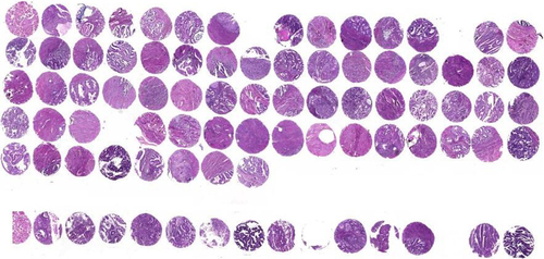
Figure S2 Immunohistochemical analysis of ER and PR expression in the glandular epithelium of endometrial polyp and endometrial cancer samples.
Abbreviations: ER, estrogen receptor; PR, progesterone receptor.
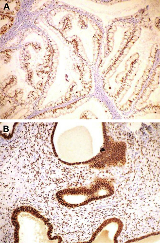
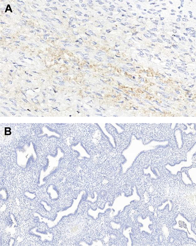
Figure S3 Immunohistochemical analysis of endothelial marker in the stroma of endometrial polyps and endometrial cancer samples.
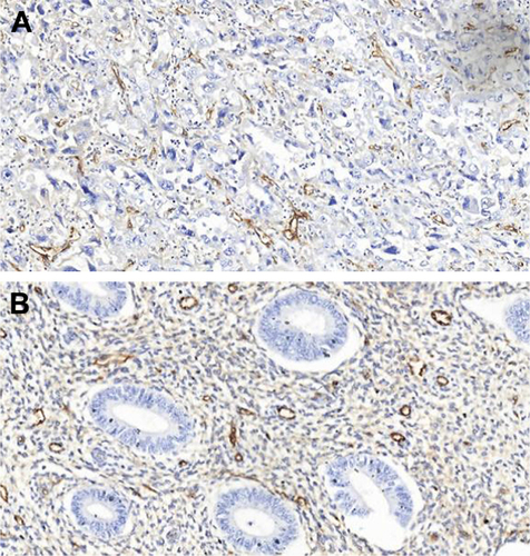
Figure S4 Immunohistochemical analysis of cell proliferative index Ki-67 nuclei expression in endometrial polyp (200×).
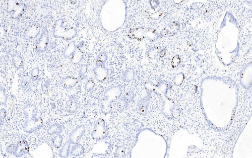
Figure S5 Immunohistochemical analysis expression of claudins 3 and 4 in endometrial polyps and endometrial cancer samples.
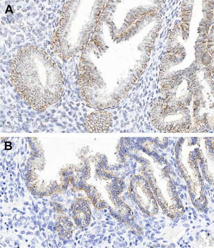
Figure S6 Immunohistochemical analysis showing nearly absence of MMP-2 and -9 expression in all groups.
Acknowledgments
This study was supported by the São Paulo Research Foundation (FAPESP, grant number 2012/17297-3).
Disclosure
The authors report no conflicts of interest in this work.