
Abstract
Background
Carcinoembryonic antigen-related cell adhesion molecule 6 (CEACAM6) is a member of CEACAM family and has been reported to be upregulated in various types of human cancer and involved in tumor progression and metastasis. However, the biological roles and clinical significances of CEACAM6 in osteosarcoma still remain to be elucidated.
Materials and methods
Real-timePCR, immunohistochemistry and Western blot analysis were used to determine CEACAM6 expression in osteosarcoma cell lines and clinical specimens. Then the clinical relevance of CEACAM6 was analyzed in osteosarcoma. The function of CEACAM6 in osteosarcoma was examined by wound-healing and cell invasion assays, and expression levels of epithelial–mesenchymal transition markers.
Results
In the present study, we found that CEACAM6 was markedly upregulated in metastatic osteosarcoma tissues when compared with the nonmetastatic osteosarcoma tissues. Upregulation of CEACAM6 was significantly associated with lung metastasis status (P=0.006) in patients with osteosarcoma. Survival analyses suggested that osteosarcoma patients with high CEACAM6 expression had a significantly shorter overall survival time and lung metastasis-free survival time than those with low CEACAM6 expression. Knockdown of CEACAM6 inhibits osteosarcoma cell migration and invasion. Moreover, silencing CEACAM6 suppressed osteosarcoma cells epithelial–mesenchymal transition.
Conclusion
Taken together, this study suggests that CEACAM6 might be a promising biomarker and a potential therapeutic target for the treatment of metastatic osteosarcoma.
Introduction
Osteosarcoma is the most common type of malignant tumor arising from bone, especially in children and adolescents, with an incidence of 4.4 per million in the USA.Citation1 Over the past 30 years, outcomes and treatment for osteosarcoma remain nearly constant. Children and young adults with localized, nonmetastatic disease have a 60%–70% chance of survival 5 years after diagnosis; however, outcomes for patients with metastatic disease are very poor, with a 5-year overall survival (OS) of 23%.Citation2–Citation4 Unfortunately, metastases are found in statistically up to 23% of osteosarcoma patients, and the lung is the most common site.Citation5,Citation6 Thus, there is an urgent need for a better understanding of the molecular mechanisms that contribute to the process of osteosarcoma metastasis and for a promising diagnostic and prognostic biomarkers for metastatic osteosarcoma.
Carcinoembryonic antigen-related cell adhesion molecule 6 (CEACAM6) is a member of the immunoglobulin supergene family.Citation7 Previous studies have revealed that CEACAM6 is involved in many crucial cellular events such as cell adhesion, migration, invasion and tumorigenicity.Citation8–Citation10 Overexpression of CEACAM6 has been observed in various types of human cancer, including colorectal cancer, breast cancer, ovarian neoplasms and intrahepatic cholangiocarcinoma, and may be associated with tumor progression and metastasis.Citation11–Citation14 Recently, Liu et al found that CEACAM6 was notably highly expressed in metastatic osteosarcoma tissues when compared with nonmetastatic osteosarcoma tissues.Citation15 However, the mechanisms and clinical significances underlying the participation of CEACAM6 in osteosarcoma metastasis still remain to be elucidated.
In the current work, we confirmed that the expression of CEACAM6 was increased in metastatic osteosarcoma tissues. Overexpression of CEACAM6 positively correlated with lung metastasis status, and predicted poorer prognosis in osteosarcoma. Moreover, silencing CEACAM6 effectively attenuated osteosarcoma cell migration and invasion. In addition, we found that CEACAM6 facilitated epithelial–mesenchymal transition (EMT) in osteosarcoma cell lines. Therefore, our research indicates that CEACAM6 plays a pivotal role in osteosarcoma progression and might serve as a promising prognostic biomarker for osteosarcoma.
Materials and methods
Cell lines
Human hFOB1.19 osteoblasts, fibroblast NIH3T3 cell lines, and osteosarcoma cell lines, including MG-63, HOS, U-2 OS, Saos-2 and 143B cells, purchased commercially from American Type Culture Collection (ATCC) (Manassas, VA, USA), were used and cultured in DMEM (Invitrogen, Carlsbad, CA, USA) supplemented with 10% fetal bovine serum (FBS; HyClone, Logan, UT, USA).
Patient information and tissue specimens
A total of 91 paraffin-embedded osteosarcoma tissues, which had been histopathologically and clinically diagnosed, were collected for this study. Fresh osteosarcoma samples, including three nonmetastatic (primary) and three metastatic osteosarcoma tissues, and two adjacent non-tumor tissues, were obtained from patients who were diagnosed with osteosarcoma. All samples were collected from the First People’s Hospital of Yunnan Province. Written informed consent forms to use these tissue samples for research purposes were obtained from all patients. This study was approved by the Ethics Committee of the First People’s Hospital of Yunnan Province according to the 1975 Declaration of Helsinki.
Total RNA extraction, reverse transcription (RT) and real-time polymerase chain reaction (PCR)
Total RNA from fresh tissues was isolated using the Trizol reagent (Invitrogen) according to instructions from the manufacturer, and the mRNA levels of CEACAM6 were evaluated by RT-PCR. First, 2 μg of total RNA was reverse transcribed to cDNA using M-MLV Reverse Transcriptase (Promega, Madison, WI, USA) according to the manufacturer’s protocol. RT-PCR analysis was performed on an ABI Prism 7500 Sequence Detection System (Applied Biosystems; Thermo Fisher Scientific, Waltham, MA, USA). The results were normalized to the expression of the housekeeping gene GAPDH. Relative expression levels were calculated as 2−([Ct of gene] – [Ct of GAPDH]), in which Ct represents the threshold cycle for each transcript. The oligonucleotide primers are listed as follows: CEACAM6 (forward, 5′-ACGTCACCCAGAATGACACA-3′; reverse, 5′-GACCATTTACCCACCACAGG-3′); GAPDH (forward, 5′-GTCTCCTCTGACTTCAACAGCG-3′; reverse, 5′-ACCACCCTGTTGCTGTAGCCAA-3′).
Plasmids, virus constructs and retroviral infection of target cells
Human CEACAM6 shRNA clone (HSH011682-CU6) was purchased from GeneCopoeia (Guangzhou, China). The plasmid transfections were performed using the Lipofectamine 3000 reagent (Invitrogen) according to instructions from the manufacturer.
Western blot analysis
Western blot analysis was performed as previously described,Citation16 using antibodies against CEACAM6 (ab154614, 1:200; Abcam, Cambridge, UK), E-cadherin (#3195, 1:1,000; Cell Signaling, Danvers, MA, USA), N-cadherin (#4061, 1:1,000; Cell Signaling), vimentin (#5741, 1:1,000; Cell Signaling), and α-tubulin (T6199, 1:2,000; Sigma-Aldrich, St Louis, MO, USA). α-Tubulin was used as the loading control.
Immunohistochemistry
Immunohistochemical (IHC) analysis was conducted as described previously.Citation17 After deparaffinization, the sections were immunohistochemically stained using anti-CEACAM6 (ab154614, 1:200; Abcam).
Evaluation of immunohistochemistry
The degree of immunostaining of formalin-fixed, paraffin-embedded sections was examined and scored independently by two pathologists. The IHC staining score was determined as previously described.Citation18 A tumor cell proportion was scored as: 0, no positive tumor cells; 1, 1%–25% positive tumor cells; 2, 25%–50% positive cells; 3, 50%–75% positive tumor cells and 4, >75% positive tumor cells. Staining intensity was scored as: 0, negative; 1, weak; 2, moderate and 3, strong. The staining index (SI) was calculated as the staining intensity score × the proportion of positive tumor cells (ranging from 0 to 12) and grouped as low (SI<6) and high (SI≥6) expression.
Wound-healing assay
MG-63 and U-2 OS cell lines were plated in 6-well plates and grown to ~90% confluence. Twenty-four hours after transfection, the cells were serum starved for 6 h. Then, a streak was created across the well using a 200 μL pipette tip. The cells were washed with PBS for three times, and medium without serum was added into the wells. The progression of migration was photographed immediately and again at 24 h after wounding. The migration distance was measured randomly at five sites perpendicular to the wound to estimate the migration ability. Each experiment was replicated three times.
Cell invasion assay
Cell invasive ability was evaluated using 24-well transwell plates (Costar; Corning Incorporated, Corning, NY, USA). Before cell seeding, the matrigel (BD Biosciences, San Jose, CA, USA) was diluted in serum-free medium at a concentration of 1 mg/mL. Hundred microliters of diluted matrigel per well was added into the top chamber and incubated for 4 h at 37°C. Twenty-four hours after transfection, 1×10⁴ cells in 200 μL of serum-free medium were placed on the top chamber. The low chamber was filled with 500 μL of DMEM supplemented with 20% FBS (Invitrogen). After 24 h of incubation, cells invading into the bottom of the inserts were fixed with methanol and stained with crystal violet. Then, the invaded cells were observed and quantified by counting in five random high-magnification fields using microscopy. Each experiment was replicated three times.
Statistical analysis
All data management and statistical analyses were carried out using the SPSS software version 17.0 (SPSS Inc., Chicago, IL, USA). Data are shown as mean ± SD. The relationship between the CEACAM6 expression and the clinicopathologic features of osteosarcoma was evaluated using the chi-square and Fisher’s exact tests. Survival curves were analyzed using the Kaplan–Meier method and the log-rank test. Other comparisons were estimated using unpaired two-sided Student’s t-test. P<0.05 was considered statistically significant in all cases.
Results
CEACAM6 is overexpressed in metastatic osteosarcoma
To explore the clinical significance and biological role of CEACAM6 in osteosarcoma, we first analyzed its expression in osteosarcoma cells and tissues. As shown in , the mRNA and protein levels of CEACAM6 were expressed at high levels in osteosarcoma cells (MG-63, HOS, U-2 OS, Saos-2 and 143B) compared with FOB or NIH3T3 cells. Notably, CEACAM6 mRNA and protein levels were significantly higher in tumors with metastasis than those in tumors without metastasis (). Similar result was obtained when analyzing the mRNA expression of CEACAM6 in osteosarcoma using publicly published dataset GSE9508 and GSE14359 (). Moreover, the expression of CEACAM6 was further assessed by IHC staining in 91 paraffin-embedded osteosarcoma tissues (). Statistical analysis confirmed that CEACAM6 was significantly upregulated in metastatic osteosarcoma.
Figure 1 CEACAM6 is overexpressed in metastatic osteosarcoma.
Abbreviations: CEACAM6, carcinoembryonic antigen-related cell adhesion molecule 6; N, adjacent non-tumor tissues; OS, overall survival; Osteob, non-neoplastic primary osteoblast cells; PCR, polymerase chain reaction.
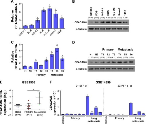
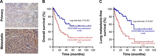
Figure 2 CEACAM6 expression is associated with the clinicopathologic characteristics of osteosarcoma.
Abbreviation: CEACAM6, carcinoembryonic antigen-related cell adhesion molecule 6.
CEACAM6 expression is associated with the clinicopathologic characteristics of osteosarcoma
We next assessed the clinical relevance of CEACAM6 in osteosarcoma. Ninety-one osteosarcoma patients, whose characteristics are listed in , were successfully followed up. Correlation analysis showed that high expression of CEACAM6 was significantly associated with lung metastasis status (P=0.006) in patients with osteosarcoma (). Moreover, Kaplan–Meier survival analyses showed that osteosarcoma patients with high CEACAM6 protein expression had a significantly shorter OS time () and lung metastasis-free survival (LMFS) time () than those with low CEACAM6 expression. Taken together, these data suggest that high CEACAM6 protein expression might contribute to osteosarcoma metastasis, leading to poor clinical outcome.
Table 1 Clinicopathologic characteristics of 91 osteosarcoma patients
Table 2 Correlation between the CEACAM6 expression and the clinicopathologic characteristics of osteosarcoma patients
CEACAM6 knockdown attenuates osteosarcoma cell migration and invasion in vitro
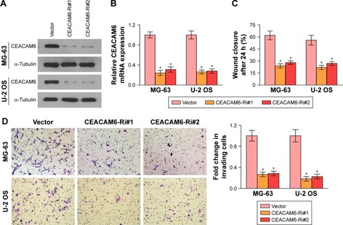
To further investigate the potential biological functions of CEACAM6 in osteosarcoma cells, we performed migration and invasion assays. MG-63 and U-2 OS cells were transduced with lentiviruses carrying either CEACAM6-siRNA or control siRNA (). As shown in , wound-healing assays indicated that silencing CEACAM6 significantly decreased the cell migration distance of MG-63 and U-2 OS osteosarcoma cell lines. Matrigel-coated tran-swell experiments revealed that CEACAM6 knockdown reduced the number of invading cells in MG-63 and U-2 OS cells (). Taken together, these results suggest that CEACAM6 knockdown attenuates the metastatic ability of osteosarcoma cells.
Figure 3 Knockdown of CEACAM6 inhibits osteosarcoma cell migration and invasion.
Abbreviations: CEACAM6, carcinoembryonic antigen-related cell adhesion molecule 6; OS, overall survival; PCR, polymerase chain reaction.
CEACAM6 facilitates EMT in osteosarcoma cell lines
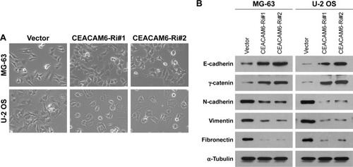
EMT is known to be associated with the metastatic ability of osteosarcoma cells.Citation19 To investigate the mechanism of CEACAM6-mediated promotion in the invasiveness and migration of osteosarcoma cells, we next sought to identify whether CEACAM6 alters the expression of EMT markers. Notably, knocked down of CEACAM6 produced a dramatic shift in morphology, from a stick-like or long spindle-shaped mesenchymal-like cell population to a short spindle-shaped or round and flat epithelial-like cell population (). Moreover, as shown in , we found that the expression levels of N-cadherin, vimentin and fibronectin were decreased, whereas E-cadherin and γ-catenin expression levels were increased, when CEACAM6 was silenced in MG-63 and U-2 OS osteosarcoma cell lines. These results imply that CEACAM6 promotes osteosarcoma cell migration and invasion by regulating the EMT pathway.
Figure 4 CEACAM6 facilitates EMT in osteosarcoma cell lines.
Abbreviations: CEACAM6, carcinoembryonic antigen-related cell adhesion molecule 6; EMT, epithelial–mesenchymal transition; OS, overall survival.
Discussion
The CEACAMs are a subgroup of the CEA family of immunoglobulin-related proteins, containing 12 different members.Citation20 Previous studies have shown that CEACAMs are involved in morphogenesis,Citation21 angiogenesis,Citation22 cell proliferation,Citation23 cell motility,Citation24 apoptosisCitation25 and regulation of cell matrix attachment.Citation26 Dysregulation of CEACAMs contributes to human cancer malignancy, including osteosarcoma.Citation8,Citation15,Citation27–Citation30 CEACAM1 was elevated in osteosarcoma patients and positively correlated with tumor size, clinical stage, tumor grade and distant metastases.Citation30 The CEA assay could be used as an important tool to prognosticate the clinical recurrence of osteosarcoma during therapy.Citation29 Similar to our results, recently, Liu et al found that CEACAM6 mRNA was notably highly expressed in metastatic osteosarcoma tissues when compared with the nonmetastatic osteosarcoma tissues.Citation15 However, its clinical significances, as well as biological functions, in osteosarcoma metastasis remain to be elucidated. Herein, we found that CEACAM6 was significantly associated with lung metastasis status (P=0.006) in patients with osteosarcoma. Moreover, Kaplan–Meier survival analyses showed that patients with high CEACAM6 expression had significantly worse OS and LMFS than those with low CEACAM6 expression, suggesting that CEACAM6 might be a promising prognostic biomarker for osteosarcoma.
Recently, more and more evidences revealed that CEACAM6 plays an important role in tumor metastasis. Kim et al discovered that high level of CEACAM6 is significantly correlated with colorectal cancer lymph nodal and distance metastasis, and promotes colorectal cancer cells invasion by regulating E-cadherin expression.Citation11 Blumenthal et al demonstrated that targeting the N and A1B1 domains of CEACAM5/CEACAM6 using antibody Fabs obviously suppressed colorectal cancer cells lung metastasis.Citation8 Moreover, CEACAM6 could promote tamoxifen-resistant breast cancer cells invasive properties, and might be an important biomarker of breast cancer metastasis.Citation31 In addition, CEACAM6 silencing impairs pancreatic adenocarcinoma cells anoikis resistance in vitro, and liver metastatic ability in vivo.Citation32 Consistent with these results, our study confirmed that CEACAM6 was upregulated in metastatic osteosarcoma. High CEACAM6 expression predicted quicker occurrence of lung metastasis event in osteosarcoma patients. Moreover, CEACAM6 gene silencing dramatically reduced osteosarcoma cells migration and invasion by regulating the EMT pathway. Taken together, these findings supported the crucial role of CEACAM6 in tumor metastasis, and it might be a useful therapeutic target against osteosarcoma metastasis.
EMT is a developmental program that guides proper cell movement during embryogenesis,Citation33 and is commonly hijacked by cancer cells in the process of metastasis, conferring on epithelial cells properties that are critical to invasion and metastatic dissemination.Citation34 The EMT program is characterized by downregulation of epithelial marker, such as E-cadherin, and upregulation of mesenchymal markers, such as N-cadherin and vimentin.Citation34 Herein, we found that E-cadherin and γ-catenin expression levels were increased, while N-cadherin, vimentin and fibronectin expression levels were decreased in CEACAM6 silencing osteosarcoma cells, suggesting that CEACAM6 promotes osteosarcoma cells metastatic properties by regulating EMT pathway. However, the molecular mechanism remains to be further investigated.
Conclusion
Our study revealed that CEACAM6 is upregulated in metastatic osteosarcoma tissues and promotes osteosarcoma cells migration and invasion by regulating the EMT pathway. CEACAM6 may serve as a predictive biomarker for poor prognosis and lung metastasis in osteosarcoma. Targeting CEACAM6 might also represent a potentially novel therapeutic strategy for osteosarcoma.
Author contributions
ZW was responsible for most of the experiments, data collection and analysis. CL and HW conducted the molecular cloning, data analysis and reviewing. CL and XY performed the immunohistochemical staining. ZM and WL supervised the project. ZW wrote the manuscript. ZM and WL designed and conceived the study. All authors contributed toward data analysis, drafting and revising the paper and agree to be accountable for all aspects of the work. All authors read and approved the manuscript.
Disclosure
The authors report no conflicts of interest in this work.
References
- MirabelloLTroisiRJSavageSAOsteosarcoma incidence and survival rates from 1973 to 2004: data from the Surveillance, Epidemiology, and End Results ProgramCancer200911571531154319197972
- Kempf-BielackBBielackSSJürgensHOsteosarcoma relapse after combined modality therapy: an analysis of unselected patients in the cooperative Osteosarcoma Study Group (COSS)J Clin Oncol200523355956815659502
- GoorinAMSchwartzentruberDJDevidasMPresurgical chemotherapy compared with immediate surgery and adjuvant chemotherapy for nonmetastatic osteosarcoma: Pediatric Oncology Group Study POG-8651J Clin Oncol20032181574158012697883
- MeyersPAHellerGHealeyJChemotherapy for nonmetastatic osteogenic sarcoma: the Memorial Sloan-Kettering experienceJ Clin Oncol19921015151370176
- MarkoTADiessnerBJSpectorLGPrevalence of metastasis at diagnosis of osteosarcoma: an international comparisonPediatr Blood Cancer20166361006101126929018
- DuchmanKRGaoYMillerBJPrognostic factors for survival in patients with high-grade osteosarcoma using the Surveillance, Epidemiology, and End Results (SEER) Program databaseCancer Epidemiol201539459359926002013
- PaxtonRJMooserGPandeHLeeTDShivelyJESequence analysis of carcinoembryonic antigen: identification of glycosylation sites and homology with the immunoglobulin supergene familyProc Natl Acad Sci U S A19878449209243469650
- BlumenthalRDHansenHJGoldenbergDMInhibition of adhesion, invasion, and metastasis by antibodies targeting CEACAM6 (NCA-90) and CEACAM5 (Carcinoembryonic Antigen)Cancer Res200565198809881716204051
- CameronSde LongLMHazar-RethinamMFocal overexpression of CEACAM6 contributes to enhanced tumourigenesis in head and neck cancer via suppression of apoptosisMol Cancer2012117423021083
- ChenJLiQAnYCEACAM6 induces epithelial-mesenchymal transition and mediates invasion and metastasis in pancreatic cancerInt J Oncol201343387788523857344
- KimKSKimJTLeeSJOverexpression and clinical significance of carcinoembryonic antigen-related cell adhesion molecule 6 in colorectal cancerClin Chim Acta2013415121922975528
- TsangJYKwokYKChanKWExpression and clinical significance of carcinoembryonic antigen-related cell adhesion molecule 6 in breast cancersBreast Cancer Res Treat2013142231132224186057
- LitkouhiBLitkouhiBFlemingEOverexpression of CEACAM6 in borderline and invasive mucinous ovarian neoplasmsGynecol Oncol2008109223423918331757
- IetaKTanakaFUtsunomiyaTKuwanoHMoriMCEACAM6 gene expression in intrahepatic cholangiocarcinomaBr J Cancer200695453254016868542
- LiuKHeQLiaoGHanJIdentification of critical genes and gene interaction networks that mediate osteosarcoma metastasis to the lungsExp Ther Med20151051796180626640552
- LiQYeLGuoWWangMHuangSPengXPHF21B overexpression promotes cancer stem cell-like traits in prostate cancer cells by activating the Wnt/beta-catenin signaling pathwayJ Exp Clin Cancer Res20173618528645312
- LiQYeLZhangXFZD8, a target of p53, promotes bone metastasis in prostate cancer by activating canonical Wnt/beta-catenin signalingCancer Lett201740216617628602974
- LiQYeLGuoWWangMHuangSPengXOverexpression of TACC3 is correlated with tumor aggressiveness and poor prognosis in prostate cancerBiochem Biophys Res Commun2017486487287828336437
- NiinakaYHaradaKFujimuroMSilencing of autocrine motility factor induces mesenchymal-to-epithelial transition and suppression of osteosarcoma pulmonary metastasisCancer Res201070229483949320978190
- KuespertKPilsSHauckCRCEACAMs: their role in physiology and pathophysiologyCurr Opin Cell Biol200618556557116919437
- YokoyamaSChenCJNguyenTShivelyJERole of CEACAM1 isoforms in an in vivo model of mammary morphogenesis: mutational analysis of the cytoplasmic domain of CEACAM1-4S reveals key residues involved in lumen formationOncogene200726557637764617546042
- HorstAKItoWDDabelsteinJCarcinoembryonic antigen-related cell adhesion molecule 1 modulates vascular remodeling in vitro and in vivoJ Clin Invest200611661596160516680193
- SingerBBScheffrahnIKammererRSuttorpNErgunSSlevogtHDeregulation of the CEACAM expression pattern causes undifferentiated cell growth in human lung adenocarcinoma cellsPLoS One201051e874720090913
- KlaileEMullerMMKannichtCSingerBBLuckaLCEACAM1 functionally interacts with filamin A and exerts a dual role in the regulation of cell migrationJ Cell Sci2005118Pt 235513552416291724
- SingerBBKlaileEScheffrahnICEACAM1 (CD66a) mediates delay of spontaneous and Fas ligand-induced apoptosis in granulocytesEur J Immunol20053561949195915909305
- MuenznerPRohdeMKneitzSHauckCRCEACAM engagement by human pathogens enhances cell adhesion and counteracts bacteria-induced detachment of epithelial cellsJ Cell Biol2005170582583616115956
- JantscheffPTerraccianoLLowyAExpression of CEACAM6 in resectable colorectal cancer: a factor of independent prognostic significanceJ Clin Oncol200321193638364614512395
- HongKPShinMHYoonSTherapeutic effect of anti CEACAM6 monoclonal antibody against lung adenocarcinoma by enhancing anoikis sensitivityBiomaterials201567324126204223
- CortesEPChuTMWangJJHolyokeDWallaceHJMurphyGPCarcinoembryonic antigen in osteosarcomaJ Surg Oncol197793257265267237
- YuHYuJRenYYangYXiaoXSerum CEACAM1 level is associated with diagnosis and prognosis in patients with osteosarcomaPLoS One2016114e015360127074014
- Lewis-WambiJSCunliffeHEKimHRWillisALJordanVCOverexpression of CEACAM6 promotes migration and invasion of oestrogen-deprived breast cancer cellsEur J Cancer200844121770177918614350
- DuxburyMSItoHZinnerMJAshleySWWhangEECEACAM6 gene silencing impairs anoikis resistance and in vivo metastatic ability of pancreatic adenocarcinoma cellsOncogene200423246547314724575
- HayEDAn overview of epithelio-mesenchymal transformationActa Anat (Basel)199515418208714286
- KalluriRWeinbergRAThe basics of epithelial-mesenchymal transitionJ Clin Invest200911961420142819487818