
Abstract
Survivin is a critical regulator of mitosis, and an inhibitor of apoptosis which is overexpressed in almost all cancers. In the current study, cell cycle profiles of normal proliferating human umbilical vein endothelial cells, prostate cancer, and lung cancer cell lines expressing varying levels of survivin and its splice variants were compared using a novel functional complementation assay. Defects in chromosome segregation and cytokinesis that were observed after depletion of endogenous survivin were not complemented by any of the survivin splice variants: survivin-2B, survivin-3B, survivin-ΔEx3, or survivin-2A when expressed exogenously at a level comparable to endogenous full-length survivin. Survivin variants were not detectable at the endogenous protein level. Cancer cells with higher levels of full-length survivin and survivin-2B expression, exhibited reduced caspase-3 activation following doxorubicin treatment and radiation. Whereas earlier studies focused on function and expression levels of survivin specific to cancer cells, the current study brings forward the essential role of survivin in normal dividing cells. Full-length survivin was found to be associated with Aurora-B kinase in the chromosomal passenger complex and depletion of survivin mimics mitotic phenotypes observed after Aurora-B kinase inhibition, in cancer as well as normal proliferating cells. Thus, our study establishes survivin as a marker of proliferation, rather than a cancer specific marker. Therefore, systemic therapeutic interventions targeting survivin will affect cancer as well as normal proliferating cells.
Supplementary figures
Figure S1 (A) Western blot showing non-specific immunoreactivity of two commonly used commercial survivin antibodies, in extracts made from several cell lines as marked. Lanes 1, 2, 3: Total extract from human umbilical vein endothelial cells, H466, and PC3 cells probed with anti-survivin rabbit monoclonal antibody (Cell Signaling: 2808), Lane 4: PC3 extracts probed with anti-survivin polyclonal antibody from Novus Biological (NB-500-201). Endogenous survivin (lower band) and non-specific signals are marked; (B) Western blot detecting full-length survivin, survivin-2B and survivin-3B at a level comparable to the endogenous survivin and actin was used as loading control; (C) Western blot detecting expression of untagged survivin and all the variants in PC3 cells. The upper panel: survivin levels 72 hours after transfection with siRNA oligonucleotide specifically targeting the endogenous survivin (knock down efficiency >95%) and the lower panel shows the levels of endogenous and introduced survivin before transfection.
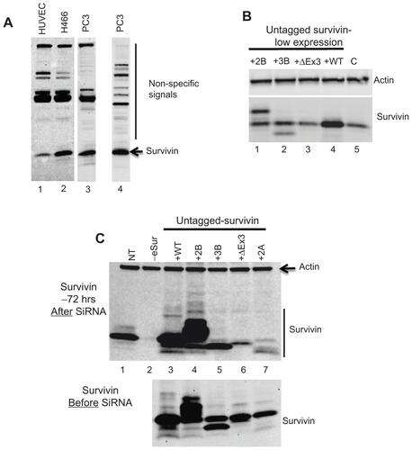
Figure S2 Flow cytometry profile of PC3 cells at 48 and 96 hours after siRNA targeting endogenous survivin showing increase in cells with polyploidy in the absence of functional endogenous survivin as marked.
Notes: (A) endogenous survivin depletion; (B) survivin 2B only; (C) survivin 3B only; (D) survivin ΔEx3 only; (E) survivin 2A only.
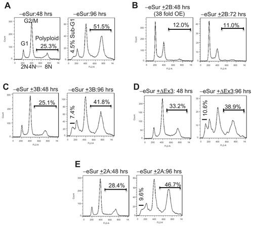
Figure S3 (A) A broader view of immunostaining showing co-localization of survivin with Aurora-B kinase in human umbilical vein endothelial cells during mitosis. Green: Survivin, Red: Aurora-B kinase, Blue: DAPI. (B–D) Immunofluorescence demonstrating predominant cytoplasmic localization of survivin variants. Human umbilical vein endothelial cells expressing respective FLAG-HA-tagged proteins were stained with HA, Aurora-B antibodies, and DAPI.
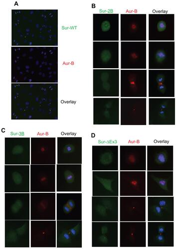
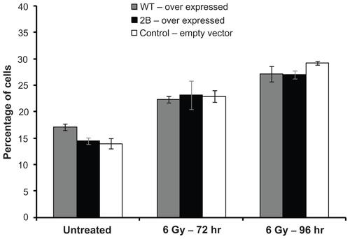
Figure S4 Subtle inhibition in radiation induced apoptosis in survivin over expressing cells detected by Annexin V-FITC based apoptosis assay. Assay was done in samples collected 96 hours after exposed to 6 Gray.
Acknowledgments
This work was supported by the NIH/NCI awards R01CA108633; RC2CA148190 and Institutional Research Funds.
Disclosure
The authors declare no conflicts of interest in this work.