
Abstract
Ameloblastoma (AB) is a common odontogenic tumor that develops in the mouth. Despite its benign nature, AB exhibits significant invasiveness leading to tumor metastasis and high postoperative recurrence rates. Studies have shown a relationship between the occurrence and development of various tumors and non-coding RNA (ncRNA). NcRNA, transcribed from the genomes of mammals and other complex organisms, are often products of alternative splicing and processing into smaller products. MicroRNA (miRNA), circular RNA (circRNA), and long non-coding RNA (lncRNA) are the main types of ncRNA. NcRNA play increasingly significant roles in the pathogenesis of human cancers, regulating their occurrence and progression as oncogenes or tumor suppressors. They are involved in tumor development and progression through alternative splicing of pre-mRNA, transcriptional regulation, mRNA stability, protein translation, and chromatin remodeling and modification. The importance of ncRNA in AB has received significant attention in recent years. However, the biological functions and mechanisms of ncRNA in AB remain largely unknown. In this review, we not only explore the functions and roles of ncRNA in AB, but also describe and envision their potential functional roles as biomarkers in AB diagnosis. In particular, we highlight the potential of miR-29a as a molecular marker for diagnosis and therapy. As promising novel therapeutic targets, the biological functions of ncRNA need further study, which is indispensable.
Keywords:
Introduction
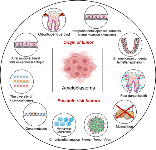
As a locally aggressive benign odontogenic epithelial tumor, ameloblastoma (AB) usually occurs in the jaw. Despite being classified as benign, the invasive growth pattern of AB can lead to severe destruction of the jaw, tooth loss, and metastasis to local lymph nodes or distant organs, such as the brain, lungs, and skin.Citation1,Citation2 AB has an alarming recurrence rate of up to 70%, and malignant transformation is possible.Citation3 Although serious complications include large jaw defects and impaired oral and facial aesthetic issues, the edge of the jaws or segmental resection remains the main treatment for AB.Citation4 Despite the discovery and investigation of various risk factors that may be trigger AB progression, such as chronic inflammation, viral infection (human tumor virus), malnutrition, individual genetic polymorphisms, and poor dental health (), no substantial progress has been made in finding non-invasive therapies to eliminate or prevent AB.Citation5,Citation6 Pivotal molecular events in AB pathogenesis include BRAF p.V600E mutations and the activation of the mitogen-activated protein kinase/extracellular signal-regulated kinase (MAPK/ERK) signaling pathways. BRAF p.V600E is a somatic missense mutation that alters the kinase domain of exon 15 (c.1799), specifically converting thymidine to adenosine (T > A), leading to the substitution of valine (V) in codon 600 with glutamate (E). Despite clinical and histopathological variations, BRAF p.V600E mutations occur in AB with a frequency ranging from 46% to 80%. Its malignant counterpart, ameloblastic carcinoma (AC), exhibits this mutation in 48% of cases. However, especially in BRAF wild-type cases, these factors alone are insufficient to explain the tumor’s complex biological behavior.Citation7–9 Both mutational variability and tumor suppressor genes such as p53 are closely related to ncRNA, making it is essential to study their role in the pathogenesis of AB.Citation10,Citation11
Figure 1 Sources of AB and possible risk factors.
So far, the exploration of markers and genetic variants has been the focus of molecular research in AB for accurate diagnosis and better prognosis prediction. Therefore, further research on the molecular level is now the scientific focus in order to explore the pathogenesis of AB.Citation12 Moreover, the role of ncRNA in AB has also attracted extensive attention.
The majority (98%) of the transcribed genome is composed of ncRNA, which are endogenous RNAs. Previously referred to as “dark matter” due to their lack of protein-coding ability, ncRNA are now recognized as important signaling molecules that regulate key cellular pathways.Citation13 They are numerous, diverse, and stable in structure, mainly including miRNA, circRNA, and lncRNA. Differences in lncRNA expression between AB and normal tissues were first reported in 2017 by Davanian et al, who also proposed that lncRNA expression may affect AB tumor size. This provides new ideas and possibilities for the diagnosis and treatment of AB.Citation14 Some findings have shown that changes in the tumor microenvironment of AB are related to the expression of miRNA.Citation12 Subsequently, scholars have proposed that miR-29a-3p may promote the invasion and metastasis of AB by targeting CTNNBIP1, thereby activating the catenin Wnt/β-catenin pathway.Citation15 In contrast to protein biomarkers, which require biopsy staining and antibody capture, ncRNA have characteristics that are conducive to the development of more sensitive PCR screening in puncture biopsies and liquid biopsies. This enables accurate and rapid diagnosis, as well as the ability to monitor tumor growth and control pathology to improve the quality of life of patients affected by such tumors. Therefore, the role of ncRNA as diagnostic and prognostic biomarkers cannot be overlooked due to their involvement in and influence on the pathogenesis of AB.
We conducted a search on AB and ncRNA in the PubMed database using keywords such as “ameloblastoma”, “AB”, “ncRNA”, and “miRNA”. The Boolean operators “AND” and “OR” were included to combine different search words. Our selection criteria focused on validated results from fresh tissue, sampled tissue, or human fluids. Our search found that while some previous works had partially explored the role of ncRNA in AB, the scope of research was too broad and mechanisms were unclear. Therefore, in this study, we focused on typical ncRNA frequently reported in search results and elaborated on their mechanisms of action in AB.
However, the clear etiology of AB, the mutation of coding genes, and the signaling molecular pathways involved in these changes have not been clearly established, requiring further exploration by researchers. Therefore, this article reviews the role of ncRNA in AB, summarizes the mechanisms and functions of ncRNA and discusses the future development direction of ncRNA in diagnostic and therapeutic research.
Overview of ncRNA
Over the past few decades, a whole class of molecules called ncRNA has been characterized as the key regulatory factor for almost all cell processes.
Biogenesis of ncRNA
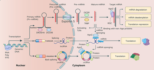
NcRNA are similar to mRNA in biological occurrence, depending on their features. Understanding the biogenesis of ncRNA is important, not only for distinguishing it from the other RNAs, but also for evaluating its functional correlations, which has profound significance.Citation16 Nucleosomal maturation, transcription, production of functional RNA, and exportation towards the cytoplasm for processing are all essential parts of the biogenesis process. RNA polymerase II/III transcribes polycistrons to produce large progenitors (pri-miRNA) with a hairpin loop structure, 5’ capping, and 3’ polyadenylation ().Citation17 Pri-mRNA pass through two processing steps: the microprocessor complex, DGCR8, by Drosha fracture identification and control pre-miRNAs. This leads to the formation of pre-miRNAs, which are then transported from the nucleus to the cytoplasm by the RAN-GTP and Exportin-5 (XPO5) proteins. Furthermore, in the cytoplasm, the RNase III endonuclease Dicer processes the progenitor molecule, enabling it to interact with the Argonaute protein (AGO-2) within the miRNA-induced silencing complex (RISC).Citation18,Citation19 In contrast, the biogenesis of lncRNA is controlled by cell type and stage-specific stimuli.Citation20 A variety of DNA components in eukaryotic genomes, including promoters, enhancers, and intergenic regions, control the transcription of different kinds of lncRNA.Citation21 The main biological processes involved include cleavage by RNase P to produce mature ends, formation of small nucleolar RNA (snoRNA) and small nucleolar ribonucleoprotein (snoRNP) complexes, and the addition of an end cap and circular structure.Citation22 In the process of synthesizing specific lncRNA, unique subnuclear structures called “paracolecules” have recently been discovered.Citation23 In general, the biosynthesis pathways and unique regulation of ncRNA are not yet fully understood. However, in the coming years, the use of various techniques, such as RNA structure mapping, chromatin separation by RNA purification (ChiRP-Seq), cross-linked immunoprecipitation (CLIP), CRISPR-Cas9 for targeted genome engineering, advanced ribosome analysis, phylogenetic lineage tracing, and genetic testing will likely enhance our understanding of the origins and applications of ncRNA.Citation24
Figure 2 Overview biogenesis and function of ncRNA in AB cells. MicroRNA (miRNA), long non-coding RNA (lncRNA), and circular RNA (circRNA) are presented along with their fundamental biogenesis and main functional mechanisms. The details are described in the text.
The role of ncRNA in disease
Accumulating evidence supports the critical role played by ncRNA in regulating glucose homeostasis and lipid metabolism. The importance of ncRNA in the development of cardiovascular diseases and metabolic diseases such as diabetes, non-alcoholic fatty liver disease, abdominal aneurysms, atherosclerosis, and obesity has been repeatedly highlighted.Citation25 Sarkar et al found that miR-34a expression activates synaptic connections, unveiling the formation mechanism of polygenic Alzheimer’s disease (AD).Citation26 Gui et al found that cerebrospinal fluid (CSF) secretions are rich in ncRNA, with significant differences between normal and neurodegenerative diseases, suggesting that CSF ncRNA can be used as early biomarkers.Citation27 This is particularly significant for neurodegenerative diseases like Parkinson’s disease and AD. A recent study found that in an epileptic rat model, after miRNA sequencing validation, the levels of miR-346 and miR-331-3p were significantly reduced. The identification of signaling pathways and targets through enrichment analysis provides a new research direction for identifying the potential mechanisms of epilepsy.Citation28 Studies have shown that some ncRNAs promote fatty acid oxidation by targeting heterogeneous ribonucleoproteins through high expression in skeletal muscle, thus playing a role in metabolic diseases such as insulin sensitivity, diabetes, and obesity.Citation29 In psoriasis, ncRNA regulate keratinocyte proliferation, differentiation, inflammation, and immune cell function.Citation30 Autoimmune skin diseases, including lichen vulgaris, pemphigus, bullous pemphigoid, and psoriasis, are closely associated with ncRNAs, which play an important role in activating T cells, stimulating keratinocyte proliferation, triggering inflammatory cascades, inhibiting cell apoptosis, modulating innate immune responses, and regulating the differentiation and function of Th17 cells.Citation31 In addition, the analysis of 21 different ncRNA involved in the malignant transformation of oral leukoplakia suggests that four of them play a potential role through epithelial–mesenchymal transition (EMT), invasion, and migration, thereby serving as potential markers for malignant transformation of oral leukoplakia.Citation32
Functional Role of ncRNA in Ameloblastoma
This review discusses the role of ncRNA in AB, validated by in vitro or in vivo studies ().
Table 1 NcRNA in Ameloblastoma
MiR-29a
The miR-29 family is one of the more common miRNA families, consisting of miR-29a, miR-29c, and mir-29b (including b-1 and b-2). These miRNA are generated from two main transcripts: the pri-miR-29a/b1 cluster on human chromosome 7q32.3 and the pri-miR-29b2/c cluster on human chromosome 1q32.2.Citation38 Several studies indicate that miR-29a may serve as a vital biomarker for predicting cancer recurrence and progression, and is closely related to the aggressiveness and prognosis of malignant tumors.Citation39 For example, it has been reported that c-Myc down-regulates miR-29a, leading to overexpression of TRAF4, which could be a mechanism promoting prostate cancer progression and bone metastasis. TRAF4 activates NF-κB through glucocorticoid-induced TNF-R (GITR). Considering the inhibitory effect of NF-κB on miR-29a transcription, there may be a positive feedback loop involving NF-κB, miR-29a, and TRAF4, leading to the progression of prostate cancer.Citation40 In addition, some scholars believe that miR-29a/miR-29b may promote the anti-apoptotic effect of myeloid leukemia cells by binding to the 1’-UTR of Bcl-3 and Mcl-2 genes, indicating potential future applications for regulating miR-29a/29b expression in the treatment of myeloid leukemia.Citation41 Some research results showed that miR-29a affected the cell response to BPDE-induced DNA damage by targeting Cdc7, suggesting that downregulation of miR-29a in lung cancer cells may contribute to BPDE-induced mutagenesis and lesion accumulation.Citation42 In the oral cavity, miR-29a is down-regulated in oral squamous cell carcinoma (OSCC) and promotes cancer invasion and anti-apoptosis by up-regulating matrix metallopeptidase 2 (MMP2).Citation43 Furthermore, a near-term study investigated the expression of miR-29a during the proliferation and apoptosis of oral cancer cells. SNHG20 can up-regulate the expression of DIX domain containing 1 (DIXDC1) through sponge miR-29a, thus promoting tumor progression in OSCC.Citation44 One study showed that miR-29a-3p directly targeted catenin beta interacting protein 1 (CTNNBIP1). In the nucleus, CTNNBIP1 inhibits the activity of the beta-catenin/TCF complex, suggesting that CTNNBIP1 may act as a tumor suppressor by inhibiting Wnt/β-catenin signaling, thereby blocking oncogenic phenotypes.Citation45 These results suggest that miR-29a-3p may promote the invasion and metastasis of AB by silencing and binding to the Wnt/β-catenin regulator CTNNBIP1, thereby activating the Wnt/β-catenin pathway.Citation15 Therefore, miR-29a-3p may serve as a potential biomarker or therapeutic target for AB.
MiR-141
As an important member of the miR-200 family, miR-141 is located on chromosome 12 in humans and chromosome 6 in mice.Citation46 MiR-141 is aberrantly expressed in many malignancies, and affects various tumor biological behaviors including EMT, proliferation, invasion, metastasis, migration, and drug resistance.Citation47 Numerous studies have reported that miR-141-3p can be a silent downstream target to regulate cancer cell migration and invasion. For example, low miR-141-3p expression, through activation of NF-kB, induces bone metastasis of prostate cancer.Citation48 MiR-141-3p also inhibits colorectal cancer cell invasion by targeting TNF receptor-associated factor 5 (TRAF5).Citation49 Furthermore, studies show that mir-141 can be inhibited by cirx-0032822 to reduce the inhibition of its target E2F transcription factor 3 (E2F3) and promote cell proliferation in head and neck squamous cell carcinoma.Citation50 In OSCC, miR-141 inhibit tumor progression by targeting PBX homeobox 1 (PBX1) through the JAK2/STAT3 pathway.Citation51 A study speculated that miR-141-3p influences AB cell metastasis and invasion by targeting neural cell adhesion molecule 1 (NCAM1), confirmed through dual-luciferase reporter gene testing. As a member of the immunoglobulin superfamily of adhesion molecules, NCAM1 plays an important role in regulating neurogenesis, neurite growth, proliferation, and cell migration.Citation52 In addition, the expression of NCAM1 in AM-1 cells was significantly inhibited by the overexpression of miR-141-3p. These findings indicate that miR-141 is involved in the development and progression of AB by suppressing NCAM1, making NCAM1 a promising therapeutic target for the treatment of AB.Citation34
MiR-524
MiR-524 is located on human chromosome 19q13.42 and is reported to act as a tumor suppressor gene in multiple types of cancers. For instance, one study found that miR-524-5p reduces breast cancer cell migration, invasion, and EMT by down-regulating FSTL1 expression.Citation53 Another study showed that by targeting lysine-free kinase 1 (WNK1), miR-524-5p could suppress angiogenesis in colon cancer cells.Citation54 In gastric cancer (GC), miR-524-5p is down-regulated, and its overexpression inhibits the development of GC, providing new prospects and ideas for the diagnosis and treatment of GC.Citation55 In OSCC, miR-524 expression is significantly decreased compared to adjacent tissue and is closely related to tumor size, lymph node metastasis, and clinical stage. MiR-524 expression also inhibits MTDH, suppressing activation of the NF-kB signaling pathway, thus inhibiting oral squamous cell migration, proliferation, and invasion.Citation56 In the tumor microenvironment, inflammatory mediators affect the tumor immune response. A recent study showed that downregulation of miR-524-5p can influence the tumor microenvironment of AB by targeting the IL-33/ST2 axis. It was also found that miR-524-5p is down-regulated in AB tissues compared to normal tissues, providing neoteric insights into the mechanisms of AB.Citation12 Therefore, understanding the role of miR-524 is crucial in studying the pathogenesis of AB.
KIAA0125
KIAA0125 (also known as FAM30A) is a lncRNA located on chromosome 14 within the heavy chain region of immunoglobulins.Citation57 Studies have found that KIAA0125 is aberrantly expressed in different cancers and diseases, functioning as a tumor suppressor. For example, it regulates the growth and metastasis of colorectal cancer through the Wnt/β-catenin pathway.Citation58 Multiple lncRNA may interact with particular miRNA clusters in a collaborative manner. In breast cancer, lncRNA KIAA0125 and MEG3 antagonize miR-150 and miR-379, respectively, to regulate the miRNA-mediated sponging interactions of almost all mRNA expressions in the cancer network.Citation59 In cervical cancer, network analysis indicated that KIAA0125 interacts with C-C motif chemokine ligand 21 (CCL21).Citation60 KIAA0125 is highly expressed in AB, potentially due to increased copy number of the KIAA0125 gene located on chromosome 14q32.33. This increased copy number has been detected in AB samples, including AM2 and AM4 sample.Citation61 In a recent study, lncRNA KIAA0125 was predicted to interact with miRNAs (miR-150-5p, miR-205-5p, miR-204-5p, and miR-135a5p) that were previously reported to be dysregulated in AB.Citation35 However, whether KIAA0125 exerts a regulatory role on these miRNAs and the role of miRNAs in AB cells remain to be elucidated.Citation62 In this context, given the strong potential of lncRNA as therapeutic targets, conducting functional experiments on KIAA0125 could advance the use of personalized treatments for recurrent lesions.
HOXC-AS5
Recent studies have found that lncRNA HOXC-AS5 is involved in transcriptional regulation, epigenetic regulation, and post-transcriptional regulation, thereby influencing processes such as stem cell maintenance, differentiation, cell proliferation, apoptosis, cell cycle regulation, metabolism, and immune modulation. Through relevant studies, Jaafari discovered how lncRNA HOXC-AS5 intervenes in tumor cell behavior, suggesting high potential for successful lncRNA HOXC-AS5 therapy research.Citation63 Timothée found that Homeobox C13 (HOXC13), which is abnormally expressed in a variety of cancers including odontogenic tumors like AB, plays a vital role in the regulation of tumor migration and proliferation.Citation64 Reducing the high expression level of HOXC13 can significantly prolong the cell cycle and suppress AB cell division.Citation65 In addition, a recent study showed that HOXC-AS5 overexpression reduces the invasion and migration ability of AB cells, suggesting that HOXC-AS5 can suppress the target gene HOXC13.Citation36 Therefore, HOXC-AS5 is a vital factor in advancing the current understanding of AB pathogenesis.
Circ_0089153
circ_0089153 is an exon which located on chromosome 9. Guan found that circRNA circ_0089153 competes with miR-198 to modulate sentrin/SUMO-specific peptidase 1 (SENP1), impacting colorectal cancer development.Citation66 Gao indicated that circ_0089153 enhanced the metastasis and proliferation of breast cancer cells through sponge miR-2467-3p/E2F6.Citation67 Coincidentally, a recent study characterized for the first time a novel circRNA in AB, circ_0089153. Screening and validation of six circRNA using high-throughput genomic technology revealed a further circRNA-miRNA-mRNA axis, namely circ_0089153/miR-608/EGFR and p53. They concluded that circ_0089153/miR-608 combined with a sponge structure down-regulates EGFR and p53, potentially influencing MAPK signaling pathways to regulate processes such as the cell cycle, cell differentiation, proliferation, and apoptosis.Citation33 These results indicate the great significance of understanding circ_0089153 in the early pathogenesis of AB and further optimizing treatment strategies.
Other ncRNA in AB may play important roles that should not be overlooked. In one study, Davanian et al found a positive correlation between lncRNA LINC340 expression and tumor lesion size.Citation14 In addition, miR-489 has been implicated in distinguishing between solid and unicystic AB.Citation35 According to another study, ENST00000512916 plays a promoting role in the cell migration, proliferation, and cell cycle progression of AM-1 cells, as well as increasing tumor growth in vivo. Interestingly, ENST00000512916 was found to mediate expression of HOXC13 in AM-1 cells, highlighting its potential as a therapeutic target against AB.Citation37
Among the above ncRNA, miR-29a-3p is most closely associated with AB. MiR-29a-3p can promote the invasion and migration of AB cells by targeting CTNNBIP1 and activating the Wnt/β-catenin signaling pathway, increasing the expression of miR-29a-3p in AB. This is of great significance for both the diagnosis and treatment of AB. Moving forward, the high expression of miR-29a-3p can be utilized for auxiliary diagnosis and provide a clear therapeutic target for targeted therapy, providing a direction for subsequent treatment strategies.
Future Expectations
Computed tomography (CT) imaging and biopsy are routine methods for the diagnosis of AB. However, tissue biopsy is an invasive assessment and requires considerable time to obtain test results. If not managed properly, the suffering of AB patients may be exacerbated. The high recurrence rate and significant tissue defects present unresolved challenges for doctors in the treatment of AB. Hence, there is an urgent need for a non-invasive and accurate detection tool to assist clinical diagnosis.
Cancer metastasis is the leading cause of cancer-related deaths. Current cancer treatments face challenges due to the lack of specificity and low bioavailability of drugs, affecting therapeutic efficacy. Despite its benign classification, AB is aggressive in nature, making tumor metastasis common, such as to local lymph nodes or distant organs including the brain, lung, and skin. Tumor-derived exosomes are involved in tumor progression and metastasis by communicating with the tumor microenvironment and are critical in establishing the pre-metastatic niche (PMN).Citation68 A recent study showed that the release of exosomes can be induced by cisplatin, while miR-29a-3p inhibits lung tumor metastasis through tumor exosomes or liposome-based nanovesicles, thereby reducing type I collagen expression in lung fibroblasts and inhibiting metastasis by disrupting the establishment of a viable PMN.Citation69 Although upregulated mir-29a-3p can promote AB invasion and metastasis by targeting genes through signaling pathways, this finding provides us with a new therapeutic concept. Another recent study revealed that miR-141, an ovarian cancer-derived exosomal miRNA, mediates tumor-stroma interactions and promotes the formation of the tumor stroma microenvironment by activating the YAP1/GROα/CXCRs signaling cascade. This provides a new approach for the treatment of ovarian cancer patients with peritoneal metastasis.Citation70 MiRNA can be used within tumor exosomes to further mediate tumor-stroma interactions by activating certain signaling cascades and promoting the formation of a matrix niche that supports tumor growth. They can also be delivered via exosomes or liposome-based nanovesicles delivery to inhibit tumor metastasis, providing new research directions and insights for the treatment of AB.
The identification of precise RNA transcripts or clusters of differentially expressed transcripts has led to insights into tumor biology and improvements in the molecular diagnosis of cancer. Prognostic and therapeutic cancer markers based on RNA have great value, because the RNA landscape of tumors determines their initiation, form, and proliferation. Technologies such as RNA sequencing and global transcriptome microarrays have provided tremendous support in expanding our view of the tumor RNA landscape. In the genome of living cells, protein-coding transcripts, which account for less than 2% of the entire genome, are the most well-defined, while up to 75% of the genome is actively transcribed into ncRNA. Emerging research suggests that ncRNA are new participants in cancer paradigms, playing a role in tumorigenesis and showing potential as biomarkers. For biomarkers used in diagnosis, prediction of disease prognosis, and treatment, durability and sensitivity are vital characteristics for effective functionality. Biomarkers offer a relatively painless method of assessment compared to invasive methods like biopsies. Recently, the feasibility of miRNA as biomarkers was demonstrated in the diagnosis of certain diseases and as therapeutic targets. Therefore, we hypothesize that they could serve as valuable biomarkers in the diagnosis of AB. However, it is important to recognize that most of the studies have been single-center retrospective studies, with many being cohort studies with poor confidence. As a result, non-overlapping and even contradictory reports are common in the literature.Citation71 Many tumor-associated ncRNA were found to be associated with the survival outcomes of multiple cancer types, including gastric cancer, breast cancer, and non-small cell lung cancer. Recent studies have shown that some AB cases contain SMO and BRAF mutations, which may sensitize them to novel small molecule therapies. The incidence of BRAF-V600E mutant AB may be as high as 40%, and BRAF-targeted therapies have shown promising results in patients with malignant BRAF-V600E mutant AB.Citation72
Defining the tumor-associated RNA landscape has a profound role for isolating RNA in tumorigenesis, enabling the identification of diagnostic and therapeutic targets to improve current AB therapy. In the context of AB, a small array studies have demonstrated that the spectrum of protein-encoding transcription contributes to the occurrence and progression of this tumor.Citation73 Traditionally, markers known to participate in cell proliferation, such as minichromosome maintenance complex component 2 (MCM2), MCM3, p16 protein, Ki-67, and CD138 have been used in immunohistochemical analyses to clarify histopathological characteristics of AB and clinical data. Protein biomarkers were not significantly associated with clinical features of AB, except for Ki-67, which showed a positively correlation with growth rate and shorter disease-free survival.Citation74,Citation75 Compared to ncRNA, protein biomarkers require biopsy slice staining and antibody capture. The characteristics of ncRNA, such as the applicability of molecular assays and bioavailability in biological fluids, thus favor the development of more sensitive PCR screening in needle biopsy and liquid biopsy. Therefore, the predictive capabilities of ncRNA surpass the currently available protein markers, enabling the monitoring and management of tumor growth to improve the quality of life of patients affected by AB.
Cytokine therapy has recently become a prominent research focus, with preliminary studies proving its efficacy. Molecular targeted therapy for AB can be used as a new auxiliary therapy. Increasing evidence suggests that dysregulated ncRNA may contribute to or be associated with AB carcinogenesis. Therefore, ncRNA holds promise as potential biomarkers for AB. By building the competitive endogenous RNA networks mediated by lncRNA, some researchers found that lncRNA can modulate miRNA expression and influence the pathogenesis of human diseases.Citation76 Diniz et al found, through the use of high-density genome-wide microarray analysis, raised levels of KIAA0125 lncRNA in AB. They predicted that this lncRNA interacts with a family of 41 miRNAs, with three miRNAs, including miR-204-5p, miR-205-5p, and miR-135a-5p, being overexpressed and miR-150-5p underexpressed in AB. In the future, KIAA0125-lncRNA may be used as a therapeutic target in the treatment of extensive and recurrent cases of AB.Citation62 CircRNA are widespread in eukaryotic transcription groups due to their resistance to ribonuclease degradation. They play important roles in several signaling pathways and regulating downstream molecules, influencing tumor occurrence, development, and other pathological processes. Hence, circRNA hold potential as biomarkers for tumor diagnosis and as therapeutic targets. Although there have been numerous preclinical studies over the years involving miRNA therapy, so far only a few miRNAs have progressed to clinical development. One of the most significant challenges in developing miRNA-based therapies is identifying the optimal miRNA candidates or targets. Other challenges include designing miRNA delivery carriers, enhancing stability of drug candidates for treatment, achieving targeted tissue specificity, and avoiding potential toxicity and off-target effects.Citation77 Many unanswered questions remain about the pathogenesis of AB. Gaining deeper insights into the role of the ncRNA-mRNA-cytokine pathway in the pathogenesis of AB will be valuable in guiding future directions for the treatment of AB. Furthermore, these findings may also be useful for advancing single-gene therapy approaches.
Conclusion
AB is a relatively common oral tumor that, despite being clinically classified as benign, exhibits high aggressiveness, with most AB cases experiencing metastases and recurrence after treatment. The current clinically effective treatment is surgery, requiring an extended excision of the patient’s jaw, which can lead to severe complications and dysfunction for patients. Many cytokines are involved in the occurrence of AB, and ncRNA may play a significant role in regulating processes such as tumor cell proliferation, differentiation, invasion, migration, and apoptosis. The regulatory pathways of ncRNA-miRNA in AB are not well understood, necessitating further research into their molecular mechanisms. We reviewed several important ncRNA studies and examined their role in various signaling pathways. This review can lay a foundation for future studies, provide potential biomarkers for the diagnosis of AB, and provide possible therapeutic targets for non-invasive therapies for AB, including drug therapy. In addition, further exploration of ncRNA therapy in in vitro and animal model studies of AB is crucial in gaining deeper insights into the pathogenesis of AB and therapeutic approaches.
Author Contributions
All authors made a significant contribution to the work reported, whether that is in the conception, study design, execution, acquisition of data, analysis and interpretation, or in all these areas. All authors have agreed on the journals to which their articles will be submitted and agree to be responsible for all aspects of their work.
Disclosure
The authors declare no conflicts of interest in this work.
Additional information
Funding
References
- Xiong G, Ouyang S, Xie N, et al. FOSL1 promotes tumor growth and invasion in ameloblastoma. Front Oncol. 2022;12:900108. doi:10.3389/fonc.2022.900108
- Bi R, Shen L, Zhu X, et al. Malignant ameloblastoma (metastatic ameloblastoma) in the lung: 3 cases of misdiagnosis as primary lung tumor with a unique growth pattern. Diagn Pathol. 2015;10(1):123. doi:10.1186/s13000-015-0367-0
- Devilliers P, Suggs C, Simmons D, et al. Microgenomics of ameloblastoma. J Dent Res. 2011;90(4):463–469. doi:10.1177/0022034510391791
- Laborde A, Nicot R, Wojcik T, et al. Ameloblastoma of the jaws: management and recurrence rate. Eur Ann Otorhinolaryngol Head Neck Dis. 2017;134(1):7–11. doi:10.1016/j.anorl.2016.09.004
- Escobar E, Peñafiel C, Gómez-Valenzuela F, et al. Cyclooxygenase-2 protein expression modulates cell proliferation and apoptosis in solid ameloblastoma and odontogenic keratocyst. An immunohistochemical study. J Oral Pathol Med. 2021;50(9):937–945. doi:10.1111/jop.13237
- Kahn MA. Ameloblastoma in young persons: a clinicopathologic analysis and etiologic investigation. Oral Surg Oral Med Oral Radiol. 1989;67(6):706–715. doi:10.1016/0030-4220(89)90013-3
- Diniz MG, Gomes CC, Guimarães BV, et al. Assessment of BRAFV600E and SMOF412E mutations in epithelial odontogenic tumours. Tumour Biol. 2015;36(7):5649–5653. doi:10.1007/s13277-015-3238-0
- Heikinheimo K, Huhtala JM, Thiel A, et al. The mutational profile of unicystic ameloblastoma. J Dent Res. 2019;98(1):54–60. doi:10.1177/0022034518798810
- Pereira NB, Pereira KM, Coura BP, et al. BRAFV600E mutation in the diagnosis of unicystic ameloblastoma. J Oral Pathol Med. 2016;45(10):780–785. doi:10.1111/jop.12443
- Mccubrey JA, Yang LV, Abrams SL, et al. Effects of TP53 mutations and mirs on immune responses in the tumor microenvironment important in pancreatic cancer progression. Cells. 2022;11(14):2155. doi:10.3390/cells11142155
- Gao Y, Xiang D, Li W, et al. BRAF(V600E) mutation-responsive miRNA-222-3p promotes metastasis of papillary thyroid cancer cells via snail-induced EMT. Front Endocrinol. 2022;13:843334. doi:10.3389/fendo.2022.843334
- Chen L, Wang G, Qiao X, et al. Downregulated miR-524-5p Participates in the Tumor Microenvironment of Ameloblastoma by Targeting the Interleukin-33 (IL-33)/Suppression of Tumorigenicity 2 (ST2) Axis. Med Sci Monit. 2020;26:e921863. doi:10.12659/MSM.921863
- Han Q, Wang M, Dong X, et al. Non-coding RNAs in hepatocellular carcinoma: insights into regulatory mechanisms, clinical significance, and therapeutic potential. Front Immunol. 2022;13:985815. doi:10.3389/fimmu.2022.985815
- Davanian H, Balasiddaiah A, Heymann R, et al. Ameloblastoma RNA profiling uncovers a distinct non-coding RNA signature. Oncotarget. 2017;8(3):4530–4542. doi:10.18632/oncotarget.13889
- Liu S, Liu D, Liu J, et al. miR −29a-3p promotes migration and invasion in ameloblastoma via Wnt/β-catenin signaling by targeting catenin beta interacting protein 1. Head Neck. 2021;43(12):3911–3921. doi:10.1002/hed.26888
- Robles V, Valcarce DG, Riesco MF. Non-coding RNA regulation in reproduction: their potential use as biomarkers. Non-Coding RNA Research. 2019;4(2):54–62. doi:10.1016/j.ncrna.2019.04.001
- Kung JT, Colognori D, Lee JT. Long noncoding RNAs: past, present, and future. Genetics. 2013;193(3):651–669. doi:10.1534/genetics.112.146704
- Statello L, Guo CJ, Chen LL, et al. Gene regulation by long non-coding RNAs and its biological functions. Nat Rev Mol Cell Biol. 2021;22(2):96–118. doi:10.1038/s41580-020-00315-9
- Beermann J, Piccoli MT, Viereck J, et al. Non-coding RNAs in development and disease: background, mechanisms, and therapeutic approaches. Physiol Rev. 2016;96(4):1297–1325. doi:10.1152/physrev.00041.2015
- Akerman I, Tu Z, Beucher A, et al. Human pancreatic β Cell lncRNAs control cell-specific regulatory networks. Cell Metab. 2017;25(2):400–411. doi:10.1016/j.cmet.2016.11.016
- Wu H, Yang L, Chen LL. The diversity of long noncoding RNAs and their generation. Trends Genet. 2017;33(8):540–552. doi:10.1016/j.tig.2017.05.004
- Ojha S, Malla S, Lyons SM. snoRNPs: functions in ribosome biogenesis. Biomolecules. 2020;10(5):783. doi:10.3390/biom10050783
- Naganuma T, Hirose T. Paraspeckle formation during the biogenesis of long non-coding RNAs. RNA Biol. 2013;10(3):456–461. doi:10.4161/rna.23547
- Salehi S, Taheri M N, Azarpira N, et al. State of the art technologies to explore long non-coding RNAs in cancer. J Cell & Mol Med. 2017;21(12):3120–3140. doi:10.1111/jcmm.13238
- Ortega R, Liu B, Persaud S J. Effects of miR-33 deficiency on metabolic and cardiovascular diseases: implications for therapeutic intervention. Int J Mol Sci. 2023;24(13):10777. doi:10.3390/ijms241310777
- Sarkar S, Jun S, Rellick S, et al. Expression of microRNA-34a in Alzheimer’s disease brain targets genes linked to synaptic plasticity, energy metabolism, and resting state network activity. Brain Res. 2016;1646:139–151. doi:10.1016/j.brainres.2016.05.026
- Gui Y, Liu H, Zhang L, et al. Altered microRNA profiles in cerebrospinal fluid exosome in Parkinson disease and Alzheimer disease. Oncotarget. 2015;6(35):37043–37053. doi:10.18632/oncotarget.6158
- Fisher RS, Acevedo C, Arzimanoglou A, et al. ILAE official report: a practical clinical definition of epilepsy. Epilepsia. 2014;55(4):475–482. doi:10.1111/epi.12550
- Liao J, Chen B, Zhu Z, et al. Long noncoding RNA (lncRNA) H19: an essential developmental regulator with expanding roles in cancer, stem cell differentiation, and metabolic diseases. Genes Dis. 2023;10(4):1351–1366.
- Shi R, Ma R, Jiang X, et al. Implications of LncRNAs and CircRNAs in psoriasis: a review. RNA Biol. 2023;20(1):334–347. doi:10.1080/15476286.2023.2223486
- Dopytalska K, Czaplicka A, Szymańska E, et al. The essential role of microRNAs in inflammatory and autoimmune skin diseases-a review. Int J Mol Sci. 2023;24(11):9130. doi:10.3390/ijms24119130
- Niklander S, Guerra D, Contreras F, et al. MicroRNAs and their role in the malignant transformation of oral leukoplakia: a scoping review. Med Oral Patologia Oral y Cirugia Bucal. 2022;27(1):e77–e84. doi:10.4317/medoral.24975
- Liu J, Qiao X, Liu J, et al. Identification of circ_0089153/miR-608/EGFR p53 axis in ameloblastoma via MAPK signaling pathway. Oral Dis. 2022;28(3):756–770. doi:10.1111/odi.13788
- Guan G, Niu X, Qiao X, et al. Upregulation of neural cell adhesion molecule 1 (NCAM1) by hsa-miR-141-3p suppresses ameloblastoma cell migration. Med Sci Monit. 2020;26:e923491. doi:10.12659/MSM.923491
- Setién-Olarra A, Marichalar-Mendia X, Bediaga NG, et al. MicroRNAs expression profile in solid and unicystic ameloblastomas. PLoS One. 2017;12(10):e0186841. doi:10.1371/journal.pone.0186841
- Li J, Zhang B, Wang B, et al. LncRNA HOXC-AS5 affects the proliferation, invasion and cell cycle of ameloblastoma cells by acting on the target gene HOXC13. Cell Mol Biol. 2022;68(5):124–134. doi:10.14715/cmb/2022.68.5.17
- Sun Y, Niu X, Wang G, et al. A novel LNCRNA ENST00000512916 facilitates cell proliferation, migration and cell cycle progression in ameloblastoma. Onco Targets Ther. 2020;13:1519–1531. doi:10.2147/OTT.S236158
- Chang TC, Yu D, Lee YS, et al. Widespread microRNA repression by myc contributes to tumorigenesis. Nature Genet. 2008;40(1):43–50. doi:10.1038/ng.2007.30
- Plaisier CL, Pan M, Baliga NS. A miRNA-regulatory network explains how dysregulated miRNAs perturb oncogenic processes across diverse cancers. Genome Res. 2012;22(11):2302–2314. doi:10.1101/gr.133991.111
- Ahmed F, Shiraishi T, Vessella RL, et al. Tumor necrosis factor receptor associated factor-4: an adapter protein overexpressed in metastatic prostate cancer is regulated by microRNA-29a. Oncol Rep. 2013;30(6):2963–2968. doi:10.3892/or.2013.2789
- Xu L, Xu Y, Jing Z, et al. Altered expression pattern of miR-29a, miR-29b and the target genes in myeloid leukemia. Exp Hematol Oncol. 2014;3(1):17. doi:10.1186/2162-3619-3-17
- Barkley LR, Santocanale C. MicroRNA-29a regulates the benzo[a]pyrene dihydrodiol epoxide-induced DNA damage response through Cdc7 kinase in lung cancer cells. Oncogenesis. 2013;2(7):e57. doi:10.1038/oncsis.2013.20
- Lu L, Xue X, Lan J, et al. MicroRNA-29a upregulates MMP2 in oral squamous cell carcinoma to promote cancer invasion and anti-apoptosis. Biomed Pharmacothe. 2014;68(1):13–19. doi:10.1016/j.biopha.2013.10.005
- Chen ZF, Wang Y, Sun LL, et al. LncRNA SNHG20 enhances the progression of oral squamous cell carcinoma by regulating the miR-29a/DIXDC1/Wnt regulatory axis. Eur Rev Med Pharmacol Sci. 2020;24(10):5436–5445.
- Tago K, Nakamura T, Nishita M, et al. Inhibition of Wnt signaling by ICAT, a novel beta-catenin-interacting protein. Genes Dev. 2000;14(14):1741–1749. doi:10.1101/gad.14.14.1741
- Luo QQ, Tian Y, Qu GJ, et al. Functional mechanism and clinical implications of miR-141 in human cancers. Cell Signalling. 2022;95:110354.
- Gao Y, Feng B, Han S, et al. The roles of MicroRNA-141 in human cancers: from diagnosis to treatment. Cell Physiol Biochem. 2016;38(2):427–448. doi:10.1159/000438641
- Huang S, Wa Q, Pan J, et al. Downregulation of miR-141-3p promotes bone metastasis via activating NF-κB signaling in prostate cancer. J Exp Clin Cancer Res. 2017;36(1):173.
- Liang Z, Li X, Liu S, et al. MiR-141-3p inhibits cell proliferation, migration and invasion by targeting TRAF5 in colorectal cancer. Biochem Biophys Res Commun. 2019;514(3):699–705. doi:10.1016/j.bbrc.2019.05.002
- Zhang S, Han J, Fu J. The circ_0032822 promotes the proliferation of head and neck squamous cell carcinoma cells through miR-141/EF3 signaling axis. Front Oncol. 2021;11:662496. doi:10.3389/fonc.2021.662496
- Cao M, Tian K, Sun W, et al. MicroRNA-141-3p inhibits the progression of oral squamous cell carcinoma via targeting PBX1 through the JAK2/STAT3 pathway. Exp Ther Med. 2022;23(1):97. doi:10.3892/etm.2021.11020
- Walmod PS, Kolkova K, Berezin V, et al. Zippers make signals: NCAM-mediated molecular interactions and signal transduction. Neurochem Res. 2004;29(11):2015–2035. doi:10.1007/s11064-004-6875-z
- Jin T, Zhang Y, Zhang T. MiR-524-5p suppresses migration, invasion, and emt progression in breast cancer cells through targeting FSTL1. Cancer Biother Radiopharm. 2020;35(10):789–801. doi:10.1089/cbr.2019.3046
- Li X, Li Z, Zhu Y, et al. miR-524-5p inhibits angiogenesis through targeting WNK1 in colon cancer cells. Am J Physiol Gastrointest Liver Physiol. 2020;318(4):G827–g39. doi:10.1152/ajpgi.00369.2019
- Zhu CY, Meng FQ, Liu J. MicroRNA-524-5p suppresses cell proliferation and promotes cell apoptosis in gastric cancer by regulating CASP3. Eur Rev Med Pharmacol Sci. 2019;23(18):7968–7977. doi:10.26355/eurrev_201909_19013
- Chang XS, Zhu J, Yang T, et al. MiR-524 suppressed the progression of oral squamous cell carcinoma by suppressing metadherin and NF-κB signaling pathway in OSCC cell lines. Arch Oral Biol. 2021;125:105090. doi:10.1016/j.archoralbio.2021.105090
- De lima DS, Cardozo LE, Maracaja-coutinho V, et al. Long noncoding RNAs are involved in multiple immunological pathways in response to vaccination. Proc Natl Acad Sci USA. 2019;116(34):17121–17126. doi:10.1073/pnas.1822046116
- Yang Y, Zhao Y, Hu N, et al. lncRNA KIAA0125 functions as a tumor suppressor modulating growth and metastasis of colorectal cancer via Wnt/β-catenin pathway. Cell Biol Int. 2019;43(12):1463–1470. doi:10.1002/cbin.11196
- Paci P, Colombo T, Farina L. Computational analysis identifies a sponge interaction network between long non-coding RNAs and messenger RNAs in human breast cancer. BMC Syst Biol. 2014;8(1):83. doi:10.1186/1752-0509-8-83
- Wang H, Zhao Y, Chen M, et al. Identification of novel long non-coding and circular RNAs in human papillomavirus-mediated cervical cancer. Front Microbiol. 2017;8:1720. doi:10.3389/fmicb.2017.01720
- Diniz MG, Duarte AP, Villacis +, et al. Rare copy number alterations and copy-neutral loss of heterozygosity revealed in ameloblastomas by high-density whole-genome microarray analysis. J Oral Pathol Med. 2017;46(5):371–376. doi:10.1111/jop.12505
- Diniz MG, Franca JA, Vilas-Boas FAS, et al. The long noncoding RNA KIAA0125 is upregulated in ameloblastomas. Pathol Res Pract. 2019;215(3):466–469. doi:10.1016/j.prp.2018.12.030
- Jaafari-Ashkavandi Z, Mehranmehr F, Roosta E. MCM3 and Ki67 proliferation markers in odontogenic cysts and ameloblastoma. J Oral Biol Craniofac Res. 2019;9(1):47–50. doi:10.1016/j.jobcr.2018.09.003
- Luo J, Wang Z, Huang J, et al. HOXC13 promotes proliferation of esophageal squamous cell carcinoma via repressing transcription of CASP3. Cancer Res. 2018;109(2):317–329. doi:10.1111/cas.13453
- Cousin T, Bobek S, Oda D. Glandular odontogenic cyst associated with ameloblastoma: case report and review of the literature. J Clin Exp Dent. 2017;9(6):e832–e6. doi:10.4317/jced.53775
- Fang G, Chen T, Mao R, et al. Circular RNA circ_0089153 acts as a competing endogenous RNA to regulate colorectal cancer development by the miR-198/SUMO-specific peptidase 1 (SENP1) axis. Bioengineered. 2021;12(1):5664–5678. doi:10.1080/21655979.2021.1967076
- Gao SL, Fan Y, Liu XD, et al. circ_0089153 exacerbates breast cancer cells proliferation and metastasis via sponging miR-2467-3p/E2F6. Environ Toxicol. 2022;37(6):1458–1471. doi:10.1002/tox.23498
- Wortzel I, Dror S, Kenific CM, et al. Exosome-mediated metastasis: communication from a distance. Dev Cell. 2019;49(3):347–360. doi:10.1016/j.devcel.2019.04.011
- Yan Y, Du C, Duan X, et al. Inhibiting collagen I production and tumor cell colonization in the lung via miR-29a-3p loading of exosome-/liposome-based nanovesicles. Acta pharmaceutica Sinica B. 2022;12(2):939–951. doi:10.1016/j.apsb.2021.08.011
- Mo Y, Leung LL, Mak CSL, et al. Tumor-secreted exosomal miR-141 activates tumor-stroma interactions and controls premetastatic niche formation in ovarian cancer metastasis. Mol Cancer. 2023;22(1):4. doi:10.1186/s12943-022-01703-9
- Solé C, Larrea E, Di Pinto G, et al. miRNAs in B-cell lymphoma: molecular mechanisms and biomarker potential. Cancer Lett. 2017;405:79–89. doi:10.1016/j.canlet.2017.07.020
- Kaye FJ, Ivey AM, Drane WE, et al. Clinical and radiographic response with combined BRAF-targeted therapy in stage 4 ameloblastoma. J National Cancer Inst. 2015;107(1):378. doi:10.1093/jnci/dju378
- Heikinheimo K, Jee KJ, Niini T, et al. Gene expression profiling of ameloblastoma and human tooth germ by means of a cDNA microarray. J Dent Res. 2002;81(8):525–530. doi:10.1177/154405910208100805
- Migaldi M, Sartori G, Rossi G, et al. Tumor cell proliferation and microsatellite alterations in human ameloblastoma. Oral Oncol. 2008;44(1):50–60. doi:10.1016/j.oraloncology.2006.12.004
- Carreón-Burciaga RG, González-González R, Molina-Frechero N, et al. Immunoexpression of Ki-67, MCM2, and MCM3 in ameloblastoma and ameloblastic carcinoma and their correlations with clinical and histopathological patterns. Dis Markers. 2015;2015:683087. doi:10.1155/2015/683087
- Wang L, Cho KB, Li Y, et al. Long noncoding RNA (lncRNA)-mediated competing endogenous RNA networks provide novel potential biomarkers and therapeutic targets for colorectal cancer. Int J Mol Sci. 2019;20(22):5758.
- Rupaimoole R, Slack FJ. MicroRNA therapeutics: towards a new era for the management of cancer and other diseases. Nat Rev Drug Discov. 2017;16(3):203–222. doi:10.1038/nrd.2016.246