
Abstract
Background
Recent studies have demonstrated that the clock gene PER1 regulates various tumor-related genes. Abnormal expressions and circadian rhythm alterations of PER1 are closely related to carcinogenesis. However, the dynamic circadian variations of PER1 and tumor-related genes at different stages of carcinogenesis remain unknown. This study was conducted to investigate the daily rhythm variation of PER1 and expression of tumor-related genes VEGF, KI67, C-MYC, and P53 in different stages of carcinogenesis.
Materials and methods
Dimethylbenzanthracene was used to establish a golden hamster model of buccal mucosa carcinogenesis. Hamsters with normal buccal mucosa, precancerous lesion, and cancerous lesion were sacrificed at six different time points during a 24-hour period of a day. Pathological examination was conducted using routine hematoxylin and eosin staining. PER1, VEGF, KI67, C-MYC, and P53 mRNAs were detected by real-time reverse transcriptase polymerase chain reaction, and a cosinor analysis was applied to analyze the daily rhythm.
Results
PER1, VEGF, C-MYC, and P53 mRNA exhibited daily rhythmic expression in three carcinogenesis stages, and KI67 mRNA exhibited daily rhythmic expression in the normal and precancerous stages. The daily rhythmic expression of KI67 was not observed in cancerous stages. The mesor and amplitude of PER1 and P53 mRNA expression decreased upon the development of cancer (P<0.05), whereas the mesor and amplitude of VEGF, KI67, and C-MYC mRNA increased upon the development of cancer (P<0.05). Compared with the normal tissues, the acrophases of PER1, VEGF, and C-MYC mRNA occurred earlier, whereas the acrophases of P53 and KI67 mRNA lagged remarkably in the precancerous lesions. In the cancer stage, the acrophases of VEGF and C-MYC mRNA occurred earlier and later, respectively, compared with the normal stage.
Conclusion
Variations in the daily rhythm characteristics of the clock gene PER1 and tumor-related genes VEGF, KI67, C-MYC, and P53 correlate with the development of cancer. Additional studies might provide new insights and methods to explore carcinogenic mechanisms and cancer treatment.
Introduction
“Circadian” is a Latin word that means “about a day”.Citation1 Living organisms on earth have evolved with an endogenous timing system, specifically the circadian clock.Citation1–Citation3 Current research has demonstrated that numerous vital physiological processes that occur in mammals in vivo, such as sleeping and waking, hormone secretion, and immune activity, exhibit a circadian fluctuation of approximately 24 hours.Citation1,Citation4–Citation7 The generation of a circadian rhythm in vivo depends on circadian variations in clock genes expression. At least 14 known core clock genes have been reported, including PER1, PER2, PER3, CRY1, CRY2, TIM, CKIε, CLOCK, BMAL1, RORs, REV-ERBs, NPAS2, DEC1, and DEC2.Citation8–Citation11 These clock genes are located in the suprachiasmatic nuclei of the anterior hypothalamus and probably in all peripheral tissue cells.Citation12–Citation14 The former is called the central circadian clock, and the latter is the peripheral circadian clock.Citation15,Citation16 Circadian rhythms in the central circadian and peripheral circadian clocks are generated by the interaction of positive and negative transcription–translation feedback loops at the molecular level. The transcriptional regulators CLOCK and BMAL1 form CLOCK/BMAL1 heterodimers, and the heterodimers subsequently bind to the E-box of the PER (PER1, PER2, and PER3) and CRY (CRY1 and CRY2) genes to activate the expression of these genes in the nuclei. These processes are the main components of the positive feedback circadian rhythm loop.Citation17,Citation18 PER and CRY proteins form heterodimers that translocate from the cytoplasm to the nucleus, where they interfere with CLOCK/BMAL1 heterodimer formation and inhibit the expressions of PER and CRY, thereby forming a negative feedback loop.Citation11,Citation19,Citation20 The clock genes generate and sustain rhythmic expressions in a 24-hour period via these feedback loops.Citation2,Citation12 The peripheral circadian clock can be regulated and influenced by the central circadian clock; meanwhile, the peripheral circadian clock can also generate and sustain a 24-hour circadian rhythm independently.Citation2,Citation16 Approximately 5%–10% of the genes of the mammal genome are regulated by clock genes,Citation3,Citation21 which exhibit 24-hour periodic expression, and these genes are known as clock-controlled genes (CCGs).Citation12,Citation22 Importantly, the circadian expression of clock genes and CCGs maintains a high level of synchronization and orderliness for normal physiological activities.Citation23,Citation24
Current reports have demonstrated that the PER1 gene is an important clock gene that exhibits circadian rhythms in normal cells.Citation25,Citation26 PER1 is a cancer suppressor gene that is downregulated in various solid tumor cells, including prostatic cancer, melanoma, and oral squamous cell carcinoma.Citation8,Citation27,Citation28 Many important cancer-related genes are CCGs; PER1 is an important clock gene that regulates many cancer-related genes. Gery et al reported that PER1 over-expression in human colon cancer cells (HCT116) increased P53 and C-MYC expression.Citation29 Soták et al demonstrated that reduced PER1 mRNA expression in mice colorectal tumors promoted the upregulation of C-MYC and downregulation of P21.Citation15
Current studies have demonstrated that the clock gene PER1 regulates numerous downstream cancer-related genes to influence cell proliferation and apoptosis as well as tumor angiogenesis; thus, PER1 is closely related to the occurrence and development of carcinoma.Citation15,Citation29,Citation30 Carcinogenesis is a process of pathological change involving multiple genes and steps. The dynamic circadian rhythm variations (including the expressions of mesor, amplitude, and acrophase) of the clock gene PER1 and cancer-related CCGs involved in the stages of carcinogenesis and the development of carcinoma are unclear. The daily profiles of the expression of clock gene PER1 and cancer-related genes VEGF, KI67, C-MYC, and P53 during three stages of carcinogenesis in a golden hamster model of buccal mucosa carcinoma were detected in this study; this research provides new concepts for further research on the mechanism of carcinogenesis and the development of cancer as well as the development of cancer therapies based on the recovery of circadian rhythms.
Materials and methods
Experimental animals
Ninety male-specific pathogen-free Syrian golden hamsters (6–7 weeks, 90–120 g) were purchased from Vital River Company (Beijing, People’s Republic of China). All experimental procedures were approved by the Experimental Animal Use and Management Committee in Experimental Animal Research Institute, Chongqing Medical University.
Establishment of a Syrian golden hamster model of buccal mucosa carcinoma by dimethylbenzanthracene
Ninety golden hamsters were randomly housed in separate cages (five hamsters per cage). The golden hamsters were synchronized for 3 weeks under 12-hour light/12-hour dark cycles (at a temperature of 24°C±1°C and humidity of 60%±10%) before the study. The hamsters’ bedding, food, and water were sterilized. In the 12-hour light/12-hour dark cycles, the time was described as hours after light onset (HALO). For example, “0 HALO” was denoted as the time to turn on the light, whereas “12 HALO” was denoted as the time to turn off the light. On the last day of the third week, five hamsters were sacrificed by cervical dislocation at each time point, including 4 HALO, 8 HALO, 12 HALO, 16 HALO, 20 HALO, and 24 HALO during a 24-hour period, and normal left buccal mucosa was simultaneously obtained from the normal group. The remaining 60 hamsters were housed under a 12-hour light/12-hour dark cycle. The hamsters were maintained in a self-made box, and their mouths were opened and painted with a 0.5% dimethylbenzanthracene acetone solution (Sigma, USA) with a No 5 flat brush on the left buccal mucosa every Monday, Wednesday, and Friday. On the last day of the sixth and 14th week after dimethylbenzanthracene painting, five hamsters were sacrificed by cervical dislocation at each time point, including 4 HALO, 8 HALO, 12 HALO, 16 HALO, 20 HALO, and 24 HALO during a 24-hour period; the left buccal diseased tissue was obtained simultaneously. The tissues obtained in the sixth week were included in the precancerous lesions group, whereas tissues obtained in the 14th week were included in the cancer group. Then, each tissue sample was divided into two parts. One portion was fixed in 4% paraformaldehyde, dehydrated, and embedded in paraffin blocks, whereas the other portion was snap-frozen in liquid nitrogen.
Pathological examination using HE staining
We obtained 4 μm thick sections from each paraffin block, and the hematoxylin and eosin (HE)-stained sections were prepared for observation under an optical microscope (Olympus BX51, Japan). Classification of histological pathology was accomplished according to the classification standard of WHO Collaborating Centre for Oral Precancerous Lesions.Citation31
Real-time RT-PCR
Real-time reverse transcriptase polymerase chain reaction (RT-PCR) was performed according to the manufacturer’s protocol. First, the total RNA was isolated using RNAiso Plus (Takara, Japan). Second, the cDNA was synthesized from 10 μL of total RNA using a Prime Script RT reagent kit (Takara). Finally, RT-PCR was performed on a real-time PCR machine (CFX96TM Real-Time PCR Detection System; BioRad, USA) in a 25 μL final volume containing 12.5 μL of 2× SYBR Premix Ex TaqTM II (Takara), 8.5 μL of RNAse-free water, 1 μL of 0.4 μmol/L forward primer and reverse primer, and 2 μL of 50 ng/μL cDNA. Specific primers for β-actin (a housekeeping gene that served as an internal control), PER1, VEGF, KI67, C-MYC, and P53 were designed using Oligo 17.0 software. The primer sequences are listed in . The following RT-PCR cycling parameters were used: 1.5 minutes at 95°C followed by 40 rounds of 10 seconds at 95°C and 30 seconds at 60°C. The data were acquired as the threshold cycle (Ct) value. The relative mRNA expression values of PER1, VEGF, KI67, C-MYC, and P53 were calculated using the 2−ΔΔCt method. Each sample was performed in triplicate to ensure the accuracy of the data.
Table 1 Primers used for real-time PCR amplification of gene expression
Statistical analysis
One-way analysis of variance and Student–Newman–Keuls test were used to analyze the differences in the mRNA expression of each gene among six time points and the differences in the mesor and amplitude of each gene in three groups with SPSS 17.0 statistical software (IBM Corporation, Armonk, NY, USA). Mean and standard deviation were used to describe the results, and a P-value of <0.05 was considered statistically significant. The daily rhythm was assessed by a single cosine test using Time Series Analysis Cosinor 6.3 software (Expert Soft Technologie Laboratory of Applied Statistics and BioMedical Computing, Richelieu, France). A value of P<0.05 indicated that the expression of the target gene showed a daily rhythm. A cosine-fitted curve was generated with SigmaPlot 10.0. The daily rhythm was characterized by the mesor, amplitude, and acrophase. The mesor is the mean value of all the statistics in a circadian fluctuation within a 24-hour period, the amplitude indicates the maximum degree above or below the mesor of the circadian fluctuation, and the acrophase represents the time point when the circadian fluctuation achieved the peak value.
Results
Pathological examination using HE staining
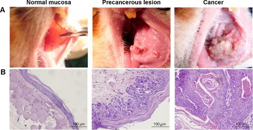
The pathological examination conducted using HE staining indicates that the 30 cases in the normal group exhibited normal buccal mucosa tissue. Among the 30 cases in the precancerous lesions group, 25 displayed moderate atypical hyperplasia, three exhibited mild atypical hyperplasia, and two exhibited severe atypical hyperplasia. The 30 cases in the cancer group exhibited squamous carcinoma tissue ().
Figure 1 The golden hamster buccal mucosa carcinogenesis model.
Abbreviation: HE, hematoxylin and eosin.
Daily rhythm variations of the clock gene PER1 mRNA in various stages of buccal mucosa carcinogenesis
Significant differences in PER1 mRNA expression were noted among the six different time points in the normal, precancerous lesions, and cancer groups (). The cosine analysis revealed that PER1 exhibited daily rhythmic expression in the normal, precancerous lesions, and cancer groups (). The cosine-fitted curves are presented in .
Figure 2 Cosine-fitted curves of PER1, VEGF, KI67, C-MYC, and P53 expression in normal buccal mucosa, precancerous lesions, and cancer tissues.
Abbreviation: HALO, hours after light onset.
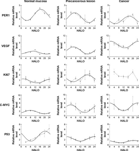
Table 2 The mRNA expression of PER1, VEGF, KI67, C-MYC, and P53 in normal buccal mucosa, precancerous lesion, and cancer at six different time points in 24 hours (mean ± SD, n=5)
The daily rhythms demonstrated the following characteristics (). 1) Mesor and amplitude: The mesor and amplitude of PER1 mRNA expression in the precancerous lesions group and cancer group were significantly reduced compared with the normal group (P<0.05). The mesor and amplitude of PER1 mRNA in the cancer group were remarkably reduced compared with the precancerous lesions group (P<0.05). 2) Acrophase: The acrophases expressed by PER1 mRNA during various stages of carcinogenesis are presented in . In the cancer group, the acrophase of PER1 mRNA was approximately identical to that observed in the normal group. In the precancerous lesions group, the acrophase of PER1 mRNA expression was 9.66 hours earlier than that in the normal group.
Table 3 The daily rhythm characteristics of PER1, VEGF, KI67, C-MYC, and P53 mRNA in normal buccal mucosa, precancerous lesion, and cancer
Daily rhythm variations of mRNA of cancer-related genes during different stages of buccal mucosa carcinogenesis
Significant differences in VEGF, KI67, and P53 mRNA were noted among the six daily time points in the normal, precancerous lesions, and cancer groups, respectively (P<0.05). C-MYC mRNA exhibited significant differences at the six daily time points in the normal and precancerous lesions groups (P<0.05); however, no differences were noted in the cancer group (P>0.05), as shown in . The cosine analysis indicated that VEGF, P53, and C-MYC mRNA, respectively, exhibited daily rhythmic expression in the normal, precancerous lesions, and cancer groups (P<0.05). KI67 mRNA exhibited daily rhythmic expression in the normal and precancerous lesions groups (P<0.05), whereas this expression pattern was not observed in the cancer group (P>0.05) ( and ).
The daily rhythms exhibited the following characteristics (). 1) Mesor: The mesor of VEGF and C-MYC mRNA expression in the precancerous lesions and cancer groups, respectively, was significantly increased compared with the normal group (P<0.05). In the cancer group, the mesor of VEGF and C-MYC mRNA was remarkably increased compared with that in the precancerous lesions group (P<0.05). The mesor of KI67 mRNA in the precancerous lesions group was remarkably increased compared with the normal group (P<0.05). The mesor of P53 mRNA in the precancerous lesions group and cancer group significantly decreased compared with the normal group (P<0.05). In the cancer group, the mesor was significantly reduced compared with the precancerous lesions group (P<0.05). 2) Amplitude: The amplitudes of VEGF and C-MYC mRNA in the precancerous lesions and cancer groups, respectively, were significantly increased compared with the normal group (P<0.05). No remarkable difference was noted between the precancerous lesions and cancer groups (P>0.05). The amplitude of KI67 mRNA in the precancerous lesions group was remarkably increased compared with the normal group (P<0.05). The amplitude of P53 mRNA in the precancerous lesions and cancer groups was significantly reduced compared with the normal group (P<0.05), and the amplitude in the cancer group was remarkably decreased compared with the precancerous lesions group (P<0.05). 3) Acrophase: The acrophases of VEGF, KI67, C-MYC, and P53 mRNA in various carcinogenesis periods are presented in . The acrophases of VEGF mRNA in the precancerous lesions and cancer groups were 3.72 hours and 8.89 hours earlier, respectively, compared with the normal group. The acrophase of KI67 mRNA in the precancerous lesions group was delayed 18.28 hours compared with the normal group. The acrophases of P53 mRNA were similar in the normal and cancer groups. In the precancerous lesions group, the acrophase was 3.4 hours later than that observed in the normal group. The acrophases of C-MYC mRNA in the precancerous lesions and cancer groups were 3.86 hours earlier and 16.15 hours later, respectively, compared with the normal group.
Discussion
Current studies have indicated that the clock gene PER1 is a cancer suppressor gene, and its circadian variations exhibit a close relationship with the occurrence and development of cancers.Citation8,Citation15,Citation27–Citation29,Citation32 Bjarnason et al reported that PER1 expression in human buccal mucosa cells exhibits a circadian rhythm.Citation25 Our previous research demonstrated decreased and circadian rhythmic PER1 expression in oral squamous cell carcinomas.Citation28,Citation33 This study reports that the expression of PER1 in three stages of oral squamous cell carcinoma exhibited daily rhythms. The mesor and amplitude of PER1 mRNA expression decreased remarkably as carcinogenesis progressed. These results demonstrate that the role of PER1 in cancer inhibition was gradually reduced, probably resulting in malignant transformation. In terms of the acrophase, the timing of PER1 mRNA expression peak values was similar in the normal buccal mucosa and carcinoma stages, and the patterns of PER1 mRNA daily rhythmic expression were similar. In the precancerous lesions stage, the acrophase of PER1 mRNA was 9.66 hours earlier than that observed in the normal buccal mucosa. The expression pattern of the daily rhythms of PER1 mRNA in the normal buccal mucosa was in contrast to that observed in precancerous lesions. The mechanism of these variations requires further study.
Carcinogenesis is dependent on tumor angiogenesis, and VEGF plays a central role in tumor-induced angiogenesis.Citation34,Citation35 Koyanagi et al reported that VEGF mRNA expression in transplanted sarcoma cells of mice exhibits circadian rhythms. In hepatic cells and plasma of mice with tumors, VEGF protein exhibits circadian rhythmic expression.Citation36 Our research found that VEGF mRNA expression exhibits daily rhythms in the three carcinogenesis stages in oral squamous cell carcinoma. The mesor and amplitude of VEGF mRNA expression were significantly elevated upon the development of cancer, thus promoting tumor angiogenesis and carcinogenesis. The acrophase of VEGF mRNA constantly progressed with the occurrence and development of cancer. The mechanism of such variations in carcinogenesis requires further exploration and serves as an important reference value for the administration of cancer chemotherapies using VEGF as a biomarker.
P53 is a cancer suppressor gene, and increased P53 expression inhibits tumor growth and induces cell apoptosis.Citation37,Citation38 KI67 and C-MYC are cell proliferation genes that promote cell proliferation via increases in their expression.Citation37,Citation39 Our research demonstrated that P53 and C-MYC mRNA exhibit daily rhythmic expression in three stages of buccal mucosa carcinogenesis. However, KI67 mRNA exhibited daily rhythmic expression in the normal and precancerous lesions groups, whereas KI67 mRNA expression was dysregulated in the cancer group. During the occurrence and development of cancer, the mesor and amplitude of P53 mRNA expression typically decreased gradually, indicating a weakened effect on cancer inhibition and apoptotic induction. KI67 and C-MYC mRNA expression increased gradually, resulting in enhanced reinforcement of cell proliferation and leading to an imbalance between cell proliferation and apoptosis. Regarding the acrophase, P53 mRNA expression was approximately identical in the normal and cancer groups; P53 mRNA expression in the precancerous lesions group lagged by 3.4 hours compared with the normal group. In contrast, the acrophase of KI67 mRNA in the precancerous lesions group was 18.28 hours later than the normal group. C-MYC mRNA expression was 3.86 hours earlier and 16.15 hours later in the precancerous lesions and cancer groups, respectively. The uncoordinated changes in the acrophases of the apoptosis gene P53 and cell proliferation genes KI67 and C-MYC probably further aggravate the imbalance between cell proliferation and apoptosis, thus promoting carcinogenesis.
Conclusion
The specific mechanisms by which the clock gene PER1 regulates and controls cancer-related genes, such as VEGF, KI67, C-MYC, and P53, and the characteristic changes in the circadian rhythms of these genes remain unclear; however, this study was the first to demonstrate the daily rhythmic expression patterns of the clock gene PER1 and cancer-related genes, such as VEGF, KI67, C-MYC, and P53, in different stages of carcinogenesis. These results indicate that the mesors, amplitudes, and acrophases of the clock gene PER1 and cancer-related genes VEGF, KI67, C-MYC, and P53 undergo substantial changes upon the development of cancer. This study provides novel ideas and methods for the exploration of carcinogenic mechanisms and cancer treatments from the perspective of the relationship between circadian variations in gene expression and carcinogenesis.
Acknowledgments
We thank Miss Ling Li for her help in preparing the manuscript and Mr Baisong Li for assisting in the statistical analysis of the data.
Disclosure
The authors report no conflicts of interest in this work.
References
- ZiekerDJenneIKoenigsrainerICircadian expression of clock- and tumor suppressor genes in human oral mucosaCell Physiol Biochem201026215516620798499
- BorgsLBeukelaersPVandenboschRBelachewSNguyenLMalgrangeBCell “circadian” cycle: new role for mammalian core clock genesCell Cycle20098683283719221497
- BozekKRelógioAKielbasaSMRegulation of clock-controlled genes in mammalsPLoS One200943e488219287494
- WulffKPorcheretKCussansEFosterRGSleep and circadian rhythm disturbances: multiple genes and multiple phenotypesCurr Opin Genet Dev200919323724619423332
- OsterHDamerowSHutRAEicheleGTranscriptional profiling in the adrenal gland reveals circadian regulation of hormone biosynthesis genes and nucleosome assembly genesJ Biol Rhythms200621535036116998155
- KellerMMazuchJAbrahamUA circadian clock in macrophages controls inflammatory immune responsesProc Natl Acad Sci USA200910650214072141219955445
- HuangXLFuCJBuRFRole of circadian clocks in the development and therapeutics of cancerJ Int Med Res20113962061206622289520
- CaoQGerySDashtiAA role for the clock gene per1 in prostate cancerCancer Res200969197619762519752089
- BjarnasonGAJordanRCSothernRBCircadian variation in the expression of cell-cycle proteins in human oral epitheliumAm J Pathol1999154261362210027418
- RanaSMahmoodSCircadian rhythm and its role in malignancyJ Circadian Rhythms20108320353609
- SavvidisCKoutsilierisMCircadian rhythm disruption in cancer biologyMol Med2012181249126022811066
- SchiblerUSassone-CorsiPA web of circadian pacemakersCell2002111791992212507418
- FuLPelicanoHLiuJHuangPLeeCThe circadian gene Period2 an important role in tumor suppression and DNA damage response in vivoCell2002111415012372299
- SchiblerURippergerJBrownSAPeripheral circadian oscillators in mammals: time and foodJ Biol Rhythms200318325026012828282
- SotákMPolidarováLErgangPSumováAPáchaJAn association between clock genes and clock-controlled cell cycle genes in murine colorectal tumorsInt J Cancer201313251032104122865596
- YooSHYamazakiSLowreyPLPERIOD2: luciferase real-time reporting of circadian dynamics reveals persistent circadian oscillations in mouse peripheral tissuesProc Natl Acad Sci USA2004101155339534614963227
- ShearmanLPSriramSWeaverDRInteracting molecular loops in the mammalian circadian clockScience200028854681013101910807566
- GekakisNStaknisDNguyenHBRole of the CLOCK protein in the mammalian circadian mechanismScience19882805369156415699616112
- KumeKZylkaMJSriramSmCRY1 and mCRY2 are essential components of the negative limb of the circadian clock feedback loopCell199998219320510428031
- SatoTKYamadaRGUkaiHFeedback repression is required for mammalian circadian clock functionNat Genet200638331231916474406
- StorchKFLipanOLeykinIExtensive and divergent circadian gene expression in liver and heartNature20024176884788311967526
- MeyerVLerchlAEvidence for species-specific clock gene expression patterns in hamster peripheral tissuesGene2014548110111125016070
- RanaSMunawarMShahidADeregulated expression of circadian clock and clock-controlled cell cycle genes in chronic lymphocytic leukemiaMol Biol Rep20144119510324190490
- FuLKettnerNMThe circadian clock in cancer development and therapyProg Mol Biol Transl Sci201311922128223899600
- BjarnasonGAJordanRCWoodPACircadian expression of clock genes in human oral mucosa and skin: association with specific cell-cycle phasesAm J Pathol200115851793180111337377
- ZhengLSeonYJMcHughJPapagerakisSPapagerakisPClock genes show circadian rhythms in salivary glandsJ Dent Res201291878378822699207
- LengyelZLovigCKommedalSAltered expression patterns of clock gene mRNAs and clock proteins in human skin tumorsTumor Biol2013342811819
- ZhaoNYangKYangGAberrant expression of clock gene period1 and its correlations with the growth, proliferation and metastasis of buccal squamous cell carcinomaPLoS One201382e5589423405233
- GerySKomatsuNBaldjyanLYuAKooDKoefflerHPThe circadian gene per1 plays an important role in cell growth and DNA damage control in human cancer cellsMol Cell200622337538216678109
- PluquetODejeansNChevetEWatching the clock: endoplasmic reticulum-mediated control of circadian rhythms in cancerAnn Med201446423324324491143
- WHO Collaborating Centre for Oral Precancerous LesionsDefinition of leukoplakia and related lesions: an aid to studies on oral precancerOral Surg Oral Med Oral Pathol197846518539280847
- LeeSDonehowerLAHerronAJMooreDDFuLDisrupting circadian homeostasis of sympathetic signaling promotes tumor development in micePLoS One201056e1099520539819
- ChenRYangKZhaoNBAbnormal expression of PER1 circadian-clock gene in oral squamous cell carcinomaOnco Targets Ther2012540340723226027
- WangCYWenMSWangHWIncreased vascular senescence and impaired endothelial progenitor cell function mediated by mutation of circadian gene Per2Circulation2008118212166217318981300
- WoodPADu-QuitonJYouSHrusheskyWJCircadian clock coordinates cancer cell cycle progression, thymidylate synthase, and 5-fluorouracil therapeutic indexMol Cancer Ther2006582023203316928823
- KoyanagiSKuramotoYNakagawaHA molecular mechanism regulating circadian expression of vascular endothelial growth factor in tumor cellsCancer Res200363217277728314612524
- GrandaTGLiuXHSmaalandRCircadian regulation of cell cycle and apoptosis proteins in mouse bone marrow and tumorFASEB J200519230430615545298
- RengarajanTNandakumarNRajendranPHaribabuLNishigakiIBalasubramanianMPD-pinitol promotes apoptosis in MCF-7 cells via induction of p53 and Bax and inhibition of Bcl-2 and NF-κBAsian Pac J Cancer Prev20141541757176224641404
- ScholzenTGerdesJThe Ki-67 protein: from the known and the unknownJ Cell Physiol2000182331132210653597