
Abstract
Background and purpose
Closed reduction and lateral-only pin fixation is one of the common treatment methods for displaced supracondylar fracture in children. However, several risk factors related to the stability have been reported. The aim of this study was to evaluate the medial comminution as a potential risk factor related to the stability after appropriate lateral-only pin fixation for Gartland type III supracondylar humerus fracture.
Methods
Sixty-seven patients with type III supracondylar fractures who were under the age of 12 years were included. Immediate postoperative and final Baumann and humerocapitellar angles were measured. Pin separation at fracture site was evaluated to estimate the proper pin placement. Presence of the medial comminution was recorded when two pediatric orthopedic surgeons agreed to the loss of cortical contact at the medial column by the small butterfly fragment or comminuted fracture fragments. Factors including age, sex, body mass index, pin number, pin separation at fracture site, and medial comminution were analyzed.
Results
Medial comminution was noted in 20 patients (29.8%). The average pin separation at fracture site was significantly decreased in patients with medial comminution compared to patients without medial comminution (P=0.017). A presence of medial comminution was associated with a 4.151-fold increase in the log odds for the Baumann angle changes of more than average difference between immediate postoperative and final follow-up angle (P=0.020).
Conclusion
When lateral-only pin fixation is applied for Gartland type III supracondylar humerus fracture in children, the medial comminution may be a risk factor for the stability because of the narrow pin separation at fracture site. We recommend additional medial pin fixation for supracondylar humerus fracture with medial comminution.
Introduction
Supracondylar humerus fracture is the most common elbow fracture in children.Citation1,Citation2 The standard treatment for displaced supracondylar fracture is closed reduction and percutaneous pin fixation, but there is controversy regarding ideal pin configuration in terms of stability and safety.Citation3–Citation6 When the pin is placed on the medial epicondyle, the stability increases but so does the risk of iatrogenic ulnar nerve injury. Although several previous clinical studiesCitation4,Citation7–Citation9 stated that there was no difference in maintenance of the reduction between cross pinning and lateral-only pinning, a previous biomechanical studyCitation10 suggested that cross-pin fixation provides greater torsional strength.
In the literature, loss of reduction has ranged from 0.3% to 0.6% after pin fixation.Citation11,Citation12 Some of the previous clinical studiesCitation4,Citation13,Citation14 reported that proper pin fixation is essential for maintaining the stability and that inappropriate lateral pin fixation is responsible for the loss of reduction. Besides surgical technique, other factors responsible for loss of reduction were reported including unstable fracture patterns such as the medial comminution, high fracture lines, or oblique fracture lines.Citation15–Citation19 In supracondylar fracture with oblique fracture line or high fracture line, lateral-only pin fixation is sometimes impossible or very difficult, so cross-pin fixation is preferred. However, in supracondylar fracture with medial comminution, lateral-only pin fixation is possible, and there is a trend toward lateral-only pin fixation.Citation20
We hypothesized that medial comminution may be a risk factor for the stability after lateral-only pin fixation. The purpose of our study is to compare the surgical outcome between supracondylar fracture with and without medial comminution and to identify whether the medial comminution is related to the stability after lateral-only pin fixation.
Methods
Ethics approval and informed consent
This was a retrospective study of children with a GartlandCitation21 type III supracondylar fracture based on radiography and medical records. All data were deidentified. This study did not need any kind of consents to participate and was approved by the Ethics Committee of Haeundae Paik Hospital (129792-2015-034).
Patients
This was a retrospective study in children with a GartlandCitation21 type III supracondylar fracture. We retrospectively reviewed prospectively collected data of 111 patients from the period between March 2010 and June 2016. The inclusion criteria were age of <12 years, >6 months follow-up, and lateral-only pin fixation. The exclusion criteria were unstable pin fixation, such as 1) failure to engage both fragments with two or more pins; 2) failure to achieve bicortical fixation; and 3) failure to achieve pin separation >2 mm at the fracture.Citation13 Sixty-seven patients were included in this study. The average age was 5 years 7 months (range, 1–11 years). Forty-three patients were boys and 24 were girls.
Surgical procedure
All fractures were fixed using the lateral-only fixation technique. First, two pins were inserted from the lateral aspect of elbow to the medial cortex. One pin was placed in the lateral column and the other pin had to be placed in the central column.Citation4,Citation14 The operator checked the stability by flexion and extension or gentle rotation. If the stability was not reliable, additional lateral fixation was added. At 3 weeks postoperative, radiographs were evaluated and the day for the removal of pin and cast was decided according to the fracture healing. After removal of the pin and cast, the patients start active daily living and range of motion exercise without passive physical therapy. Final follow-up radiography was performed at 6 months postoperative.
Variables
For radiographic analyses, the immediate postoperative and final follow-up anteroposterior (AP) and lateral radiographs were used. All AP radiographs were obtained with the upper arm flat on the cassette, the forearm in supine position and elbow at 45° flexion, because the elbow could not achieve full extension immediately after surgery, and after taking the cast off. Radiologic indices were measured by two orthopedic surgeons and averaged. The Baumann angle was calculated on the AP radiograph using the method of Williamson et al.Citation22 The angle between the humeral shaft and the capitellum on a lateral radiograph defined as the humerocapitellar angle.Citation16 Pin separation at fracture site was defined as the length between medial and lateral pin divided by the humerus width at the fracture site ().Citation18,Citation23 Medial comminution was defined as a lack of cortical contact on the medial column by small butterfly fragment or comminuted small fragments after operation,Citation18,Citation19 and the presence or absence of medial comminution was recorded when two pediatric orthopedic surgeons agreed ().
Figure 1 Pin separation was calculated using the formula: (B/A) × 100.
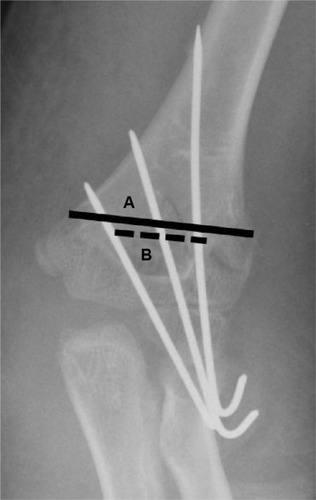
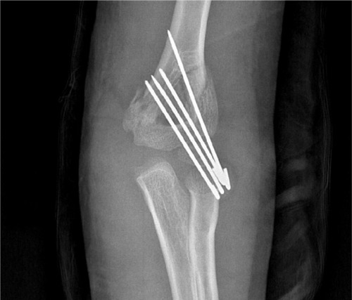
Figure 2 Examples of medial comminution and the lack of cortical contact on medial column in anteroposterior radiography.
Statistics
Statistical analyses were performed using SAS software (version 9.1; SAS Institute, Cary, NC, USA). Measurement reliability for the Baumann angle, humerocapitellar angle, and pin separation at fracture site was expressed as intraclass correlation coefficient, which ranged from 0.763 to 0.869. Independent t-test was used to compare continuous data between supracondylar fracture with medial comminution (Group I) and supracondylar fracture without medial comminution (Group II). Chi-squared test was used to compare categorical data. There was very small difference between immediate postoperative angle and final follow-up angle in Baumann angle and humerocapitellar angle. We defined the significant angle change during fracture healing period as a change of more than average difference between immediate postoperative angle and final follow-up angle. Multivariable logistic regression analysis was done to find factors related to the significant change of Baumann angle in age, sex, body mass index (BMI), pin number, pin separation at fracture site, and medial comminution. Values are presented as the mean ± SD, and the level of significance was defined at P<0.05.
Results
All patients showed complete union without significant complication. Average time from injury to operation was 25 ± 7 hours and average operation time was 32 ± 17 min. Average cast fixation period was 29.2 ± 4.5 days. One patient had a distal radius fracture on the same side. Two patients needed an open reduction. Before the operation, one patient showed median nerve palsy with radial nerve palsy, one patient had anterior interosseous nerve palsy, and two patients had radial nerve palsy. They all completely recovered without additional surgery. Two patients had superficial pin site infection, but it was improved after pin removal and bony union.
Usually, three Kirshner wires (range, 2–4) were used. Average pin separation at fracture site was 36.8% ± 6.8%. Average postoperative Baumann angle was 72.6° ± 5.3° and final Baumann angle was 71.8° ± 4.7°. Average Baumann angle difference was 4.0° ± 2.2°. All differences were <10°. Average postoperative humerocapitellar angle was 33.9° ± 11.3° and final humerocapitellar angle was 32.6° ± 9.9°. Average humerocapitellar angle difference was 1.3° ± 3.6°.
Medial comminution was noted in 20 patients. Twenty patients were classified as Group I and others were classified to Group II. There was no significant difference between groups, except the pin separation at fracture site (P=0.017) (). In multivariable logistic regression that evaluate the significant Baumann angle change (Nagelkerke R2 0.195), only the medial comminution was related to the Baumann angle change (OR =4.151 [95% CI 1.249–13.800], P=0.020) ().
Table 1 Demographic data of Groups I (patients with medial comminution) and II (patients without medial comminution)
Table 2 Factors related to the changes in Baumann angle using multivariable logistic regression
Discussion
In 2001, Skaggs et alCitation8 compared the cross-pinning and lateral pinning in Gartland type II and III fractures, and they recommend “do not use the routine cross pin”, because there was no difference in the maintenance of reduction. However, the maintenance of reduction after pinning for supracondylar humerus fracture is still surgeon’s concern in terms of the higher reliability of fixation. In a quantitative study,Citation7 the rate of displacement after lateral-only pin fixation was 0.7%. A recent meta-analysis showed an overall loss of reduction rate of 4% for lateral entry-pin constructs and 2% for cross-pin constructs, although there was heterogeneity in data.Citation24 However, to our knowledge, there has been no study about the risk factor for the loss of reduction after lateral-only pin fixation. We evaluated the medial comminution as a potential risk factor for the stability after lateral-only pin fixation.
The more important reason for the preference of the lateral-only pin fixation is avoidance of iatrogenic ulnar nerve injury. Topping et alCitation9 found no loss of reduction in both methods and one ulnar nerve injury in cross-pinning. In a randomized prospective study with 52 patients, Kocher et alCitation4 stated that there was no difference in loss of reduction and iatrogenic ulnar nerve injury between lateral and cross-pinning. The rate of iatrogenic ulnar nerve injury from cross-pinning was 3.4% in a systematic reviewCitation12 and also the relative risk factor was 0.30 in another meta-analysis.Citation6 Another study reported an 8-fold increase in the ulnar nerve injury after cross-pinning.Citation25 In our series, there was no iatrogenic ulnar nerve injury, like the results of previous study with lateral-only pin fixation.Citation14 The lateral-only fixation method is better than cross-pinning for the prevention of iatrogenic ulnar nerve injury.
A recent prospective study showed no iatrogenic injury after medial pin fixation and stated the importance of the preventive technique with a small incision on medial side and elbow extension during medial pin fixation.Citation26 However, the technical points are similarly important in lateral-only fixation in terms of stability. Previous studies proposed a principle that distal humerus is separated into lateral, central, and medial columns, and that the lateral pin should engage at least two different columns.Citation4,Citation14 If the lateral fixation rule is followed, supracondylar humerus fractures can be treated stably without the risk of ulnar nerve injury. We have been followed the lateral fixation rule and aimed to identify risk factors related to the stability after accurate lateral-only pin fixation. Sankar et alCitation13 highlighted three technical reasons for loss of reduction, and these were exclusion criteria in this study. Recently, the pin spread has been mentioned as an important factor associated with preventing loss of reduction.Citation18,Citation23 Pennock et alCitation23 reported 4.2% loss of reduction rate with a definition of 10° differences. However, they reported no loss of reduction in 36% pin spread, which is similar to our 36.8% pin spread. In our series, the lateral pin fixation was technically appropriate.
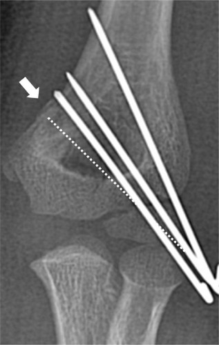
In this study, medial comminution was evaluated as the risk factor for the stability after lateral-only pin fixation. De Boeck et alCitation17 described the importance of medial column collapse. They reported cases of patients with medial column impaction as a cause of cubitus varus and the reason for this is that the injury is easily misdiagnosed as a simple supracondylar fracture with minimal displacement, requiring no reduction. Medial column is also important in the stability after lateral-only fixation because of the bicortical fixation.Citation13 In terms of stability, considering the need for bicortical fixation when treating supracondylar fractures, medial comminution should be considered as an important issue, because that make pin spread narrow (). In our study, the pin spread at fracture site in patients with medial comminution was significantly decreased compared to those in patients without medial comminution.
Figure 3 Due to medial comminution (white arrow), medial pin is more vertical than ideal position (dotted line).
Medial comminution was also reported as a factor related to the loss of reduction in the previous study,Citation18 although they compared cross-pinning and lateral-only pinning. The effect of medial column comminution to reduction loss was also demonstrated by biomechanical testing.Citation19 Testers described the lack of support on medial column was related to the reduction loss. We selected cases that thoroughly followed the stable lateral pin fixation rule, and all the Baumann angle differences were below 10°. So, we tried to find any factor related to the Baumann angle change of more than average difference, and medial comminution was noted as a risk factor in the logistic regression analysis, although the difference was very small. If supracondylar humerus fracture with medial comminution was fixed with lateral-only pin fixation, there is a chance of Baumann angle change until bone union. So, more stable fixation than lateral-only pin fixation will be better, and we recommend additional medial pin fixation.
Our study has several limitations. This was a retrospective study including lateral-only pin fixation. Bahk et alCitation16 reported that fractures with substantial degrees of obliquity (coronal obliquity >10° or sagittal obliquity >20°) may be more prone to rotational or extension malunion and should receive the most stable pin construct, whether it be a cross-pin configuration or a third lateral pin. We did not evaluate the fracture line, however, medial comminution may be a risk factor for the stability after lateral-only fixation in any fracture pattern. For pin fixation, we excluded cases with unstable pin fixation that were described by Sankar et alCitation13 and this may have led to another selection bias. But, because of this selection, we can conclude that the only medial comminution is related to the stability after stable lateral-only pin fixation.
Conclusion
When lateral-only pin fixation is applied for Gartland type III supracondylar humerus fracture, medial comminution may be a risk factor of the stability because of the narrow pin separation at fracture site. Supracondylar humerus fracture with medial comminution should be fixed more carefully, and we recommend additional medial pin fixation.
Data availability
The datasets used and/or analyzed during the current study are available from the corresponding author on reasonable request.
Author contributions
KBP and YCK conceived and designed this research. JHK and KBP acquired the data. KBP and YHK drafted the manuscript. KBP revised the manuscript. All authors read and approved the final manuscript. All authors contributed toward data analysis, drafting and revising the paper and agree to be accountable for all aspects of the work.
Acknowledgments
This work was supported by a grant from Research year of Inje University 2015 (20140648).
The authors wish to thank Dong-Su Jang, MFA (medical illustrator) for his help with the electronic artwork, and Min-Kyung Oh, PhD for her help with the statistical analysis.
Disclosure
The authors declare that they have no conflicts of interest in this work.
References
- FarnsworthCLSilvaPDMubarakSJEtiology of supracondylar humerus fracturesJ Pediatr Orthop19981838429449099
- OtsukaNYKasserJRSupracondylar fractures of the humerus in childrenJ Am Acad Orthop Surg19975192610797204
- GastonRGCatesTBDevitoDMedial and lateral pin versus lateral-entry pin fixation for Type 3 supracondylar fractures in children: a prospective, surgeon-randomized studyJ Pediatr Orthop20103079980621102204
- KocherMSKasserJRWatersPMLateral entry compared with medial and lateral entry pin fixation for completely displaced supracondylar humeral fractures in childrenJ Bone Joint Surg Am20078970671217403790
- OzturkmenYKaramehmetogluMAzboyIClosed reduction and percutaneous lateral pin fixation in the treatment of displaced supracondylar fractures of the humerus in childrenActa Orthop Traumatol Turc20053939640316531696
- ZhaoJ-GWangJZhangPIs lateral pin fixation for displaced supracondylar fractures of the humerus better than crossed pins in children?Clin Orthop Relat Res20134712942295323653099
- MaityASahaDRoyDSA prospective randomised, controlled clinical trial comparing medial and lateral entry pinning with lateral entry pinning for percutaneous fixation of displaced extension type supracondylar fractures of the humerus in childrenJ Orthop Surg Res20127622335830
- SkaggsDLHaleJMBassettJKaminskyCKayRMToloVTOperative treatment of supracondylar fractures of the humerus in childrenJ Bone Joint Surg Am200183573574011379744
- ToppingREBlancoJSDavisTJClinical evaluation of crossed-pin versus lateral-pin fixation in displaced supracondylar humerus fracturesJ Pediatr Orthop1995154354397560029
- ZiontsLEMcKellopHAHathawayRTorsional strength of pin configurations used to fix supracondylar fractures of the humerus in childrenJ Bone Joint Surg Am19947622532568113261
- BashyalRKChuJYSchoeneckerPLDobbsMBLuhmannSJGordonJEComplications after pinning of supracondylar distal humerus fracturesJ Pediatr Orthop20092970474820104149
- BrauerCALeeBMBaeDSWatersPMKocherMSA systematic review of medial and lateral entry pinning versus lateral entry pinning for supracondylar fractures of the humerusJ Pediatr Orthop20072718118617314643
- SankarWNHebelaNMSkaggsDLFlynnJMLoss of pin fixation in displaced supracondylar humeral fractures in children: causes and preventionJ Bone Joint Surg Am200789471371717403791
- SkaggsDLCluckMWMostofiAFlynnJMKayRMLateral-entry pin fixation in the management of supracondylar fractures in childrenJ Bone Joint Surg Am200486470270715069133
- AltonTBWernerSEGeeAOClassifications in brief: the Gartland classification of supracondylar humerus fracturesClin Orthop Relat Res201547373874125361847
- BahkMSSrikumaranUAinMCPatterns of pediatric supracondylar humerus fracturesJ Pediatr Orthop20082849349918580360
- De BoeckHDe SmetPPendersWDe RydtDSupracondylar elbow fractures with impaction of the medial condyle in childrenJ Pediatr Orthop1995154444487560031
- ReisogluAKazimogluCHanayEAgusHIs pin configuration the only factor causing loss of reduction in the management of pediatric type III supracondylar fractures?Acta Orthop Traumatol Turc201751343827956078
- SilvaMKnutsenARKalmaJJBiomechanical testing of pin configurations in supracondylar humeral fractures: the effect of medial column comminutionJ Orthop Trauma201327527528022932754
- SinikumpuJJPokkaTSirviöMSerloWGartland Type II supracondylar humerus fractures, their operative treatment and lateral pinning are increasing: a population-based epidemiologic study of extension-type supracondylar humerus fractures in childrenEur J Pediatr Surg201727545546127936484
- GartlandJJManagement of supracondylar fractures of the humerus in childrenSurg Gynecol Obstet1959109214515413675986
- WilliamsonDMCoatesCJMillerRKColeWGNormal characteristics of the Baumann (humerocapitellar) angle: an aid in assessment of supracondylar fracturesJ Pediatr Orthop19921256366391517426
- PennockATCharlesMMoorMBastromTPNewtonPOPotential causes of loss of reduction in supracondylar humerus fracturesJ Pediatr Orthop20143469169724590332
- WoratanaratPAngsanuntsukhCRattanasiriSAttiaJWoratanaratTThakkinstianAMeta-analysis of pinning in supracondylar fracture of the humerus in childrenJ Orthop Trauma201226485321909033
- AbbottMDBuchlerLLoderRTCaltoumCBGartland type III supracondylar humerus fractures: outcome and complications as related to operative timing and pin configurationJ Child Orthop20148647347725381182
- Kwak-LeeJKimREbramzadehESilvaMIs medial pin use safe for treating pediatric supracondylar humerus fractures?J Orthop Trauma20142821622124045433