Abstract
Management of a “difficult airway” remains one of the most relevant and challenging tasks for anesthesiologists and pulmonary physicians. Several conditions, such as inflammation, trauma, tumor, and immunologic and metabolic diseases, are considered responsible for the difficult intubation of a critically ill patient. In this case report we present the case of a 46-year-old male with postintubation tracheal stenosis. We will focus on the method of intubation used, since the patient had a “difficult airway” and had to be intubated immediately because he was in a life-threatening situation. Although technology is of utter importance, clinical examination and history-taking remain invaluable for the appropriate evaluation of the critically ill patient in everyday medical life. Every physician who will be required to perform intubation has to be familiar with the evaluation of the difficult airway and, in the event of the unanticipated difficult airway, to be able to use a wide variety of tools and techniques to avoid complications and fatality.
Introduction
Failed or difficult endotracheal intubation is a significant cause of morbidity and mortality during anesthesia.Citation1 It has been estimated that inability to successfully manage a difficult airway has been responsible for as many as 30% of deaths attributable to anesthesia. Waiting too long before manipulation of the airway could increase the partial pressure of the volatile anesthetic in the body and result in apnea and bradycardia. The reported incidence of difficult intubation is one in every 65 patients. Fiberoptic bronchoscopes and laryngeal mask airways have contributed to a large extent to the management of difficult airway.Citation2 Several methods have been introduced to identify patients who are in danger of difficult intubation before the initiation of anesthesia.Citation3,Citation4 Not all cases can be identified before anesthesia, however, and many cases of difficult intubation arise after trying to find the vocal cords by direct laryngoscopy once unconsciousness has been induced and the skeletal muscles relaxed. The actual difficulties surrounding intubation can only be determined by grading the exposure of the vocal cords by conventional direct laryngoscopy.Citation5
A number of devices are available to manage the difficult airway, including flexible fiberoptic bronchoscopes, the rigid optical stylet, light wands, rigid fiberscopes, the Bullard™ laryngoscope, (BL, Gyrus ACMI, Southborough, MA), the Augustine Scope, (Augustine Biomedical + Design, Eden Prarie, MN), the intubation laryngeal mask airway, and the visualized endotracheal tube.Citation6 Flexible fiberoptic bronchoscopy is immensely useful for the critical-care doctor in the management of difficult tracheal intubations (DTIs), evaluation of the upper airway, verification of endotracheal tube placement, repositioning or checking of patency of endotracheal tubes, changing of endotracheal tubes, placement of double lumen tubes, and placement of endobronchial blockers. The flexible fiberoptic intubation bronchoscope gives the competent practitioner the unparalleled opportunity to secure almost any difficult airway encountered.Citation7 The flexible fiberoptic bronchoscope is regarded as the gold standard in planning predicted difficult airway management, but numerous hours of training are necessary to optimize control of this device.Citation8–Citation10
Case report
A 46-year-old man was admitted to the emergency department with exertional dyspnea, wheezing, and respiratory acidosis. Three days earlier he had been discharged from a tertiary hospital after being hospitalized for 15 days due to a labor accident. He had sustained bilateral pneumothorax, hemithorax, and rib fractures. Upon his admission at that time he was intubated for 5 (out of 15) days. He was discharged in a generally good condition. The patient was a lifetime nonsmoker and he was not receiving any medications. Nevertheless, he had morbid obesity, with a body mass index of >45.
On admission the patient had tachypnea (respiratory rate 35 breaths/minute), inspiratory stridor, and tachycardia (heart rate 135 beats per minute); use of accessory respiratory muscles was noticed and he was unconscious, responding only to painful stimuli. His arterial blood gas revealed respiratory acidosis: partial pressure of oxygen 48 mmHg, partial pressure of carbon dioxide 75 mmHg, pH 6.80, and bicarbonate 43 mmol/L, with on-air fraction of inspired oxygen of 21%. A chest X-ray revealed blunting of the left costophrenic angle and evidence of tracheal stenosis (). Therefore, a computed tomography scan of the thorax was performed and revealed narrowing of the trachea at the level of the thyroid gland ().
Figure 1 (A) Chest X-ray upon admission. (B) Computed tomography scan of neck upon admission. (C) Computed tomography scan of neck post-laser intervention and systemic treatment. (D) Bronchoscopic findings demonstrating (web-like) fibrotic stenosis.
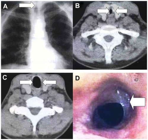
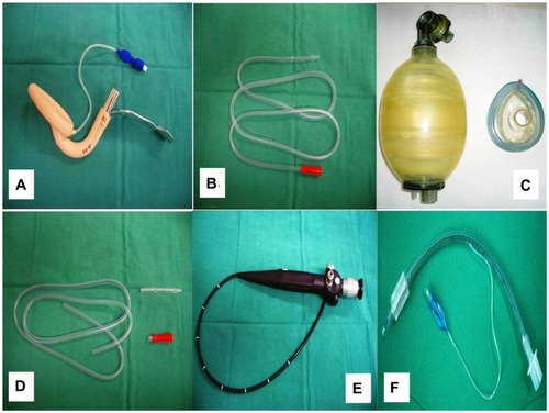
Figure 2 (A–C) Application of the Levin tube through the bronchoscope (steps). (D) Demonstration of the laryngeal mask with the bronchoscope and Levin tube. (E) Levin tube within the laryngeal steel handle mask. (F) Endotracheal tube with the Levin tube inserted as a guide.
Upon discharge the patient was given painkillers and anticoagulant treatment with low-molecular-weight heparin. Systemic corticosteroids (methylprednisolone) and inhaled treatment with nebulizer (bronchodilators ipratropium bromide and budesonide) were administered.
The patient was ventilated with an Ambu face mask (DIGAS GEORGE & Co., Thessaloniki, Greece) during his transport to the hospital and during the radiologic examination. Noninvasive ventilation was applied with a bilevel positive airway pressure model of inspiratory positive airway pressure of 12 and expiratory positive airway pressure of 6, and titration was applied according to the arterial blood gas, but the patient continued to have respiratory acidosis. Attempting to intubate the patient to sustain ventilation revealed edema of the oropharyngeal structures.
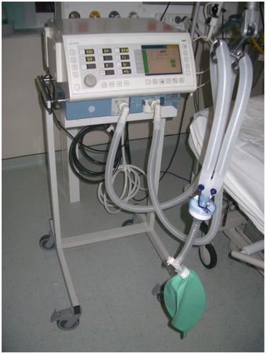
Since all attempts to intubate with lighted stylet failed, we decided to apply a laryngeal supraglottic steel handle mask. The patient was admitted into the intensive care unit and, with the use of a fiberoptic bronchoscope (model 11301ABN1, Storz insertion cord diameter 2.8 mm, insertion cord length 500 mm, working channel 1.2 mm; Karl Storz GmbH & Co. KG, Tuttlingen, Germany), an endotracheal tube of 7.5 mm (high-volume, low-pressure; Well Lead Medical Co, Guangzhou, China) was inserted. A red stomach tube (Levin’s type) of 18 (Fr/Ch) or 6.7 mm (size OD; Well Lead Medical Co) was firstly applied throughout the bronchoscope and was inserted into the laryngeal mask to be used as a guide for the endotracheal tube that we intended to use ( and ). The fiberoptic bronchoscope revealed a membranous web-like stenosis of the trachea (). The Levin tube was placed and the fiberoptic bronchoscope was removed. The endotracheal tube was then applied with the Levin tube as a guide. All laboratory findings were normal. The patient was ventilated with an Evita 2 Dura ventilator (Dräger Medical GmbH, Lübeck, Germany) (). We attributed the formation of the membranous tissue and consequently the tracheal stenosis to the former intubation period. Endoscopic treatment with laser incision and systematic steroid administration provided the solution (). The arterial blood gas on air after the endoscopic treatment was partial pressure of oxygen 82 mmHg, partial pressure of carbon dioxide 37 mmHg, pH 7.42, and bicarbonate 22 mmol/L.
Figure 3 (A) Laryngeal steel handle mask. (B) Levin tube. (C) Ambu face mask. (D) Levin tube with the edges cut off. (E) Bronchoscope. (F) Endotracheal tube.
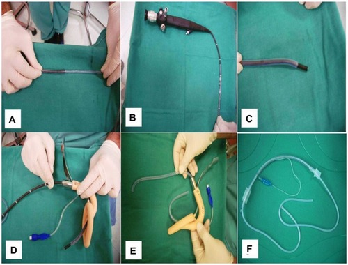
Figure 4 Evita 2 Dura (Dräger, Medical GmbH, Lübeck, Germany).
Discussion
The difficult airway has been defined as “the clinical situation in which a conventionally trained anesthetist experiences difficulty with mask ventilation of the upper airway, tracheal intubation, or both.”Citation10 DTI accounts for 17% of respiratory-related injuries and results in significant morbidity and mortality.Citation1 In fact, up to 28% of all deaths associated with anesthesia are due to the inability of a mask to ventilate or intubate.Citation1 The American Society of Anesthesiologists defines a difficult airway as the existence of clinical factors that complicate both ventilation administered through a face mask and intubation performed by an experienced person. The difficult airway algorithm of the American Society of Anesthesiologists was developed to guide clinicians in the management of the patient who is either predicted to have a difficult airway or whose airway cannot be adequately managed after induction of anesthesia.Citation10
Though the American Society of Anesthesiologists’ taskforce did not attempt to enumerate the features that identify those patients who may prove difficult to manage, it did recognize that an airway evaluation should be performed. Difficult ventilation is defined as the inability of a trained anesthesiologist to maintain oxygen saturation >90% using a face mask, with a goal of oxygen fraction of 100%. Difficult intubation is defined as the need for more than three attempts for intubation of the trachea or more than 10 minutes to achieve it, a situation that occurs in between 1.5% and 8% of general anesthesia procedures.Citation11,Citation12 Greater degree of difficulty in intubation is associated with greater incidence and severity of complications.Citation13
Up to 30% of anesthetic deaths can be attributed to a compromised airway.Citation14 This has generated the need for highly predictive tests for the identification of an airway with assumed intubation difficulty, to be applicable in all anesthetic and surgical procedures.Citation10,Citation15
There are several factors that may cause tracheal stenosis, including traumatic conditions, inflammatory diseases, benign and malignant lesions, collagen vascular diseases, and congenital conditions. Of these causes the leading cause of tracheal stenosis still continues to be endotracheal intubation, despite technological improvements such as the introduction of high-volume low-pressure cuffs and better patient care. Local inflammation and ischemia caused by an endotracheal tube can result in the upregulation of the fibrinolytic pathway, including C and S proteins locally, resulting in the creation of membranous-like stenosis.Citation16 Two studies, the first by Spittle and BeavisCitation16 and the second by Spittle and McCluskey,Citation17 present data that elucidate the underlying mechanism when a cuff pressure greater than 30 mmHg exceeds a critical point in the mucosal capillary perfusion pressure, causing mucosal ischemia leading to ulceration, chondritis of the tracheal cartilages, and, ultimately, development of irreversible fibrotic tissue. The endotracheal tube cuff causes circumferential erosion of the mucosa, which heals with a concentric (web-like) stenosis. It has been reported that based on the length of tracheal stenosis, the depth of the tracheal wall involvement, and the presence or not of tracheomalacia, postintubation tracheal stenosis falls into three categories: short, “complex”, and “pseudoglottic”.Citation18
Assessment of a difficult airway begins with a comprehensive medical history, and physical and regional examination. There are several key elements for the clinician to check: (1) variations in normal anatomy; (2) pathologic conditions; (3) a small mouth opening; (4) protruding upper teeth; (5) a large tongue; and (6) immobility of the head, neck, and jaw. Variations in “normal” anatomy and characteristic airway anatomy resulting from pathologic conditions can result in problems despite proper positioning and equipment ( and ).
Table 1 Most valuable scales/distances used in the prediction of difficult airway
Table 2 Congenital and acquired compromising conditions
Moreover, several conditions have been reported to pre-dispose patients to difficult airway intubation. These conditions include infections, trauma, obesity, endocrine factors, foreign body, tumors, inflammatory conditions, congenital problems, and physiologic conditions ().Citation10 Difficult airway intubation can result in numerous complications (). Infections such as epiglottitis, abscesses, croup, bronchitis, and pneumonia can affect airway management.Citation19 Radiological methods such as a computed tomography scan or a lateral neck radiograph may be helpful as an initial management of the underlying condition and should be tried, where possible.Citation10 Trauma also alters the airway structures. The ABC (airway, breathing, and circulation) rule should be followed in this situation. Indications for tracheal intubation include protection of the airway, airway obstruction, positive pressure ventilation, tracheal toilet, and a decreased level of consciousness. Alternatively, orotracheal intubation may be contraindicated or may not be possible in the patient with massive facial, laryngeal, or tracheal trauma.Citation20 A surgical airway may be necessary instead.Citation1 Moreover, obesity (body mass index ≥ 25 kg/m2) alters respiratory pathophysiology and distorts upper airway anatomy.Citation5,Citation21
Table 3 Difficult airway complications
In addition, patient age >55 years and lack of teeth has also been associated with DTI.Citation25 Finally, acromegaly, tumors, gastric reflux, and pregnancy are included among the factors predisposing a patient to DTI, either through morphologic and anatomic differentiations or pathogenic mechanisms of the underlying condition.Citation26–Citation28
Failure to intubate the trachea occurs in one in 2000 patients in the nonobstetric population and one in 300 patients in the obstetric population.Citation22 The need for equipment other than a direct laryngoscope may also help define DTI, although devices such as the gum elastic bougie (introducer) may or may not be viewed as part of standard technique. Therefore, the intubation difficulty scale is used, incorporating seven variables to calculate a score. An intubation difficulty scale score of 5 has been used to define DTI and, in a large study, occurred in 8% of patients.Citation23,Citation24
No single airway test can provide a high index of sensitivity and specificity for prediction of difficult airway. Therefore, a combination of multiple tests is used. The grading tools and scales most commonly used to assess difficult intubation are presented in .Citation13 They provide accuracy in assessment of preoperative stable patients. Emergency patients are more difficult to assess because of coexisting stress factors, hypoxemia, hypotension, and hypertension, and require intubation under less than optimal conditions. Furthermore, some patients with a difficult airway will remain undetected despite the most careful preoperative airway evaluation. Thus, anesthesiologists must always be prepared with a variety of preformulated and practiced plans for airway management in the event of an unanticipated difficult airway ().
Table 4 Accuracy indexes of prognostic tests for difficult airway
The most widely used scale is the Mallampati test, which originally categorized patients into three grades according to the ability to visualize the soft palate, fauces, uvula, and anterior and posterior pillars.Citation3 In 1987, Samsoon and Young added a fourth grade.Citation22 Cormack and Lehane also provided a grading system of four grades according to exposure of the larynx at laryngoscopy.Citation5 Their classification also underwent modification by Cook, who subdivided grade II into IIa and IIb, and grade III into IIIa and IIIb.Citation29 Wilson developed a scoring system that was based on body weight (<90 kg, 90–110 kg, >110 kg), head and neck movement, jaw movement, mandibular recession, and the presence or absence of protruding (“buck”) teeth.Citation30 Arne et alCitation31 developed a scoring system with seven individual predictive factors, including not only anatomical factors and scales but clinical symptoms and pathologies associated with difficult intubation and history of difficult intubation. Sensitivity ranged from 90% to 94%, and specificity was 66% and 96% for cancer and general surgery, respectively; nevertheless the positive predictive value was low (34%). In conclusion, the Mallampati test was more accurate than the other predictive factors. The LEMON airway assessment method () was assessed in two studies, which showed that patients in the difficult intubation group scored higher than those in other groups.Citation32,Citation33
Table 5 The LEMON assessment method
Despite the advances in available devices, most airway practitioners tend to resort to the surgical airway approach when facing difficulty in intubation, although the ACLS (advanced cardiac life support) guidelines include a variety of alternatives when tracheal intubation is not achieved.Citation10 There are two key parameters for the management of the difficult airway: (1) practitioner experience, and (2) clinical setting. During the last 10 years, many researchers agreed that the flexible fiberoptic endoscope is the single most useful tool when facing a difficult airway. Direct visualization of the upper airway, vocal cords, and tracheal placement ensures correct placement of the endotracheal tube. In some studies, the success rates were as high as 93.9%.Citation11,Citation34 The simplest and easiest approach to intubating using a flexible endoscope is that the larynx is in view and the bronchoscope passes through it. Then the operator can rotate the scope and bend its tip when navigating through a difficult airway. This method of intubation allows the practitioner to have a wide variety of choices regarding whether to intubate with the patient awake or asleep, or whether to use an oral or nasal pathway. Most experts agree that awake intubation in an informed adult patient is the safest choice, using local anesthesia and sedation when necessary, whereas children are more difficult to intubate awake. Awake intubation provides for spontaneous respiration and maintenance of upper airway tone.Citation35,Citation36
Conclusion
Despite the variety of prediction tests for the difficult airway, none can provide an accurate assessment, so every patient has to be considered as possibly having a difficult airway upon performing an intubation. A combination of scales could be used for early identification of difficult airway intubation with higher sensitivity and specificity results. In most cases the airway has to be maintained for a long period of time with adequate oxygenation and ventilation, and the intubation attempts have to be minimized to avoid injury and complications. Although the conventional laryngoscopic technique remains the standard with a high success rate, every physician who will be required to perform intubation has to be familiar with the process of evaluating a difficult airway and, in the event of the unanticipated difficult airway, be able to use a wide variety of tools and techniques to avoid complications and fatality. The flexible fiberoptic bronchoscope is the gold standard to predict difficult airway and to ensure tube position. Limitations involve purchasing and maintenance costs and skill development.
Acknowledgments
All authors contributed equally to the preparation of the manuscript.
Disclosure
Written informed consent was obtained from the patient upon discharge for publication of this case report and all accompanying images. The authors report no conflicts of interest in this work.
References
- BenumofJLManagement of the difficult adult airway. With special emphasis on awake tracheal intubationAnesthesiology1991756108711101824555
- XieZZChenJJScamellRWGonzalezMAAn interactive multimedia training system for advanced cardiac life supportComput Methods Programs Biomed199960211713110505967
- MallampatiSRGattSPGuginoLDA clinical sign to predict difficult tracheal intubation: a prospective studyCan Anaesth Soc J19853244294344027773
- RedickLFThe temporomandibular joint and tracheal intubationAnesth Analg19876676756763605679
- CormackRSLehaneJDifficult tracheal intubation in obstetricsAnaesthesia19843911110511116507827
- KitamuraTYamadaYDuHLHanaokaKEfficiency of a new fiberoptic stylet scope in tracheal intubationAnesthesiology19999161628163210598603
- ColeAFMallonJSRolbinSHAnanthanarayanCFiberoptic intubation using anesthetized, paralyzed, apneic patients. Results of a resident training programAnesthesiology1996845110111068624004
- RosenblattWHWagnerPJOvassapianAKainZNPractice patterns in managing the difficult airway by anesthesiologists in the United StatesAnesth Analg19988711531579661565
- BokhariABenhamSWPopatMTManagement of unanticipated difficult intubation: a survey of current practice in the Oxford regionEur J Anaesthesiol200421212312714977343
- American Society of Anesthesiologists Task Force on Management of the Difficult AirwayPractice guidelines for management of the difficult airway: an updated report by the American Society of Anesthesiologists Task Force on Management of the Difficult AirwayAnesthesiology20039851269127712717151
- LeeAFanLTGinTKarmakarMKNgan KeeWDA systematic review (meta-analysis) of the accuracy of the Mallampati tests to predict the difficult airwayAnesth Analg200610261867187816717341
- PaixADWilliamsonJARuncimanWBCrisis management during anaesthesia: difficult intubationQual Saf Health Care2005143e515933302
- Orozco-DiazEAlvarez-RiosJJArceo-DiazJLOrnelas-AguirreJMPredictive factors of difficult airway with known assessment scalesCir Cir201078539339921219809
- SalimiAFarzaneganBRastegarpourAKolahiAAComparison of the upper lip bite test with measurement of thyromental distance for prediction of difficult intubationsActa Anaesthesiol Taiwan2008462616518593650
- ShigaTWajimaZInoueTSakamotoAPredicting difficult intubation in apparently normal patients: a meta-analysis of bedside screening test performanceAnesthesiology2005103242943716052126
- SpittleCSBeavisSEPost-intubation tracheal stenosisHosp Med20016215411211466
- SpittleNMcCluskeyALesson of the week: tracheal stenosis after intubationBMJ200032172671000100211039970
- BissonABonnettePel KadiNBTracheal sleeve resection for iatrogenic stenoses (subglottic laryngeal and tracheal)J Thorac Cardiovasc Surg199210448828871405685
- BrookIMartinWJBacterial colonization in intubated newbornsRespiration19804063233287012971
- ChacurFHVilella FelipeLMFernandesCGLazzariniLCThe total face mask is more comfortable than the oronasal mask in noninvasive ventilation but is not associated with improved outcomeRespiration201182542643021846957
- BrodskyJBLemmensHJBrock-UtneJGVierraMSaidmanLJMorbid obesity and tracheal intubationAnesth Analg200294373273611867407
- SamsoonGLYoungJRDifficult tracheal intubation: a retrospective studyAnaesthesia19874254874903592174
- AdnetFBorronSWRacineSXThe intubation difficulty scale (IDS): proposal and evaluation of a new score characterizing the complexity of endotracheal intubationAnesthesiology1997876129012979416711
- AdnetFRacineSXBorronSWA survey of tracheal intubation difficulty in the operating room: a prospective observational studyActa Anaesthesiol Scand200145332733211207469
- KheterpalSHanRTremperKKIncidence and predictors of difficult and impossible mask ventilationAnesthesiology2006105588589117065880
- RameshBVVinodNMurugesanKPharyngeal airway changes following mandibular setback surgeryIndian J Dent Res200516414715016761707
- el-GanzouriARMcCarthyRJTumanKJTanckENIvankovichADPreoperative airway assessment: predictive value of a multivariate risk indexAnesth Analg1996826119712048638791
- SchmittHBuchfelderMRadespiel-TrogerMFahlbuschRDifficult intubation in acromegalic patients: incidence and predictabilityAnesthesiology200093111011410861153
- CookTMA new practical classification of laryngeal viewAnaesthesia200055327427910671848
- WilsonMEPredicting difficult intubationBr J Anaesth19937133333348398510
- ArneJDescoinsPFusciardiJPreoperative assessment for difficult intubation in general and ENT surgery: predictive value of a clinical multivariate risk indexBr J Anaesth19988021401469602574
- ReedMJDunnMJMcKeownDWCan an airway assessment score predict difficulty at intubation in the emergency department?Emerg Med J20052229910215662057
- SoyuncuSEkenCCeteYBektasFAkcimenMDetermination of difficult intubation in the EDAm J Emerg Med200927890591019857405
- HeideggerTGerigHJUlrichBSchniderTWStructure and process quality illustrated by fibreoptic intubation: analysis of 1612 casesAnaesthesia200358873473912859463
- KoernerIPBrambrinkAMFiberoptic techniquesBest Pract Res Clin Anaesthesiol200519461162116408537
- KlaftaJMFlexible tracheal tubes facilitate fiberoptic intubationAnesth Analg1994796121112127978458