Abstract
A congenital anomaly, optic nerve pit is often associated with serous retinal detachment involving macula. Long standing serous detachment leads to outer retinal atrophy and decrease in visual sensitivity. Recently, spectral-domain optical coherence tomography (OCT) has been reported to demonstrate a communication between the optic nerve sheath and the subretinal space. Vitreous cavity is proposed as an alternate source of fluid for accumulation in the subretinal space. We imaged a patient with optic nerve pit with Spectralis OCT and report the findings seen including the presence of an area of peripapapillary retinal atrophy, due to the spontaneous resolution of associated long-standing retinal detachment.
Introduction
A congenital anomaly, optic nerve pit is often (50%) associated with serous retinal detachment which can lead to vision loss over time.Citation1 The advent of optical coherence tomography (OCT) has provided evidence in support of a schisis cavity progressing to involve the outer retinal layers in patients with optic nerve pits.Citation2–Citation5 Our case report depicts atrophy of the outer retina in a patient with an optic nerve pit highlighted on spectral-domain OCT (SD-OCT) and fundus autoflourescence (FAF) imaging. Functional microperimetry (MP-1) confirmed loss of visual function in the affected area.
Case report
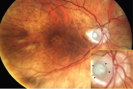
An 80-year-old male presented for a routine follow up for dry age-related macular degeneration (AMD). Best-corrected visual acuity was stable at 20/30 and 20/40, respectively. Anterior segment examination was significant for pseudophakia OU and otherwise unremarkable. Fundus examination revealed the presence of a temporal optic nerve pit OD (), with pigmentary changes and fine drusen in the macula OU.
Figure 1 Color fundus photograph of the right eye, depicting an optic nerve pit with peripapillary atrophy. Mild pigmentary changes are present in the macula. Arrow heads point to the margins of the pit.
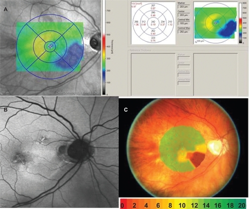
SD-OCT (Spectralis; Heidelberg Engineering, Heidelberg, Germany) sections through the pit demonstrate excavation in the optic nerve (). In addition, an area of outer retinal atrophy is noted adjacent to the optic nerve pit with preservation at the fovea (). No communication between the pit and the adjacent retina was identified on high resolution scanning. Retinal thickness mapping using automated analysis highlighted an area of retinal thinning that extended inferior temporally from the pit (). Hyperflourescence was present on FAF (Spectralis; Heidelberg Engineering) imaging in the area of retinal thinning (). MP-1 (Nidek, Gamagori, Japan) revealed significant reduction in threshold in this region, with preserved function at the fovea and sparing of retina superiorly ().
Figure 2 A) Combined infrared imaging and SD-OCT (spectralis; Heidelberg engineering, Germany) demonstrate excavation of the optic nerve with an adjacent area of outer retinal atrophy. Arrow heads pointing to the excavation seen on spectralis OCT. B) spectralis OCT scan demonstrates loss of the photoreceptor outer segments with preservation of the underlying retinal pigment epithelium. Arrow indicating the point where inner segment–outer segment junction of the photoreceptor becomes discontinues indicating atrophy. RPE is seen as a dark continuous band.
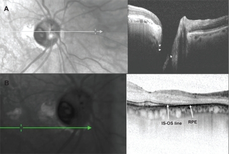
Figure 3 A) Retinal thickness mapping demonstrates thinning of the retina in the paillomacular bundle, extending inferior temporally. B) Fundus autoflourescence shows hyperfluorescence in the area of retinal thinning. C) Microperimetry (MP-1) demonstrates loss of threshold corresponding to the area of thinning. The blue dots are a fixation map of the patient while doing the test. Color codes indicate visual sensitivity in decibels.
Discussion
Sixty-three percent of optic nerve pits are temporal with the natural history of associated serous maculopathy resulting in vision loss over time.Citation1 In one report, 80% of patients had a final visual acuity of 20/200 or worse, with an average follow-up of nine years.Citation2 Subsequently, OCT findings have illustrated the connection between the serous detachment and optic nerve pit and confirmed the presence of a schisis cavity, which has been proposed to develop secondarily.Citation3–Citation5
In our case, Spectralis OCT showed preservation of photoreceptor outer segments at the fovea which correlated with normal threshold values on microperimetry and preserved Snellen visual acuity. Outer retinal thinning, marked by discontinuity of the inner segment–outer segment (IS–OS) line, was seen adjacent to the optic nerve pit, in an area of prior serous detachment. A recent case report describes retinal nerve fiber layer thinning (RNFL) in the papillomacular bundle in a young patient without corresponding changes on Goldman visual field testing.Citation5 However, data from MP-1 testing, in our case, confirm the relationship between photoreceptor loss and scotoma development, with preservation of the RNFL in this region.
The source of the schisis fluid has been debated over the years. It has been hypothesized that vitreous is a source of the schisis fluid and vitrectomy has been advocated as a treatment modality to collapse the schisis.Citation6,Citation7 However, in our case, we could not neither demonstrate any vitreous syneresis overlying the pit nor observe any kind of communication between the pit and the subretinal space.
FAF imaging revealed hyperflourescence in the area of the scotoma, indicating the accumulation of lipofuscin.Citation8 Spectralis OCT did not demonstrate defects in the underlying retinal pigment epithelium, suggesting the pathology is limited to the retina.
In conclusion, our report describes consecutive outer retinal atrophy in vivo with corresponding reduction in functional testing but preserved central vision in a patient with an optic disc pit.
Disclosures
The authors report no conflicts of interest in this work.
References
- SobolWMBlodiCFFolkJCWeingeistTALong-term visual outcome in patients with optic nerve pit and serous retinal detachment of the maculaOphthalmology19909711153915422255526
- KrivoyDGentileRLiebmannJMImaging congenital optic disc pits and associated maculopathy using optical coherence tomographyArch Ophthalmol199611421651708573019
- LincoffHSchiffWKrivoyDRitchROptic coherence tomography of optic disk pit maculopathyAm J Ophthalmol199612222642668694098
- SchneiderMGeitzenauerWAhlersCGolbazISchmidt-ErfurthUThree-dimensional imaging of an optic disk pit using high resolution optical coherence tomographyEur J Ophthalmol200919232132319253259
- MeyerCHRodriguesEBSchmidtJCCongenital optic nerve head pit associated with reduced retinal nerve fibre thickness at the papillomacular bundleBr J Ophthalmol200387101300130114507771
- HirakataAOkadaAAHidaTLong-term results of vitrectomy without laser treatment for macular detachment associated with an optic disc pitOphthalmology200511281430143516024082
- GeorgalasIPetrouPKoutsandreaCPapaconstadinouDLadasIGotzaridisEOptic disc pit maculopathy treated with vitrectomy, internal limiting membrane peeling, and gas tamponade: a report of two casesEur J Ophthalmol200919232432619253260
- Schmitz-ValckenbergSFleckensteinMGöbelAPEvaluation of autofluorescence imaging with the scanning laser ophthalmoscope and the fundus camera in age-related geographic atrophyAm J Ophthalmol2008146218319218514607