

Abstract
Aim: Sequence-specific CpG methylation of eukaryotic promoters is an important epigenetic signal for long-term gene silencing. We have now studied the methylation status of African swine fever virus (ASFV) DNA at various times after infection of Vero cells in culture. Methods & results: ASFV DNA was detectable throughout the infection cycle and was found unmethylated in productively infected Vero cells as documented by bisulfite sequencing of 13 viral DNA segments. Conclusion: ASFV DNA does not become de novo methylated in the course of infection in selected segments spread across the entire genome. Thus DNA methylation does not interfere with ASFV genome transcription. Lack of de novo methylation has previously been observed for free intracellular viral DNA in cells permissively infected with human adenoviruses, with human papillomaviruses and others.
Graphical abstract
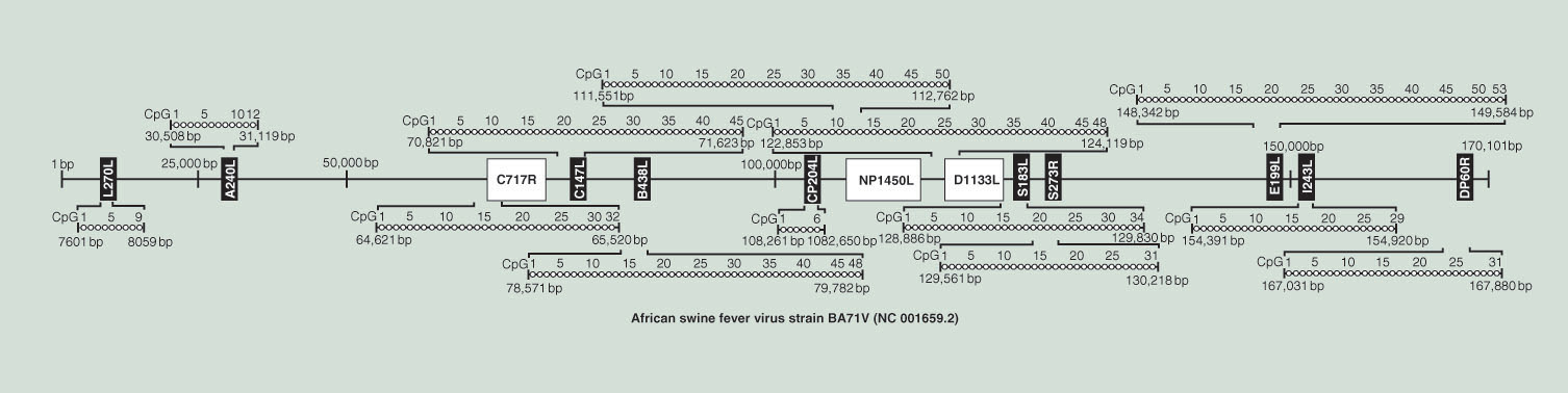
African swine fever virus (ASFV) is an important animal pathogen in many countries. In basic research on numerous different viruses, profound analyses of the molecular genetics of the virus have proved of paramount importance to understand its biological and medical characteristics. In this report, we describe the analysis of the methylation status of ASFV DNA after the infection of monkey Vero cells in culture. By applying the bisulfite sequencing technique, the gold standard in work on DNA methylation, no evidence was found for the presence of methylated cytosine residues in the BA71V strain of the ASFV genome in Vero cells. A selection of genome sites spread across the entire genome was analyzed; 5-methylcytosine (5-mC) residues were not found. The graph presents a map of the 170,101 base pair genome of the BA71V strain in which the bisulfite-analyzed genome segments and the viral genes therein have been indicated. The strings of open squares (□) lined up by brackets to the investigated genome sections symbolically depict unmethylated 5-mC residues. Thus, these genome segments have remained unmethylated throughout virus infection. We have analyzed 7.25% of all the methylation-sensitive CpG sites and 8.55% of the genes in the ASFV genome. Although unlikely, we cannot rule out the possibility that pockets of methylated sequences might exist somewhere in the genome.
Supplementary Data
To view the supplementary data that accompany this paper please visit the journal website at: https://www.tandfonline.com/doi/suppl/10.2217/epi-2017-0131
Author’s contributions
S Weber and A Hakobyan did all the experimental work, were involved in the planning of experiments and in the interpretation of the results. H Zakaryan and W Doerfler designed the project and many of the experiments. They also wrote the manuscript. S Weber and A Hakobyan have contributed equally to this work.
Acknowledgments
S Weber and W Doerfler are indebted to the Institute for Virology Erlangen University Medical School for their continued support of W Doerfler’s senior research group. S Weber and W Doerfler are grateful to the staff of the sequencing facility of the Institute of Human Genetics, University Hospital Erlangen for nucleotide sequence determinations.
Financial & competing interests disclosure
We are indebted to the Staedtler Foundation in Nürnberg, Germany for their support (WW/eh 01/15) to W Doerfler. We also thank the RA MES State Committee of Science, Armenia (16YR1F064) for a grant to H Zakaryan. H Zakaryan is also associated with the Department of Medical Biology, Yerevan State Medical University, 0025, Yerevan, Armenia. The authors have no other relevant affiliations or financial involvement with any organization or entity with a financial interest in or financial conflict with the subject matter or materials discussed in the manuscript apart from those disclosed.
No writing assistance was utilized in the production of this manuscript.