

Abstract
Aim:
It remains a challenge to accurately identify malignancy of thyroid nodules when biopsy is indeterminate. The authors aimed to investigate the abnormal DNA methylation signatures in papillary thyroid cancer (PTC) compared with benign thyroid nodules (BTNs).
Methods:
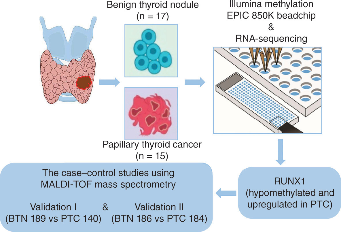
The authors performed genome profiling by 850K array and RNA sequencing in early-stage PTC and BTN tissue samples. The identified gene was validated in two independent case–control studies using mass spectrometry.
Results:
Hypomethylation of RUNX1 in PTC was identified and verified (all odds ratios: ≥1.50). RUNX1 methylation achieved good accuracy in differentiating early-stage PTC from BTNs, especially for younger women.
Conclusion:
The authors disclosed a significant association between RUNX1 hypomethylation and PTC, suggesting RUNX1 methylation as a potential biomarker for companion diagnosis of malignant thyroid nodules.
Graphical abstract
Data availability statement
All data for this study are in a formal, shared and widely applicable language and are assigned a globally unique and persistent identifier (DOI). In addition, the data are open, free and accessible, with rich descriptions, detailed provenance and compliance with domain-relevant community standards. The authors confirm that all data and reagents involved in the results and conclusions are fully represented in the article, figures and tables.
Results
Discovery & validation of significant hypomethylation & high expression of the RUNX1 gene in early-stage PTC cases compared with BTN patients
In the discovery study, the authors performed epigenome-wide screening using 850K BeadChip array and whole transcriptome screening using RNA sequencing in parallel in the fresh-frozen tissue samples of 15 early-stage PTC and 17 BTN. By overlapping differentially methylated and expressed genes, a CpG site, cg01725383, located at the 5′ untranslated region of the RUNX1 gene was identified (). Compared with BTN subjects, the CpG site cg01725383 located in the RUNX1 gene showed significant hypomethylation in early-stage PTC cases (the median and IQRs of RUNX1 methylation values: 0.68 [0.55–0.77] in BTN and 0.32 [0.21–0.46] in PTC; FDR-corrected p = 0.002; A). It is well known that the alteration in DNA methylation can affect gene expression. Indeed, compared with BTN subjects, the mRNA levels of RUNX1 in PTC cases were significantly increased (the median and IQRs of RUNX1 mRNA values: 0.67 [0.46–0.94] in BTN and 4.51 [3.62–5.13] in PTC; FDR-corrected p = 1.41E-07; B). Moreover, there was a significant inverse correlation between the methylation levels of cg01725383 and the mRNA levels of RUNX1, with a Spearman correlation coefficient of -0.78 (FDR-corrected p = 1.24E-07; C). Immunohistochemical results of human thyroid tissue showed that the RUNX1 protein was significantly deposited in patients with papillary thyroid cancer, and the RUNX1-positive area was significantly higher than that in BTN subjects (D). Consistently, the TCGA results illustrated that RUNX1 expression was significantly higher in the 512 patients with thyroid cancer than in the 337 normal healthy controls (FDR-corrected p = 4.77E-07; E). In addition, the qPCR result showed that the mRNA level of the RUNX1 gene was obviously upregulated in PTC cases compared with BTN subjects (F). Similarly, western blot results demonstrated that the RUNX1 protein was significantly overexpressed in PTC cases compared with BTN subjects (G & H). In summary, compared with BTN subjects, the RUNX1 gene was significantly hypomethylated and highly expressed in early-stage PTC patients, suggesting that the RUNX1 gene may play a crucial role as an oncogene in the occurrence and development of PTC.
Furthermore, the authors identified several other CpG sites with significantly different methylation patterns between PTC and BTN groups (Supplementary Table 2). The genes containing these CpG sites are integral to key signaling pathways, including the DDHD domain family (e.g., DDHD2), TGF-β (e.g., TGF-β1/2, SMAD3/4), WNT (e.g., WNT1/5, β-catenin), BMP (e.g., BMP4/6, MAPK6), EMT (e.g., vimentin, Twist1/2, ZEB1) and PI3K/AKT/mTOR (e.g., AKT1/2, mTOR1) pathways. Additionally, there was a significant positive correlation between the mRNA levels of several genes within the aforementioned signaling pathways and the mRNA levels of RUNX1. Moreover, the authors observed statistically significant differences in the mRNA expression levels of these genes between the PTC and BTN groups (Supplementary Table 3).
Validation of the association between RUNX1 hypomethylation & PTC in FFPE tissues by two independent case–control studies
The validation of the RUNX1 methylation difference between early-stage PTC cases and BTN subjects was carried out with FFPE tissue samples in two independent case–control studies (validation I and validation II; ). A 196 bp amplicon containing the cg01725383 site and flanking CpG sites was designed for further validation by MALDI-TOF mass spectrometry (A & Supplementary Table 4). No known single nucleotide polymorphisms or CpG sites were found within the primer sequence. Within the amplicon, the EpiTYPER system measured the methylation levels of four CpG sites and produced four distinguishable mass peaks; cg01725383 was referred to as CpG_1.
(A) Schematic diagram of the target amplicon within the RUNX1 gene. A 196 bp amplicon covering four differentially methylated CpG sites was located at the 5′ untranslated region of the RUNX1 gene (Chr21: 35,259,768–36,259,964, build GRCh37/hg19, defined by the University of California, Santa Cruz, Genome Browser). (B & C) Box plots for DNA methylation levels of the four CpG sites in the RUNX1 amplicon in benign thyroid nodule (BTN) and papillary thyroid cancer (PTC) detected by mass spectrometry in validation I (B) and validation II (C). Furthermore, validation I and validation II were combined and stratified by the age of 55 years old, gender, tumor size or tumor stage, respectively. (D–I) Box plots for DNA methylation levels of the four CpG sites in the RUNX1 amplicon in BTN and PTC detected by mass spectrometry in the age <55 years old group (D), age ≥55 years old group (E), male group (F), female group (G), tumor size (H) and tumor stage (I), respectively. All false discovery rate-corrected p-values were calculated by logistic regression with covariates adjusted.
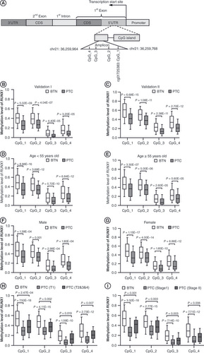
In validation I, consisting of 140 early-stage PTC cases and 189 age- and gender-matched BTN subjects, CpG_1 presented the most significantly reduced methylation levels in PTC cases compared with BTN subjects (methylation values of BTN and PTC: 0.41 vs 0.25). Similarly, CpG_2, CpG_3 and CpG_4 were hypomethylated in PTC cases compared with BTN subjects (methylation values of BTN and PTC, respectively: 0.39 vs 0.27, 0.08 vs 0.03, 0.15 vs 0.08; B & ). Furthermore, there was a significant association between RUNX1 hypomethylation and early-stage PTC. After adjusting for age and gender, the ORs per 10% reduced methylation of all CpG sites in the RUNX1 amplicon ranged from 1.50 to 1.96 (all FDR-corrected p ≤ 4.40E-05; ).
Table 2. Association between RUNX1 methylation and early-stage papillary thyroid cancer in two validation studies.
Consistent results were obtained in validation II, consisting of 184 early-stage PTC cases and 186 age- and gender-matched BTN subjects. The authors observed that the RUNX1 gene was significantly hypomethylated in PTC cases compared with BTN subjects. CpG_1 exhibited the most significantly reduced methylation levels in PTC cases compared with BTN subjects (methylation values of BTN and PTC: 0.62 vs 0.41). Similarly, CpG_2, CpG_3 and CpG_4 were hypomethylated in PTC cases compared with BTN subjects (methylation values of BTN and PTC, respectively: 0.52 vs 0.38, 0.23 vs 0.14, 0.35 vs 0.21; C & ). Binary logistic regression analyses revealed that hypomethylation of RUNX1 had robust associations with early-stage PTC. Covariate-adjusted ORs ranged from 1.72 to 2.13 per 10% reduction in methylation of each RUNX1 CpG site (all FDR-corrected p ≤ 2.38E-11; ).
Combination analysis of the association between FFPE tissue-based RUNX1 hypomethylation & early-stage PTC stratified by age & gender
The authors next investigated whether the association between RUNX1 hypomethylation and early-stage PTC was influenced by age and gender. To avoid the possible bias due to small sample size, the subjects in two validations were combined (324 PTC cases vs 375 BTN subjects) and binary logistic regression analysis was performed in gender- or age-stratified groups.
All participants were stratified by the age of 55 years old, which is a cutoff threshold for tumor staging of PTC cases [Citation22]. For the age <55 years group, all four CpG sites in the RUNX1 amplicon showed significantly decreased methylation levels in PTC cases compared with BTN subjects (D & ). After adjusting for gender, the ORs per 10% reduced methylation of the four CpG sites ranged from 1.62 to 1.89 (all FDR-corrected p ≤ 5.72E-10; ). Consistently, for the age ≥55 years group, the four CpG sites were significantly hypomethylated in PTC cases more than in BTN subjects (E & ). After adjusting for gender, the ORs per 10% reduced methylation of the four CpG sites ranged from 1.51 to 1.86 (all FDR-corrected p ≤ 5.60E-05; ). Compared with the age ≥55 years group, RUNX1 methylation and OR values showed a greater difference between BTN subjects and PTC cases in the age <55 years group, indicating that the RUNX1 hypomethylation was a great risk factor for younger people.
Table 3. Association between RUNX1 methylation and early-stage papillary thyroid cancer stratified by age or gender after combining validation I and validation II.
When stratified by gender, for males, all four CpG sites i the RUNX1 amplicon in PTC cases showed significantly decreased methylation levels compared with BTN subjects (F & ). After adjusting for age, the ORs per 10% reduced methylation of the four CpG sites ranged from 1.44 to 1.86 (all FDR-corrected p ≤ 0.001; ). Similarly, for females, the four CpG sites were significantly hypomethylated in PTC cases more than in BTN subjects (G & ). After adjusting for age, the ORs per 10% reduced methylation of the four CpG sites ranged from 1.61 to 1.85 (all FDR-corrected p ≤ 1.85E-10; ). Compared with the male group, RUNX1 methylation levels and OR values showed a greater difference between BTN subjects and PTC cases in the female group, indicating that the RUNX1 hypomethylation was a great risk factor for females.
Combination analysis of the correlation between RUNX1 methylation & the clinical characteristics of early-stage PTC
Next, 324 PTC cases combining two validations were stratified by clinical characteristics, and RUNX1 methylation differences were analyzed between subgroups by nonparametric tests. There was no significant correlation between RUNX1 methylation and tumor length, tumor size, lymph node involvement or tumor stage (all FDR-corrected p > 0.05; Supplementary Table 5). In spite of that, all four CpG sites in the RUNX1 amplicon showed significantly decreased methylation levels in PTC cases with T1 or at stage I compared with BTN subjects (H & I & ). After adjusting for age and gender, the ORs per 10% reduced methylation of the four CpG sites ranged from 1.50 to 1.66 for PTC cases with T1 and 1.48 to 1.59 for PTC cases at stage I (all FDR-corrected p ≤ 7.14E-10; ). All these results suggested that RUNX1 methylation is more effective in differentiating early-stage PTC cases with T1 or at stage I from BTN patients, although the accuracy of this conclusion might be limited by the small sample size of PTC cases with T2,3,4 or at stage II.
Table 4. Association between RUNX1 methylation and early-stage papillary thyroid cancer stratified by tumor size or tumor stage after combining validation I and validation II.
Validation of the clinical value of RUNX1 hypomethylation in distinguishing early-stage PTC from BTN
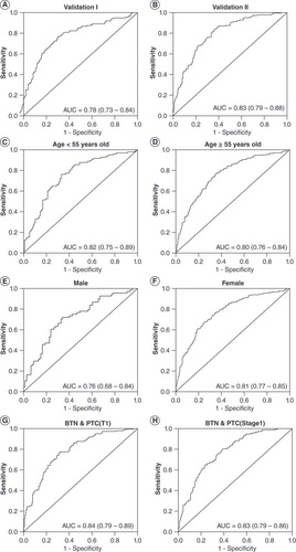
To evaluate the potential clinical application of RUNX1 hypomethylation as a biomarker of PTC cases, ROC curve analysis was performed and binary logistic regression analysis was conducted. First, the clinical efficacy of RUNX1 hypomethylation was assessed in two independent case–control studies (validation I and validation II), and the area under the ROC curve (AUC) was 0.78 (95% CI: 0.73–0.84) and 0.83 (95% CI: 0.79–0.88), respectively (A & B). Next, validation I and validation II were combined and stratified to investigate the effect of age, gender and clinical characteristics on the clinical value of RUNX1 hypomethylation. The results showed that the AUC (95% CI) was 0.82 (0.75–0.89) in subjects <55 years old and 0.80 (0.76–0.84) in subjects ≥55 years old (C & D). The AUC (95% CI) was 0.76 (0.68–0.84) and 0.81 (0.77–0.85) in male and female subjects, respectively (E & F). Furthermore, the AUC (95% CI) was 0.84 (0.79–0.89) in BTN subjects and PTC cases with T1, and it was 0.83 (0.79–0.86) in BTN subjects and PTC cases at stage I (G & H). Together, the AUC results demonstrated that RUNX1 hypomethylation based on FFPE tissue samples has high credibility and accuracy in distinguishing early-stage PTC from BTN, especially for younger women and PTC cases with T1 or at stage I.
The methylation levels of four CpG sites within the RUNX1 gene were generated a prediction probability. (A & B) Receiver operating characteristics curve analyses for the discriminatory power of RUNX1 prediction probability to distinguish papillary thyroid cancer (PTC) cases from benign thyroid nodule (BTN) subjects in validation I (A) and validation II (B). In addition, validation I and validation II were combined and stratified by the age of 55 years old, gender, tumor size or tumor stage, respectively. (C–H) Receiver operating characteristics curve analyses for the discriminatory power of RUNX1 prediction probability to distinguish PTC cases from BTN subjects in the age <55 years group (C), the age ≥55 years group (D), the male group (E), the female group (F), the tumor size group (G) and the tumor stage group (H), respectively. All the above 95% CIs of the area under the curve were calculated by logistic regression with covariates adjusted.
Discussion
In clinical practice, it is necessary to explore objective and stable biomarkers for the accurate diagnosis of thyroid nodules, which can not only prevent the overdiagnosis and overtreatment of BTN patients but also provide timely treatment for patients with nodules of malignant potential. Recently, the relationship between DNA methylation and thyroid cancer has been studied intensively and attracted wide attention [Citation17]. However, most reports have focused on cancerous and para-cancerous tissues, while only a few studies have involved the identification of benign and malignant thyroid tumors, mostly based on candidate genes [Citation25,Citation26]. In addition, these studies have shortcomings such as small sample size and poor systematic research content. The present study systematically investigated the value of tissue DNA methylation in the differential diagnosis of thyroid nodules in the Chinese population, thereby providing new evidence for the potential clinical application of methylated genes as cancer biomarkers. Furthermore, this study has reliable methods, rigorous design and sufficient sample size. The combined analysis of 850K BeadChip array and RNA sequencing in fresh-frozen tissue samples of early-stage PTC cases and BTN subjects revealed a strong negative correlation between reduced DNA methylation and increased mRNA of RUNX1. Subsequently, RUNX1 hypomethylation in PTC was validated by mass spectrometry in two independent studies with FFPE samples from a total of 699 BTN and PTC subjects.
RUNX proteins are a family of transcription factors with highly conserved sequences, which are involved in cell growth, proliferation and differentiation [Citation27]. RUNX1 is also known as AML1, and its abnormal expression or mutation can lead to human leukemia [Citation28]. Expression of RUNX1 is regulated by DNA methylation and post-translational modifications (e.g., phosphorylation, acetylation and ubiquitination) [Citation29]. First identified in acute myeloid leukemia, RUNX1 has also been found to play contrary roles in different solid tumors. For example, RUNX1 acts as a tumor suppressor gene in hepatocellular carcinoma and gastric cancer but as an oncogene in non-small-cell lung cancer and endometrial cancer [Citation30–33]. Additionally, RUNX1, an important transcription factor, has been implicated in the regulation of the TGF-β, WNT/β-catenin and BMP signaling pathways [Citation34]. A recent study has demonstrated upregulation of RUNX1 in PTC tissues compared with adjacent normal tissues, and the expression levels of RUNX1 were related to PTC stage. Knockdown of RUNX1 significantly reduced the malignancy of PTC cells and resulted in significant differential expression of downstream circRNAs, miRNAs and mRNAs. Further bioinformatic analyses revealed that the iron channel-related pathways, glycosaminoglycan synthesis and transcriptional disorder-related signaling pathways, including EMT, PI3K/AKT/mTOR and MAPK pathways, were enriched [Citation35]. Chu et al. found high expression of circRUNX1 in PTC, which promotes PTC progression and metastasis by sponging miR-296-3p and regulating DDHD2 expression [Citation36]. The RNA-sequencing results from the present study have confirmed the high expression of RUNX1 in PTC. Moreover, the mRNA levels of the potential downstream DDHD2 and the genes within the TGF-β, WNT/β-catenin, BMP, EMT and PI3K/AKT/mTOR signaling pathways exhibited a significant increase in PTC compared with BTN. The RNA sequencing results further illuminated a close and positive correlation between RUNX1 and many key genes within these aforementioned signaling pathways, the TGF-β, WNT/β-catenin and BMP pathways in particular. These noteworthy correlations and differences could offer valuable insights into the identification and comprehension of potential targets of RUNX1 in PTC initiation and metastasis. In addition, RUNX2 and RUNX3, which are in the same family as RUNX1, can significantly induce the proliferation and activation of human skin tissue-resident CD8+ memory T cells, enhance immune response and tumor immune monitoring, thus obviously improving the overall survival rate of melanoma patients, and can be used as biomarkers for diagnosis and prognosis [Citation37]. Previous studies have shown that abnormal RUNX1 methylation was implicated in tumor formation and malignant progression. For example, hematopoietic stem cells acquire a survival advantage by RUNX1 hypomethylation in familial leukemia [Citation38]; abnormal methylation in the RUNX family is negatively correlated with immune cell infiltration in breast cancer [Citation39]. So far, abnormal methylation of RUNX1 has not been reported in thyroid-related diseases. Here, the authors found that RUNX1 methylation was significantly reduced and its mRNA was significantly increased in early-stage PTC cases, suggesting that RUNX1 may act as an oncogene in the progression of PTC.
DNA methylation is an early and concomitant event during cancer progression [Citation40]. It has been well documented that cancer is a disease closely associated with aging and may have similar abnormalities in DNA methylation, mainly including hypomethylation in oncogene promoter regions or widespread hypermethylation in the gene body [Citation16,Citation41]. Due to the influence of sex hormones, there are inherent differences in DNA methylation at CpG sites in certain genes between males and females [Citation42,Citation43]. In China, the incidence of TC increases significantly along with aging and reaches its peak at 55 years old, and the incidence is three-times higher in women than in men [Citation44,Citation45]. Therefore, both age and gender are key factors in the formation and malignant progression of TC [Citation46]. The present study revealed that the association between RUNX1 hypomethylation and PTC cases was more obvious in subjects younger than 55 years and females in age- and gender-stratified analyses, suggesting that RUNX1 hypomethylation was a very important risk factor for PTC cases in young woman.
Clinically, tumor length, tumor size, tumor stage and lymph node involvement are strongly associated with malignant progression and poor prognosis of PTC patients [Citation47,Citation48]. In this study, no significant correlation was observed between RUNX1 methylation and malignant progression of PTC. Interestingly, the authors found that differences in RUNX1 methylation between PTC cases with T1 or at stage I and BTN subjects were more significant than those between PTC cases with T2&3&4 or at stage II and BTN subjects. Besides, RUNX1 methylation achieved good accuracy in differentiating early-stage PTC cases with T1 or at stage I from BTN subjects. Although these results might be less credible due to the small sample size of PTC patients with larger tumor size or at advanced stages, the findings suggested that the modulation of RUNX1 methylation may play different roles in the initiation and progression of PTC and further indicated the complex epigenetic regulation in cancer. The association between RUNX1 hypomethylation and PTC malignant progression needs further evaluation in future studies involving more subjects.
Both benign and malignant thyroid tumors have abnormal cell proliferation, DNA mutations or epigenetic changes, and large tumors are often accompanied by abundant angiogenesis and energy consumption [Citation49]. Nevertheless, benign tumors do not invade neighboring tissue or metastasize and can be safely monitored with no or minimal treatment, whereas malignant tumors are usually aggressive and have a chance of recurrence and metastasis after resection of the focus, seriously threatening the life and health of patients [Citation50,Citation51]. Therefore, accurately identifying the malignancy of thyroid tumors, especially those with indeterminate cytology, is essential for avoiding overtreatment and controlling tumor metastasis, which could benefit from supportive biomarkers. In this study, the authors identified RUNX1 methylation with high credibility and good accuracy in differentiating early-stage PTC cases from BTN subjects, suggesting it as a potential biomarker to aid the diagnostic process [Citation51,Citation52]. Future studies with larger sample size are warranted to further evaluate the clinical value of RUNX1 methylation in assessing different subtypes and the aggressiveness of thyroid cancer. The function and underlying molecular mechanisms of RUNX1 hypomethylation and overexpression in the malignant progression of thyroid cancer need further investigation. These results demonstrate a new tumor-driven indicator and provide novel insight into the etiology of PTC.
Conclusion
This study revealed significant hypomethylation and overexpression of RUNX1 in early-stage PTC cases compared with BTN subjects in a genome-wide study and further validated the altered RUNX1 methylation in two independent case–control studies by mass spectrometry. Our findings suggested that RUNX1 methylation might be used as a novel biomarker to distinguish malignant from benign thyroid nodules.
DNA methylation is an important event in the initiation and progression of thyroid diseases.
It is a great challenge to accurately identify the properties of thyroid nodules in clinical practice.
RUNX1 is significantly hypomethylated in early-stage papillary thyroid cancer (PTC) cases.
RUNX1 is highly expressed in early-stage PTC cases.
RUNX1 methylation achieves good accuracy in distinguishing the characteristics of thyroid nodules.
RUNX1 hypomethylation is an important risk factor for PTC in young women.
RUNX1 methylation is a novel biomarker to distinguish the malignancy of thyroid nodules.
RUNX1 may play a crucial role in the development of PTC.
Supplementary data
To view the supplementary data that accompany this paper please visit the journal website at: www.tandfonline.com/doi/suppl/10.2217/epi-2023-0338
Author contributions
J Li, C Jiang and R Yang conceived and designed this study; J Li and Y Yin wrote the main manuscript and prepared figures and tables; J Li, H Huang and M Li conducted major experiments; H Li and M Zhang were responsible for data interpretation and analysis; J Li drafted and R Yang revised the manuscript. All authors reviewed the manuscript.
Financial disclosure
This study was financed by the research funding of Nanjing Medical University and the research funding of the Jiangsu Province (grant no. 20182020). The authors have no other relevant affiliations or financial involvement with any organization or entity with a financial interest in or financial conflict with the subject matter or materials discussed in the manuscript apart from those disclosed.
Competing interests disclosure
The authors have no competing interests or relevant affiliations with any organization or entity with the subject matter or materials discussed in the manuscript. This includes employment, consultancies, honoraria, stock ownership or options, expert testimony, grants or patents received or pending or royalties.
Writing disclosure
No writing assistance was utilized in the production of this manuscript.
Ethical conduct of research
The studies were approved by the ethics committee of the Nanjing Medical University. Informed consent was obtained from all patients.