

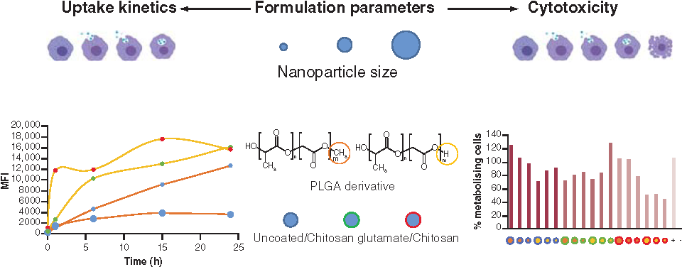
Abstract
Aim: This research aims to identify important formulation parameters for the enhancement of nanoparticle (NP) uptake and decreasing the cytotoxicity in macrophages. Materials & methods: Fluorescent poly(lactic-co-glycolic acid) (PLGA) nanocarriers were characterized for size distributions, zeta potential and encapsulation efficiency. Incubation time, size class, PLGA derivative and chitosan derivative were assessed for uptake kinetics and cell viability. Results: The major determining factor for enhancing cellular uptake were chitosan coatings, combined with acid-terminated PLGA and small NP size. Moreover, cytotoxicity was more favorable for small, chitosan glutamate-coated, acid-terminated PLGA NPs compared with its plain chitosan-coated counterparts. Conclusion: Chitosan glutamate has been shown to be a valuable alternative coating material for acid-terminated PLGA NPs to efficiently and safely target macrophages.
Graphical abstract
Supplementary data
To view the supplementary data that accompany this paper please visit the journal website at: www.tandfonline.com/doi/suppl/10.2217/nnm-2020-0317
Author contributions
This study was performed as a collaboration between the laboratory of Pharmaceutical Technology and Biopharmacy and the laboratory of Microbiology, Parasitology and Hygiene of the University of Antwerp. SV Hees, P Delputte and F Kiekens conceived and designed the study protocol. SV Hees and K Elbrink were responsible for the production and characterization of the NPs, as well as the phagocytosis experiments, the collecting, analysis and interpretation of the data and finally the drafting or revision of the manuscript. Therefore, SV Hees and K Elbrink are equal contributors to this work and share first authorship. M De Schryver substantially contributed to this work through the design and optimization of the uptake experiments and the critical revision of the drafted manuscript. Finally, P Delputte and F Kiekens also gave their insightful, critical input on the drafted manuscript.
Financial & competing interests disclosure
The authors have no relevant affiliations or financial involvement with any organization or entity with a financial interest in or financial conflict with the subject matter or materials discussed in the manuscript. This includes employment, consultancies, honoraria, stock ownership or options, expert testimony, grants or patents received or pending, or royalties.
No writing assistance was utilized in the production of this manuscript.
Ethical conduct of research
The authors state that they have obtained institutional review board approval from Universiteit Antwerpen for the research described.
Acknowledgments
The authors wish to thank all staff of The Laboratory of Microbiology, Parasitology and Hygiene of the University of Antwerp for their technical assistance and expertise during experiments. We would like to acknowledge P-B Feijens for the isolation of macrophages, NV Pelt for support during the resazurin assay and S Thys from the laboratory of Cell Biology and Histology at the University of Antwerp for providing the scanning electron microscopy pictures. We especially want to thank E Fransen of the University of Antwerp (STATUA) for the statistical analysis of the uptake kinetics data. Images of cells in the graphical abstract were created with BioRender.com. Finally, we wanted to dedicate this paper to our colleague, Prof. Dr S Apers, who passed away much too early on 5 February 2017.