Abstract
Background: Radiologic imaging is increasingly utilized as supplemental material in preclinical gross anatomy courses, but few studies have investigated its utility as a fully integrated instructional tool.
Aims: Establish the benefit of a teaching method that simultaneously correlates cadaveric and radiologic structures for learning human anatomy.
Method: We performed a mixed-methods randomized controlled trial and one-way cross-over study comparing exam grades and subjective student perception in a gross anatomy course. The intervention consisted of daily direct correlation small group sessions in which students simultaneously identified and correlated radiologic and cadaveric structures. The control method utilized identical laboratory and teaching conditions but students did not simultaneously correlate structures. Spatial relationships of structures within each respective media (gross or radiologic) were emphasized in both groups.
Results: No significant differences in radiology, gross, or written exam scores were observed between the intervention and control groups. The cross-over group preferred the intervention and control methods equally. The correlation teaching sessions ranked equally with active dissection as the most important instructional components of the course.
Conclusion: Direct, simultaneous correlation of radiologic and cadaveric structures did not affect exam scores or student preference but helped students understand anatomical concepts in comparison with other course components.
Introduction
The movement to utilize radiologic imaging in anatomy instruction continues to grow internationally. Calls for undergraduate medical students to have deeper anatomical understanding (Miller et al. Citation2002; Fitzgerald et al. Citation2008; Mukhtar et al. Citation2009) and more exposure to radiology (Squire Citation1969; Subramaniam et al. Citation2005) have led to a myriad of teaching methods that merge the two fields.
The most commonly reported methods to incorporate radiology with anatomy instruction include concurrent radiology lectures (Sullivan et al. Citation1987; Squire Citation1989; Erkonen et al. Citation1992), small group learning with (Forrester Citation1971) and without (Tegtmeyer et al. Citation1974; Whitley Citation1977) formal instructors, and radiologic images of de-identified patients in the dissection laboratory (Squire et al. Citation1975; Reidy et al. Citation1978; Bassett & Squire Citation1985; Turmezei et al. Citation2009). Others include problem-based learning (Navsa et al. Citation2004; Subramaniam et al. Citation2004; Subramaniam Citation2006), ultrasound workshops using students and human models (Teichgraber et al. Citation1996; Wittich et al. Citation2002), and radiologic imaging of dissection laboratory cadavers (McNiesh et al. Citation1983; Hisley et al. Citation2008). Notably, no reported methods to our knowledge formally teach cadaveric and image structures simultaneously – in the same time and space – with an emphasis on relationships between the same structures of different representations.
The ability to mentally toggle between the three- and two-dimensional representations of structures in cadavers and radiologic imaging requires mental rotation skills and conceptual understanding (Squire Citation1969). Both components have been shown to improve anatomical knowledge. For example, mental rotation activities unrelated to anatomy have been shown to quantitatively improve anatomy exam scores (Hoyek et al. Citation2009). In addition, studying spatial relationships of structures yields deeper conceptual understanding and improved anatomy exam scores (Mattick & Knight Citation2007; Pandey & Zimitat Citation2007).
Thus, we hypothesize that simultaneous formal instruction of cadaveric and gross structures will result in improved anatomical knowledge compared with traditional teaching methods. In particular, we expect improved anatomical knowledge will be reflected by higher exam scores and enhanced student preference for this method.
Methods
Study setting and participants
All protocols for this study were granted exemption status by the institutional review board, and written informed consent was obtained from all participants. All study participation was voluntary, blinded to instructors, and had no relationship with course grades.
Participants were recruited from the 102-member 2008 first-year class of a university medical school during the human anatomy course. The anatomy course and our study spanned two academic quarters. The gross laboratory component of the course lasted 3 hours each day. Five Medical Scientist Training Program students and one PhD student who began the course early were excluded from analysis.
Study design
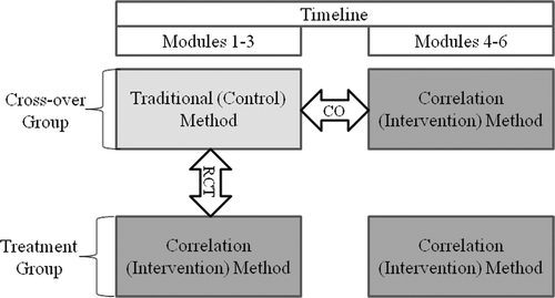
Mixed methods comprised a prospective, single-blinded, randomized controlled trial, a one-way cross-over design, and a survey were utilized. Students were randomized into two groups: cross-over and intervention. The cross-over group experienced the traditional teaching method for the first half of the course and the intervention method for the second half. The intervention group experienced only the intervention method for the entire course. Thus, the first half of the study was a randomized controlled design and was followed in the second half by a one-way cross-over design with the control group (). A randomized trial with a control group for the entire course was considered but rejected by the authors out of ethical concern for completely withholding a presumed beneficial intervention from students. A full cross-over design in which the intervention group experienced the control teaching method during the second half of the course was also considered. However, we anticipated that students may carry concepts of the intervention method into the control method phase, thus providing additional confounders to the data set.
Students were informed that various teaching methods would be utilized and studied during the course, but they were blinded to which components were being studied and which method they were experiencing. Control and cross-over groups were separated into two different sections of the dissection laboratory, divided by a wall, to maintain the integrity of the assigned teaching method. Four students were assigned to each cadaver table, and all students actively dissected.
Instruction methods
For the intervention teaching method, second- and fourth-year medical student teaching assistants (TA's) reviewed 15–20 pre-specified structures in both radiographic images and cadavers every day for 20 minutes during dissection in groups of approximately 10 students. Image structures were labeled and displayed on LCD panels within viewing distance of each cadaver, and cadaveric structures were dissected in the laboratory by students just before the image/cadaver correlation sessions.
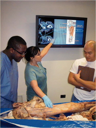
During the correlation sessions, students simultaneously identified structures on both an image and a cadaver during TA instruction, making note of the three-dimensional spatial relationships and tissue densities in both. Correlation between visualizing structures in the cadaver and in the images was emphasized (). For example, students were often asked to match an axial image to its approximate location in a cadaver, then describe how the image would appear differently as image slices were taken superiorly or inferiorly to the aforementioned image.
Figure 2. Demonstration of direct correlation of radiologic and cadaveric structures during daily teaching assistant small group teaching sessions.

The traditional method was identical to the intervention method with respect to images, structures identified, length of instruction, day and time, and instructors. However, no reference or correlation to cadavers was made. References to models were permitted in both intervention and traditional groups. Image structures were chosen for clinical relevance and image clarity by radiology faculty. Image modalities included computed tomography, magnetic resonance imaging, magnetic resonance angiography, X-ray imaging, angiography, sonography, and echocardiography. TA's rotated table assignments weekly and were monitored daily for adherence to the appropriate teaching method.
Data analysis
Multiple quantitative and qualitative measurements were used to best characterize various component impacts on learning, defined as exam scores, participant perception of integration and understanding of anatomy, and participant instructional method preference. Comparisons of demographic data between intervention and traditional groups utilized chi-square, Fisher's exact test, and t-tests, as appropriate.
Exams for each of six total body regions were composed of a radiology section, a gross anatomy section, and a written section. Radiology exams were composed entirely of new images that had never been previously seen by the students. Participant grades for each section type were compared between the traditional and intervention methods experienced during the randomized controlled phase of the study (first half of the course). To control for confounders, a mixed three-way ANOVA was performed. The randomization group (intervention vs. control) served as the between-subjects variable, and exam-type (radiology, gross, and written exams) and body region (thorax, abdomen, and pelvis.) served as the within-subjects variables, completed by all students. A power analysis assuming a moderate effect size (Murphy & Myors Citation2004) of f = 0.25, α ≤ 0.05, and 1 -β = 0.80 demonstrated a necessary sample N = 98 (Faul et al. Citation2007). Thus, variation in exam characteristics was accounted.
In a course conclusion survey, all participants were asked to rank (1 through 6, 1 = highest) the course's laboratory components with respect to influence on anatomical integration and understanding. Components included gross dissection, image/cadaver correlation, small group anatomy reviews, small group radiology reviews, a novel radiology study guide (Phillips et al. Citation2012), and guest physician clinical presentations. Average ranks were analyzed with a one-way ANOVA and compared with Tukey's post hoc test to account for multiple comparisons. During the same survey, participants in the cross-over group, which experienced both the traditional and intervention methods, were asked their preferences of the methods with regard to their personal definitions of learning and course goals. A power analysis for a two-tailed exact binomial goodness of fit test for a large effect size (g = 0.25, α ≤ 0.05, 1 − β = 0.80) demonstrated a required minimum sample size, N = 30 (Faul et al. Citation2007). Class size limited the ability to detect a smaller effect size, but the comparison was deemed to be nonetheless worthwhile to observe the extent of impact of the intervention.
All data was entered into Excel 2007 (Microsoft Corporation, Seattle, WA) and calculated with SPSS version 18 (Statistical Package for the Social Sciences Corporation, Chicago, IL).
Results
Of the 96 students in the 2008 first-year class of medical students who met inclusion criteria, 89 students responded to the survey, yielding a 93% response rate and all consented for analysis of their exams. The cross-over group was composed of 48 respondents (54% of total) and the intervention group of 41 respondents. No significant demographic differences were observed between groups ().
Table 1 Demographics of cross-over and intervention group participants
There was not a significant main effect of intervention vs. traditional teaching method on exam scores, F (1, 94) = 0.211, p = 0.647. Significant main effects on exam grades were observed for exam type, F (2, 188) = 195.38, p < 0.001, and for body region, F (2, 188) = 4.59, p < 0.001. Assumptions of sphericity were not met for the interaction effect of exam type and body region, Mauchly's test χ2(9) = 41.16, p < 0.001. A Greenhouse-Geisser correction (ε = 0.84) demonstrated a significant interaction effect, F (3.36, 315.89) = 57.60, p < 0.001.
Ranked course components differed significantly in their contributions to anatomy integration and understanding, F (5,484) = 37.7, p < 0.001. Students reported that cadaver dissection and cadaver/image correlation (intervention method) were equally the most helpful laboratory components (mean rank 2.37 vs. 2.62, respectively, p = 0.865). Small group anatomy review sessions in classrooms without cadavers led by staff members were equally as helpful as the image/cadaver correlations (3.04 vs. 2.62, respectively, p = 0.440), but not dissection (3.04 vs. 2.37, respectively, p = 0.036). The remaining laboratory components and their rankings are described in .
Table 2 Ranked comparison by students of the human morphology laboratory course components with respect to influence on integration and understanding of anatomy
Participants in the cross-over group, who experienced both intervention and traditional teaching methods, were asked their personal preferences regarding the use of radiology in the anatomy course. Of 43 total responses, 21 participants (49%) preferred the approach that ‘emphasizes gross anatomy correlations with radiological images’. The remaining 22 participants (51%) preferred the approach that ‘emphasizes anatomy and includes radiology instruction as supplemental material’. The approaches were preferred equally (exact binomial test, p = 1.000).
Discussion
An instructional method that simultaneously and directly correlated radiographic and cadaveric structures was found to be generally equivalent to the traditional method as measured by exams and student preference, in contrast to our initial hypothesis. Notably, the correlation instructional method was also reported by students to be one of the most influential components of the course with respect to integration and understanding of anatomy, equivalent to dissection.
Exam performance
In a previous study that provided passive access to radiographic imaging during dissection, students reported that direct instruction and additional labeling would further potentiate the use of radiographic imaging in the anatomy laboratory (Turmezei et al. Citation2009). In addition, there is a long-held belief and evidence that mental rotation ability and spatial reasoning are necessary for both radiologic and gross anatomical comprehension (Squire Citation1969; Squire et al. Citation1975; Folan & de Montfort Supple Citation1986; Terrell Citation2006; Khalil et al. Citation2008). Moreover, a series of self-guided radiology atlas study modules that emphasized spatial relationships found a significant improvement in radiology and gross practical exam scores (Phillips et al. Citation2012). All suggest that direct, simultaneous instruction should enhance anatomical comprehension. The similar exam scores we observed between the control and intervention groups may provide clarity to the current concepts of instructional methods for the complex spatial relationships of human anatomy.
First, the equivalent exam scores may suggest that inanimate models are as effective as cadavers for image and physical structure correlation instruction since both the control and intervention groups were permitted to correlate image structures directly with models in the laboratory. It is plausible that the metacognitive lessons from the visualization process are more important than the specific type of three- and two-dimensional structures used to learn the process. This possibility is supported by the finding of Hoyek et al. (Citation2009) that students who utilized mental rotation exercises that were unrelated to anatomy performed better on anatomy exams.
Alternatively, the equivalent scores may reflect an abundance of anatomy course components so that a single part contributed relatively little that was unique to the overall learning experience. The spatial reasoning objectives may have been sufficiently addressed in other course components. This possibility has important implications since contemporary gross anatomy courses are often composed of a multitude of components that have not only the potential to be mutually reinforcing but also distracting (Sugand et al. Citation2010).
It is additionally possible that the students who were motivated to grasp the deeper anatomical understanding offered by the intervention would have grasped the information without direct instruction. Smith and Mathias demonstrated that individual students choose different levels of learning approaches: deep, strategic, and superficial (Smith & Mathias Citation2007). Thus, it is plausible that students in both control and intervention groups who implicitly chose deep approaches gained the conceptual understanding by their own course of study, regardless of the intervention. Those students who chose superficial or strategic approaches may have limited their ability to benefit from the intervention.
Perceived influence of the teaching method on learning anatomy
The equal ranking of active dissection and post-dissection cadaver/image correlation contributes to the extensive debate around the value of dissection versus prosection. Our findings support the mixed literature that suggests the two environments are essentially equal in their instructional potential and that students learn from both (McLachlan & Patten Citation2006; Winkelmann Citation2007; Winkelmann et al. Citation2007). It is notable that the image/cadaver correlation instruction was only 20 minutes each day, considerably less than the remaining 2 hours 40 minutes of the dissection laboratory. The nonetheless equivalent ranking may be due to the timing of the correlation method at the end of the dissection laboratory during which students had been reviewing identities and locations of gross structures. There are no reported studies to our knowledge that assess the timing efficacies of radiologic interventions in gross anatomy (before, during, or after introduction to gross structures), and our observations suggest the need exists. These conjectures must be taken in the context that our study did not address why the shorter correlation instruction was as valuable to students as the extensive process of dissection, nor did it delineate physical dissection and study of completed dissections. Nonetheless, the observation is quite remarkable and suggests that direct correlation instruction on previously dissected cadavers promotes more focused instruction and learning than active dissection.
Additional influence
Direct correlation instruction of radiologic and cadaveric structures may bear merit beyond our end-points. The full extent of the correlation method's impact may yet to be observed because it cannot be realized until students begin clinical work. For example, the transfer gap between preclinical knowledge and clinical use is growing in recognition, and preclinical laboratories that incorporate clinical applications may help reduce the gap (Wilson et al. Citation2009). Direct correlation between cadavers of the preclinical laboratory with the corresponding radiology that will be used by the majority of students on a daily basis in their clinical practices may buffer the knowledge gap in ways that are difficult to measure.
Limitations
Our study is subject to several limitations. Of particular practical significance, the anatomy TA's were not openly supportive of the study concept nor the additional work to prepare for daily radiology instruction. Multiple studies have shown that the success of an educational intervention is heavily dependent on the behavior of the instructors (Kearney et al. Citation1991; Burroughs Citation2007; Gorzelsky Citation2009; Gunn Citation2010). This in itself could also explain some equivocal findings. A second limitation was the study's attenuated power for small effect sizes. However, we consider a medium effect size to be the minimum of practical significance in this setting.
Conclusion
In conclusion, our study demonstrated that direct correlation of radiologic and cadaveric structures to teach anatomy results in similar exam scores and student preference but plays an essential role in students’ understanding and integration of anatomy with respect to other course components. Future studies may be directed at the underlying reasons for student preference and the high importance students placed on the direct correlation sessions.
Acknowledgments
We thank the students, faculty, and staff of the Pritzker School of Medicine, whose dedication to continuously improving medical education pedagogies made this study possible. We are also grateful to Jim O’Reilly, PhD, for instructional support, Lorenzo Pesce, PhD, for consultation of statistical analyses, Cara V. Phillips for data compilation and graphics support, Charlene Sheridan for administrative support, and Kelly Ledbetter for graphics support. We thank Erin Mullarkey, Tunde Yerokun, and Kevin Choo, all students at the University of Chicago, for demonstrating the correlation teaching method in .
Financial support: This study was supported internally by the Pritzker School of Medicine and the Department of Radiology, both of the University of Chicago, Chicago, IL.
Declaration of interest: The authors report no conflicts of interest. The authors alone are responsible for the content and writing of the article. This study was internally funded by the Department of Radiology and Office of Medical Education, both of the University of Chicago.
References
- Bassett LW, Squire LF. Anatomy instruction by radiologists. Invest Radiol 1985; 20(9)1008–1010
- Burroughs NF. A reinvestigation of the relationship of teacher nonverbal immediacy and student compliance – Resistance with learning. Commun Educ 2007; 56(4)453–475
- Erkonen WE, Albanese MA, Smith WL, Pantazis NJ. Effectiveness of teaching radiologic image interpretation in gross anatomy. A long-term follow-up. Invest Radiol 1992; 27(3)264–266
- Faul F, Erdfelder E, Lang AG, Buchner A. G*Power 3: A flexible statistical power analysis program for the social, behavioral, and biomedical sciences. Behav Res Methods 2007; 39(2)175–191
- Fitzgerald JE, White MJ, Tang SW, Maxwell-Armstrong CA, James DK. Are we teaching sufficient anatomy at medical school? The opinions of newly qualified doctors. Clin Anat 2008; 21(7)718–724
- Folan JC, de Montfort Supple M. Visual memory and auditory recall in anatomy students. Med Educ 1986; 20(6)516–520
- Forrester D. Teaching anatomy through radiology. A new challenge requiring new techniques. Radiology 1971; 100(3)561–565
- Gorzelsky G. Working boundaries: From student resistance to student agency. Coll Compos Comm 2009; 61(1)64–84
- Gunn CL. Exploring MATESOL student "Resistance" to reflection. Lang Teach Res 2010; 14(2)208–223
- Hisley KC, Anderson LD, Smith SE, Kavic SM, Tracy JK. Coupled physical and digital cadaver dissection followed by a visual test protocol provides insights into the nature of anatomical knowledge and its evaluation. Anat Sci Educ 2008; 1(1)27–40
- Hoyek N, Collet C, Rastello O, Fargier P, Thiriet P, Guillot A. Enhancement of mental rotation abilities and its effect on anatomy learning. Teach Learn Med 2009; 21(3)201–206
- Kearney P, Plax T, Burroughs N. An attributional analysis of college students' resistance decisions. Comm Educ 1991; 40(4)325–342
- Khalil MK, Paas F, Johnson TE, Su YK, Payer AF. Effects of instructional strategies using cross sections on the recognition of anatomical structures in correlated CT and MR images. Anat Sci Educ 2008; 1(2)75–83
- Mattick K, Knight L. High-quality learning: Harder to achieve than we think?. Med Educ 2007; 41(7)638–644
- McLachlan JC, Patten D. Anatomy teaching: Ghosts of the past, present and future. Med Educ 2006; 40(3)243–253
- McNiesh LM, Madewell JE, Allman RM. Cadaver radiography in the teaching of gross anatomy. Radiology 1983; 148(1)73–74
- Miller SA, Perrotti W, Silverthorn DU, Dalley AF, Rarey KE. From college to clinic: Reasoning over memorization is key for understanding anatomy. Anat Rec 2002; 269(2)69–80
- Mukhtar Y, Mukhtar S, Chadwick SJ. Lost at sea: Anatomy teaching at undergraduate and postgraduate levels. Med Educ 2009; 43(11)1078–1079
- Murphy KR, Myors B. Statistical power analysis: A simple and general model for traditional and modern hypothesis tests2nd. Lawrence Erlbaum Associates, Mahwah, NJ 2004
- Navsa N, Boon JM, L'Abbé LN, Greyling LM, Meiring JH. Evaluation of clinical relevance of a problem-orientated head and neck module. SADJ 2004; 59(3)113–117
- Pandey P, Zimitat C. Medical students' learning of anatomy: Memorisation, understanding and visualisation. Med Educ 2007; 41(1)7–14
- Phillips AW, Smith SG, Ross CF, Straus CM. Improved performance in medical student gross anatomy through radiographic imaging. 2012, 10. Acad Radiol. Epub ahead of print, 26 April 2012; doi 1016/j.acra.2012.03.011
- Reidy J, Williams J, Dilly N, Fraher J. The learning of radiological anatomy by medical students. Clin Radiol 1978; 29(5)591–592
- Smith CF, Mathias H. An investigation into medical students' approaches to anatomy learning in a systems-based prosection course. Clin Anat 2007; 20(7)843–848
- Squire LF. Perception related to learning radiology in medical school. Radiol Clin North Am 1969; 7(3)485–497
- Squire LF. On teaching radiology to medical students: Challenges for the nineties. AJR Am J Roentgenol 1989; 152(3)457–463
- Squire LF, Twersky N, Pais JM, Becker JA. More effective devices for teaching undergraduate radiology. Radiology 1975; 117(1)63–65
- Subramaniam R, Hall T, Chou T, Sheehan D. Radiology knowledge in new medical graduates in New Zealand. N Z Med J 2005; 118(1224)U1699
- Subramaniam RM. Problem-based learning: Concept, theories, effectiveness and application to radiology teaching. Australas Radiol 2006; 50(4)339–341
- Subramaniam RM, Scally P, Gibson R. Problem-based learning and medical student radiology teaching. Australas Radiol 2004; 48(3)335–338
- Sugand K, Abrahams P, Khurana A. The anatomy of anatomy: A review for its modernization. Anat Sci Educ 2010; 3(2)83–93
- Sullivan DC, Effmann EL, Chen JT. Experience with an alternative curriculum. Invest Radiol 1987; 22(3)246–249
- Tegtmeyer CJ, Keats TE, Pullen EW, Langman J. The teaching of roentgen anatomy to medical students: A self-instructional approach. J Med Educ 1974; 49(5)455–456
- Teichgraber UK, Meyer JM, Poulsen Nautrup C, von Rautenfeld DB. Ultrasound anatomy: A practical teaching system in human gross anatomy. Med Educ 1996; 30(4)296–298
- Terrell M. Anatomy of learning: Instructional design principles for the anatomical sciences. Anat Rec B New Anat 2006; 289(6)252–260
- Turmezei TD, Tam MD, Loughna S. A survey of medical students on the impact of a new digital imaging library in the dissection room. Clin Anat 2009; 22(6)761–769
- Whitley J. Effectiveness of small-group self-instruction in radiographic anatomy. Invest Radiol 1977; 12(6)486–487
- Wilson AB, Ross C, Petty M, Williams JM, Thorp LE. Bridging the transfer gap: Laboratory exercise combines clinical exposure and anatomy review. Med Educ 2009; 43(8)790–798
- Winkelmann A. Anatomical dissection as a teaching method in medical school: A review of the evidence. Med Educ 2007; 41(1)15–22
- Winkelmann A, Hendrix S, Kiessling C. What do students actually do during a dissection course? First steps towards understanding a complex learning experience. Acad Med 2007; 82(10)989–995
- Wittich CM, Montgomery SC, Neben MA, Palmer BA, Callahan MJ, Seward JB, Bruce CJ. Teaching cardiovascular anatomy to medical students by using a handheld ultrasound device. JAMA 2002; 288(9)1062–1063
