
Abstract
Benign breast disease (BBD) is very common among women in their fertile age, but its correlation with breast reproductive function remains unclear. Our study aimed to investigate the relation between BBD and breast-feeding. We collected data on 105 women with BBD and 98 controls, focusing on their reproductive history and breast-feeding. We analysed data by R (version 2.12.1) considering p < 0.05 as significant. The results showed that fibroadenoma represented the most frequent BBD (55%), followed by fibrocystic changes (19%), intraductal papilloma (6%) and inflammatory breast disorders (5%). The mean age was 31.5 years (± 6.1), BMI 21.2 kg/m² (± 3.4) and age at menarche 13.0 years (± 1.5). Duration of breast-feeding was not significantly different between controls and BBD types (p = NS). Selecting women with fibroadenoma breast-feeding duration directly correlated with the number of benign lesions (p < 0.05), which remains significant also by multivariate analysis. It was concluded that there seemed to be no difference in breast-feeding among BBDs types, but lactation may influence the number of fibroadenomas. Moreover, prospective studies would better define the correlation between lactation and BBDs.
Introduction
The vast majority of lesions that occur in the breast are benign, but much concern is given to malignant ones because breast cancer is the most common malignancy in women in Western countries (Caleffiet al. Citation2004; Fitzgibbons et al. Citation1998; Sarnelli and Squartini Citation1991). The most frequent benign breast disease (BBD) is fibroadenoma (FA) (Dent and Cant Citation1989), which undergoes abnormal growth during the fertile age, mostly due to the oestrogen exposure, influenced by age, parity and hormonal status (Anderson et al. Citation1982).
Breast palpable mass and breast pain are the most frequently reported symptoms by women with BBD, sometimes affecting their quality of life.
With the use of modern imaging of the breast and the extensive use of needle biopsies, the diagnosis of BBD can be accomplished without surgery in the majority of patients.
The term BBD encompasses a heterogeneous group of lesions that may present a wide range of symptoms or may be detected as incidental microscopic findings. The incidence of benign breast lesions begins to rise during the 2nd decade of life and peaks in the 4th and 5th decades, as opposed to malignant diseases, for which the incidence continues to increase after menopause, although at a less rapid pace (Kabat et al. Citation2010; Shaaban et al. Citation2002; Pfeifer et al. Citation1999; Fitzgibbons et al. Citation1998; Morrow Citation1992; London et al. Citation1992; McDivitt et al. Citation1992; Sarnelli and Squartini Citation1991; Kelsey and Gammon Citation1990).
It is known that breast-feeding and pregnancy are protective against malignant breast disease and are risk factors for BBD (Ma et al. Citation2010; Minami et al. Citation1998). In the literature, it is not clarified if there is a correlation between breast-feeding and its duration with BBD. Moreover, we have only little information about BBD aetiology and correlation to women's reproductive function, despite its high prevalence in the young female population. The objective of our study is to investigate the possible correlation between BBD and breast-feeding.
Materials and methods
The series in this study consisted of women who presented to the Senology Outpatients Facility in our Department of Surgery during 2008, and a random control group of women who delivered in our Clinic of Obstetrics and Gynecology during 2008. This study was conducted according to the declaration of Helsinki, and with internal review board approval.
Women were referred to our outpatients facility complaining of breast pain, discomfort or incidental clinical findings. Among 355 women with a diagnosis of BBD, we chose to include those in their fertile age, under 40 years, with a histological diagnosis of BBD or a confirmed BI-RADS 1–2 (at least twice). We stated these selection criteria to minimise the possible inclusion of malignant pathologies. A total of 105 BBD and 98 controls were available for data analysis.
Information on reproductive history (age at menarche, age at first birth, age at last birth, history of abortion, parity, history of lactation for the last and previous children); medical history (histories of benign breast disease and gynaecological disease and family history of breast cancer); and other information (e.g. age, BMI, use of contraceptive or any other hormonal therapy) was collected by telephone interview, at routine outpatients visits or consulting clinical files of women affected by BBDs. In the control group, we collected the following information: age, parity, breast-feeding and its duration.
Women affected by BBD were regrouped into two major categories according to their parity (nullipara and non-nullipara). We then divided the non-nulliparous group of women who breast-fed, into two sub-groups based on breast-feeding duration. In particular, we took into consideration as duration cut-off the 3rd quartile of cumulative breast-feeding duration (corresponding to 20 months) and the 3rd quartile of breast-feeding duration per child (i.e. 13 months).
We evaluated breast size with a scale ranging from 1 to 8: 1 = 65 cm; 2 = 70 cm; 3 = 75 cm; 4 = 80 cm; 5 = 85 cm; 6 = 90 cm; 7 = 95 cm and 8 = 100 cm. We considered gynaecological age as the difference between the chronological age and the age at menarche. We included in the category of inflammatory breast disorders, only women who had been referred to the surgeon consultant for the following reasons: acute mastitis; granulomatous mastitis; foreign body reactions and recurring sub-areolar abscess. In the category of other BBDs, we grouped the following instances: isolated cysts; diabetic fibrous mastopathy; lipomas and adenomas. We defined BBDs as previously reported (Guray and Sahin Citation2006).
Data were analysed by R (version 2.12.1), considering p < 0.05 as significant. Monovariate analysis was performed by t-test, one way ANOVA, Wilcoxon test or Kruskal–Wallis test, in case of continuous variables, and χ2-test or Fisher's exact test, in case of categorical variables. Monovariate linear regression analysis and monovariate and multivariate logistic regressions were also performed.
Results
We included 105 women, younger than 40 years of age, affected by BBD, and 98 controls without BBD, and reported the characteristics of the population affected by BBD (). The mean women's age was 31.5 years (± 6.1); age at menarche 13.0 years (± 1.5); and gynaecological age 18.4 years (± 6.4) (). In the control group, the mean age was 32.3 years (± 4.5), and no significant difference was observed in comparison with the BBD group (p = NS).
Table I. Population characteristics.
The most common BBD was FA (55%), followed by fibrocystic changes (19%); isolated mastalgia (10%); intraductal papilloma (6%); inflammatory breast disorders (5%) and other BBDs (5%). Selecting only non-nullipara, FA and fibrocystic changes remained the most prevalent BBDs, but inflammatory breast disorders were significantly more common (8%) than in nullipara (3%) (). We found a mean BMI of 21.2 kg/m² (± 3.4) and a mean breast size of 2.8 (± 1.0), that significantly influenced BMI itself (effect 0.119; CI.95 0.051–0.185; p < 0.05).
Table II. BBDs prevalence in the general population, in nullipara and in pluripara women.
We had 43 women (41%) with a positive familial history of breast pathology, which was malignant in 31 cases (72%) and benign in the other 12 cases (28%). In our study, 63 women (60%) used contraception and 44 (41%) had at least one pregnancy, with a mean age at first pregnancy of 26.5 years (± 5.4). The 86% of these mothers breast-fed for a mean cumulative time of 14.8 months (± 13.1) and a mean time for each single pregnancy of 9.7 months (± 8.9).
The chronological and the gynaecological age results were significantly greater in women who had ever had a pregnancy (p < 0.05), and show a direct correlation with parity.
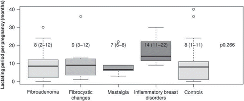
We found a non-significant difference in breast-feeding duration between BBDs and controls (); but we saw a non-significant longer breast-feeding duration in women who suffered from inflammatory breast disorders ().
Figure 1. Comparison of breast-feeding duration among BBDs and controls (median, interquartile range and Kruskal–Wallis test).
Considering only FA, which represents a benign proliferative breast disorder without atypia, among women who breast-fed, there is a direct correlation between the breast-feeding duration and the number of benign lesions (p < 0.05) ( and ), which remains significant also by multivariate logistic regression analysis ().
Table III. Comparison of nullipara with pregnant women who cumulatively breast-fed more or less than 20 months, and who breast-fed more or less than 13 months/child.
Table IV. Correlation between ≥ 2 locations of fibroadenoma and breast-feeding duration by mono- and multivariate logistic regression.
Discussion
Duration of breast-feeding it was not significantly different between controls and BBD types, but duration of lactation is significantly correlated to the number of lesions in FA.
In our population, the most common BBDs were FA and fibrocystic changes. However, according to the literature, inflammatory breast disorders were significantly more common among women who had a pregnancy than in nullipara, being usually managed with antibiotics and, in the case of abscess, with percutaneous drainage with good aesthetic results (Romero et al. Citation2007; Amir and Pakula Citation1991).
Mastalgia is reported by 47% of women, and represents an isolated symptom in 10% of cases, in the absence of any other evident breast disorder. In their fertile age, a great number of women are affected by mastalgia, probably depending on a combination of hormonal stimuli on the mammary gland, which cause pain by periodically increasing breast tension (Goyal Citation2011).
Focusing on the correlation between BBD and breast-feeding, our data reveal no significant differences based on breast-feeding duration (). However, selecting only women with fibroadenoma, there seems to be a direct correlation between the breast-feeding duration and the number of benign lesions, which remains significant also by multivariate analysis.
To our knowledge, we describe this correlation for the first time. In the previous literature, we found a positive correlation between lactation duration and BBDs of proliferative type (Minami et al. Citation1998), but not between lactation history and BBDs in general (Minami et al. Citation1998).
We know that hyperplastic lobules, histologically identical to clinical FA, are present so commonly as to be regarded as normal, and can probably be found in all breasts if they are sought with sufficient care. All the cellular elements of FA are normal, and epithelium and myoepithelium maintain a normal relationship (Courtillot et al. Citation2005), but FAs are a proliferative benign breast disease and present a small increase in breast cancer (BC) risk (Santen and Mansel Citation2005; Minami et al. Citation1998).
Nulliparity and increased age at first birth were clearly associated with hormone receptor-positive tumours, but not with triple-negative BC (Yang et al. Citation2011). Among women with invasive BC, higher parity and the absence or short duration of breast-feeding were independently associated with triple-negative BC (Shinde et al. Citation2010). Therefore, even if parity is a known protective factor against BC in general (Yang et al. Citation2011; Ma et al. Citation2010), it seems to play a role, as well as lactation, in triple-negative BC and BBD development. In our opinion, a better understanding of the influence of pregnancy on those pathologies could give us more information on their aetiologies.
Despite the retrospective nature of our study, the detailed information available gave us the opportunity to explore new and interesting correlations in the field of BBD and pregnancy. We also point out that, to give a strong answer to our question, a prospective study is required.
Conclusion
In conclusion, lactation may influence the number of fibroadenomas, but we need more information to better clarify the aetiologies of BBD of proliferation type. In fact, we need to better understand why people affected by some BBD will develop cancer more frequently than the general population. Moreover, prospective studies would better define the correlation between lactation and BBDs in terms of disease evolution and impact on breast-feeding and that is of paramount importance for the newborn.
Declaration of interest: The authors report no conflicts of interest. The authors alone are responsible for the content and writing of the paper.
References
- Amir LH, Pakula S. 1991. Nipple pain, mastalgia and candidiasis in the lactating breast. Australian and New Zealand Journal of Obstetrics and Gynaecology 31:378–380.
- Anderson TJ, Ferguson DJ, Raab GM. 1982. Cell turnover in the ‘resting’ human breast: influence of parity, contraceptive pill, age and laterality. British Journal of Cancer 46:376–382.
- Caleffi M, Filho DD, Borghetti K, Graudenz M, Littrup PJ, Freeman-Gibb LA . 2004. Cryoablation of benign breast tumors: evolution of technique and technology. Breast 13:397–407.
- Courtillot C, Plu-Bureau G, Binart N, Balleyguier C, Sigal-Zafrani B, Goffin V . 2005. Benign breast diseases. Journal of Mammary Gland Biology and Neoplasia 10:325–335.
- Dent DM, Cant PJ. 1989. Fibroadenoma. World Journal of Surgery 13: 706–710.
- Fitzgibbons PL, Henson DE, Hutter RV. 1998. Benign breast changes and the risk for subsequent breast cancer: an update of the 1985 consensus statement. Cancer Committee of the College of American Pathologists. Archives of Pathology and Laboratory Medicine 122:1053–1055.
- Goyal A. 2011. Breast pain. Clinical evidence. Available at: http://clinicalevidence.bmj.com/ceweb/conditions/woh/0812/0812-get.pdf (Accessed 29 August 2011).
- Guray M, Sahin AA. 2006. Benign breast diseases: classification, diagnosis, and management. Oncologist 11:435–449.
- Kabat GC, Jones JG, Olson N, Negassa A, Duggan C, Ginsberg M . 2010. A multi-center prospective cohort study of benign breast disease and risk of subsequent breast cancer. Cancer Causes and Control 21: 821–828.
- Kelsey JL, Gammon MD. 1990. Epidemiology of breast cancer. Epidemiologic Reviews 12:228–240.
- London SJ, Connolly JL, Schnitt SJ, Colditz GA. 1992. A prospective study of benign breast disease and the risk of breast cancer. Journal of the American Medical Association 267:941–944.
- Ma H, Henderson KD, Sullivan-Halley J, Duan L, Marshall SF, Ursin G . 2010. Pregnancy-related factors and the risk of breast carcinoma in situ and invasive breast cancer among postmenopausal women in the California teachers study cohort. Breast Cancer Research 12:R35.
- McDivitt RW, Stevens JA, Lee NC, Wingo PA, Rubin GL, Gersell D. 1992. Histologic types of benign breast disease and the risk for breast cancer. The cancer and steroid hormone study group. Cancer 69:1408–1414.
- Minami Y, Ohuchi N, Taeda Y, Fukao A, Hisamichi S. 1998. Risk factors for benign breast disease according to histopathological type: comparisons with risk factors for breast cancer. Japanese Journal of Cancer Research 89:116–123.
- Morrow M. 1992. Pre-cancerous breast lesions: implications for breast cancer prevention trials. International Journal of Radiation Oncology, Biology, Physics 23:1071–1078.
- Pfeifer JD, Barr RJ, Wick MR. 1999. Ectopic breast tissue and breast-like sweat gland metaplasias: an overlapping spectrum of lesions. Journal of Cutaneous Pathology 26:190–196.
- Romero C, Lombardía J, Almenar A, Calvo P, Fandiño E, Aso S . 2007. [Diagnosis and treatment of benign breast lesions during pregnancy]. Radiologia 49:255–261.
- Santen RJ, Mansel R. 2005. Benign breast disorders. New England Journal of Medicine 353:275–285.
- Sarnelli R, Squartini F. 1991. Fibrocystic condition and ‘at risk’ lesions in asymptomatic breasts: a morphologic study of postmenopausal women. Clinical and Experimental Obstetrics and Gynecology 18:271–279.
- Shaaban AM, Sloane JP, West CR, Moore FR, Jarvis C, Williams EMI . 2002. Histopathologic types of benign breast lesions and the risk of breast cancer: case-control study. American Journal of Surgical Pathology 26:421–430.
- Shinde SS, Forman MR, Kuerer HM, Yan K, Peintinger F, Hunt KK . 2010. Higher parity and shorter breastfeeding duration: association with triple-negative phenotype of breast cancer. Cancer 116:4933–4943.
- Yang XR, Chang-Claude J, Goode EL, Couch FJ, Nevanlinna H, Milne RL . 2011. Associations of breast cancer risk factors with tumor subtypes: a pooled analysis from the breast cancer association consortium studies. Journal of the National Cancer Institute 103:250–263.