

Dear Sir,
We were pleased to read the recent case report on cervical schistosomiasis (Al-Baghdadi et al. Citation2014). We would like to add a few important points illustrated by a case presenting with cervical sandy patches. In our experience, this is a common presentation of the disease in endemic areas.
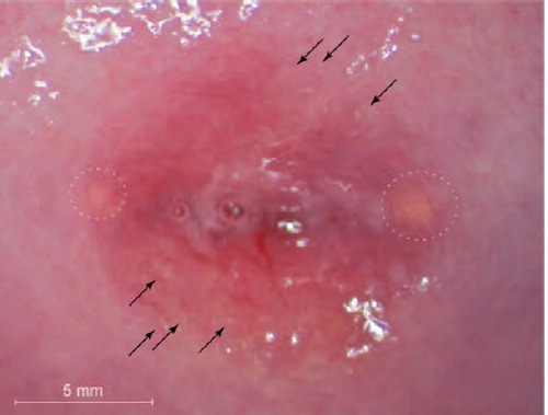
Schistosoma haematobium ova have been found in all genital organs and are characterised by so-called sandy patches with yellow colour on mucosal surfaces on colposcopy (Kjetland et al. Citation2012). The mucosal cervical lesions are largely unknown, often subtle and likely to go undiagnosed. However, from our current work in an endemic area in South Africa, we find that sandy patches are very common if looked for by colposcopy. A typical case may present with scattered, single grains of a yellow colour in the cervical mucosa (). The lesions are often surrounded by branched, reticular blood vessels of uneven calibre.
Figure 1. Female genital schistosomiasis lesions. This figure shows an image from a photocolposcopic examination. Single grains are seen in the ectocervical mucosa (arrows point at examples). Nabothian cysts are also seen in the transformation zone (dashed circles). The image was captured using an Olympus OCS 500 colposcope with a mounted Olympus E-420 10 megapixel (MP) single lens reflex camera. Urine microscopy as well as urine PCR was negative for S. haematobium ova. However, Schistosoma PCR in cervico-vaginal lavage was positive. STI tests were negative except for HPV and HIV. The Pap smear showed LSIL.
It has been hypothesised that vulvar tumours caused by S. haematobium are more often seen in the young, however these presentations are rare and no studies to-date have adequately tested for differential diagnoses (Hegertun et al. Citation2013).
Al-Baghdadi et al. (Citation2014) suggest that a biopsy serves as the definitive diagnostic tool. However, the biopsy must be used with caution in many endemic areas due to the co-endemicity with HIV; the iatrogenic lesion may increase the risk of HIV acquisition until the wound heals. Urine microscopy for ova is not a reliable diagnostic tool for genital lesions (Kjetland et al. Citation2012). The Pap smear has been found to have low sensitivity for FGS (Poggensee et al. Citation2001). PCR may be a useful test, but is likely to be negative in chronic lesions where calcified, dead ova are predominant and parasite DNA is absent (Pillay et al. Citation2014; Kjetland et al. Citation2009). However, such lesions may still be a risk factor for HIV infection (Jourdan et al. Citation2011). Therefore, gynaecological examination with colposcopy is currently the preferred diagnostic method. shows that large magnification may be necessary to see the subtle manifestations of female genital schistosomiasis; lesions are easily overlooked.
The clinician working in an endemic area or with immigrants and travellers should be aware that the diagnosis is easily missed. Sandy patches may also be an important differential diagnosis to other infectious and cancerous lesions (Kjetland et al. Citation2012). Unnecessary treatment and biopsies may be the consequence. Praziquantel is the recommended treatment for schistosomiasis, but the effect on genital lesions is still unknown (Kjetland et al. Citation2012).
Declaration of interest: The authors report no conflicts of interest. The authors alone are responsible for the content and writing of the paper.
References
- Al-Baghdadi O, Samarasinghe A, Wissa I. 2014. Cervical schistosomiasis. Journal of Obstetrics and Gynaecology 34:206.
- Hegertun IE, Sulheim Gundersen KM, Kleppa E, Zulu SG, Gundersen SG, Taylor M et al. 2013. S. haematobium as a common cause of genital morbidity in girls: a cross-sectional study of children in South Africa. PLoS Neglected Tropical Diseases 7:e2104.
- Jourdan PM, Holmen SD, Gundersen SG, Roald B, Kjetland EF. 2011. HIV target cells in Schistosoma haematobium-infected female genital mucosa. American Journal of Tropical Medicine and Hygiene 85: 1060–1064.
- Kjetland EF, Hove RJ, Gomo E, Midzi N, Gwanzura L, Mason P et al. 2009. Schistosomiasis PCR in vaginal lavage as an indicator of genital Schistosoma haematobium infection in rural Zimbabwean women. American Journal of Tropical Medicine and Hygiene 81:1050–1055.
- Kjetland EF, Leutscher PD, Ndhlovu PD. 2012. A review of female genital schistosomiasis. Trends in Parasitology 28:58–65.
- Pillay P, Taylor M, Zulu SG, Gundersen SG, Verweij JJ, Hoekstra P et al. 2014. Real-time polymerase chain reaction for detection of Schistosoma DNA in small-volume urine samples reflects focal distribution of urogenital Schistosomiasis in primary school girls in KwaZulu Natal, South Africa. American Journal of Tropical Medicine and Hygiene 90: 546–552.
- Poggensee G, Sahebali S, Van Marck E, Swai B, Krantz I, Feldmeier H. 2001. Diagnosis of genital cervical schistosomiasis: comparison of cytological, histopathological and parasitological examination. American Journal of Tropical Medicine and Hygiene 65:233–236.