Dear Sir,
Thank you so much for according the opportunity to respond to the letter by Drs. Mano et al. I appreciate the interest shown by them in my technique.
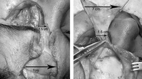
Drs. Mano et al. present an interesting but untenable hypothesis about survival of our flap. We have a true axial flap with an identifiable vessel in its pedicle. The supratrochlear vessels are always confirmed preoperatively using a hand-held Doppler probe. Such a loud and distinct signal cannot be picked up from a random blood supply as suggested by Drs. Mano et al. In this island, any input from the branches of ipsilateral superficial temporal vessel has also been severed. The inclusion of the periosteal cuff and the frontogaleal muscle in the flap ensures that the axiality is maintained even up to 10 cm from the supraorbital margin.Citation1 Our flap has a very narrow pedicle of about 1.5 cm around the vessel and this facilitates its smooth passage into the defect (). In the unlikely event of both supratrochlear and supraorbital vessels having been sacrificed because of surgery, merely depending upon the random blood supply would be perilous. In such a scenario, alternative reconstructive options have to be explored.
FIGURE 1 Showing 7 cm wide flap designed on a narrow 1.5 cm pedicle containing the vessels (left). The flap being easily transferred into the defect (right).
An island of skin as used by us helps blanking the orbit and provides more stable wound cover as compared to a skin graft on the pericranium.
Declaration of interest: The author reports no conflict of interest.
REFERENCES
- Fukuta K, Potparic Z, Sugihara T, Rachmiel A, Forté RA, Jackson IT. A cadaver investigation of the blood supply of the galeal frontalis flap. Plast Reconstr Surg 1994;94:794–800.