
Abstract
Purpose: A model of in vivo porcine kidneys is used to clarify the characteristics of laparoscopic microwave ablation (MWA) of renal tissue. Materials and methods: Six pigs were utilised for the experiment using 18G water circulating-cooling microwave needles. The operating frequency was 2450 MHz, and the independent variables were power (50–60 W) and time (300–600 s). The kidneys were dissociated laparoscopically and ablated with a single or double needle at different power/time combinations and depths of puncture. Changes in the kidneys were carefully observed. Specimens of the ablated lesions were stained with haematoxylin-eosin (H&E) to evaluate the pathological features. Results: Thirty-four thermoablations were applied. The effective ablation shape was similar to a chestnut. The ablated lesions could be divided into three zones: carbonization zone, coagulation zone, and inflammatory reaction zone. The ablation zone enlarged with increasing power and time. When combined with two needles, the maximum diameter of the ablated lesions significantly increased. Pathological results indicated that renal tissues of the carbonisation zone were thoroughly necrotic. Coagulative necrosis was observed in the coagulation zone. No ‘skipped’ areas were noted in any ablation zone. The structure of the inflammatory reaction zone was integrated, and interstitial small blood vessels were highly expanded and congested with infiltrated inflammatory cells. Conclusions: MWA achieved excellent effects in this porcine model. It can be safely and effectively used in renal tissue. For patients with poor physical condition or small renal masses (<4 cm), we can refer to these data and select the appropriate combinations to obtain satisfactory therapeutic efficacy.
Introduction
Kidney cancer is one of the most common cancers of the urinary system. Due to improvements in health awareness and the widespread use of imaging examinations, the detection rate of kidney cancer continues to increase [Citation1,Citation2]. According to the literature, from 2001 to 2010 a total of 342,501 patients were diagnosed with kidney cancer in the USA, and the incidence of newly diagnosed small renal masses has increased by approximately 2.5–3% annually [Citation3,Citation4]. Additionally, because of our ageing society, patients with multiple co-morbidities or chronic renal failure have increased significantly [Citation5,Citation6]. Research has indicated that 22% of patients who present for surgery with a ‘normal’ baseline serum creatinine level have chronic kidney disease (CKD) stage III or greater. Moreover, in patients 70 years or older, 40% have CKD stage III [Citation7]. These cumulative data highlight the importance of renal preservation and as a result have altered the diagnosis and treatment of stage T1 renal tumours over the past 20 years [Citation8,Citation9]. Previously, solid kidney tumours encountered in the clinic were often considered malignant and radical nephrectomy was actively considered; however, now we have realised that there are substantial differences in the biological characteristics of these tumours and radical nephrectomy may damage renal function. This new concept has deeply affected the therapeutic area. To conserve renal units in response to a decline in renal function, nephron-sparing surgery has become the mainstream therapy for early kidney cancer [Citation10–12]. In 2012, Kim et al. [Citation13] published a systematic review and meta-analysis on the comparative effectiveness for the survival and renal function of partial and radical nephrectomy for localised renal tumours. The findings suggested that partial nephrectomy conferred a survival advantage and a lower risk of severe chronic kidney disease after surgery for localised renal tumours. In addition, other studies also suggested that nephron-sparing surgery was superior to radical nephrectomy in the treatment of localised renal cell carcinoma, considering the post-operative recovery of renal function, oncological effects, non-cancer-related mortality and quality of life [Citation14–16].
In the continued evolution towards a minimally invasive approach for nephron-sparing surgery, ablative technologies such as radiofrequency ablation (RFA), cryoablation, microwave ablation (MWA), laser interstitial thermal therapy, and high-intensity focused ultrasound (HIFU) have recently been added to the urologist’s alternatives for the treatment of small renal masses [Citation17–21]. In 2014, Katsanos et al. [Citation22] reported a systematic review to provide a meta-analysis of clinical trials comparing thermal ablation with surgical nephrectomy for small renal tumours (mean size 2.5 cm). The results indicated that thermal ablation of small renal masses produced oncological outcomes similar to surgical nephrectomy and was associated with significantly lower overall complication rates and a lower decline in renal function (p < 0.05). Among them, RFA and cryoablation were applied to the treatment of renal tumours earlier. In recent years, MWA has been used in the clinical treatment of renal tumours. Compared with other energy ablations, MWA has many unique properties. The most important is that microwaves can penetrate various types of tissues and non-metallic materials, even those with low electrical conductivity, high impedance, or low thermal conductivity, including water vapour, charred or desiccated tissues and powdered tissues [Citation23,Citation24]; however, RFA, laser and ultrasound have different penetrations based on organisation and are especially affected by ablated tissues. For example, bone and lung are two types of tissue that have been associated with suboptimal outcomes with RFA due to high baseline impedance. RF power delivery is limited by desiccation in the ablation zone and water vaporisation as the temperature approaches 100 °C because these factors increase the tissue impedance and inhibit further effects [Citation25–27]. Radiofrequency energy is likely to decay rapidly with increases in distance and is more suitable for coagulating tissues or ablating small tumours. When the temperature of the surrounding tissues increases, the application of laser and ultrasound is also inhibited. Laser energy, which is closely related to the wavelength, scatters and attenuates rapidly in tissues; however, microwave energy is not affected by such factors and is suitable for different organisations and thus is more attractive than other energy therapies. Secondly, microwave energy has been shown to ablate tissues up to and around large vessels of approximately 10 mm and create larger ablation zones in high perfusion areas. Yet, high perfusion rates in vessels greater than 3 mm limit the effectiveness of radiofrequency ablation [Citation28–31]. In addition, microwaves are highly conducive to the use of multiple applicators and do not require grounding pads or other ancillary components [Citation32]. However, there are also disadvantages with MWA. As is known, microwaves have the ability to deliver large amounts of power, produce faster heating and generate high temperatures, thus making it difficult to monitor and assess their effects, control the ablation size and operate accurately.
Currently, basic research on the MWA of tumours has mainly focused on the liver [Citation33–36]. As the organisational structure of the liver is different from the kidney, the application of MWA parameters for the liver to the kidney is controversial. Compared with laparoscopic ablation, there are high risks for imaging-guided percutaneous ablation with tumours located in special positions, such as damage to adjacent organs, or incomplete ablation. Although Hope et al. [Citation37] and Bartoletti et al. [Citation38] have reported an in vivo MWA study with porcine kidneys by laparotomy, open surgery creates enormous trauma in patients, and laparoscopic approaches may be more appropriate. In addition, laparoscopic ablation does not require renal artery clamping which may induce warm ischaemia reperfusion injury, and is thus superior to laparoscopic partial nephrectomy. Based on the above, to provide experimental evidence for the clinical application of laparoscopic MWA of small renal tumours, we undertook an experimental in vivo study using a porcine model assisted by laparoscopy. The characteristics, pathological features and ablation diameter with a single or double needle at different power/time combinations and depths of puncture were explored.
Materials and methods
The experimental protocols were approved by the Ethics Committee of Beijing Chaoyang Hospital affiliated with Capital Medical University. Six female pigs (SPF level, China Agricultural University, strain II) weighing 30–40 kg were used in the study. The laparoscopic system (VISERA ELITE OTV-S190) was provided by Olympus Animal Experimental Centre, Beijing. The microwave instruments were from Nanjing KangYou company, including the power source, microwave generator (Model: KY-2000), MWA needles, connecting cable and cooling system. The working frequency is 2450 MHz, and the microwave irradiation power output is 0–100 W. MWA used a 1.1-cm active tip antenna and worked with a continuous wave. The temperature measuring needles were made of medical stainless steel and thermal resistor (Model: KY-CWZ-180). MWA of renal tissues was assisted with a laparoscope.
The pigs were fasted for 12 h preoperatively. The operation was performed under general anaesthesia induced with ketamine (20 mg/kg) and xylazine (0.1 mg/kg) and then the pigs were maintained in deep anaesthesia with isoflurane (2–4%). The pigs lay on the operating table to receive skin preparation with routine disinfection. A 10-mm skin incision was operated above the umbilicus, establishing a pneumoperitoneum and maintaining the pressure at 12 KPa, then punctured with a 10-mm trocar to observe the inner organs with a 30° microscope. Under the laparoscopic observation, another two 10-mm trocars were punctured at 4 cm under the costal margin along the rectus abdominis, and an ultrasonic scalpel, separation clamp or titanium clamp could be operated through these tunnels; a 5-mm trocar was placed at the intersection of the umbilical horizontal line and right anterior axillary line, and a separation plier could be used in this tunnel. Both of the kidneys were isolated from the adjacent organs, with no renal vascular pedicle clamping. A single or double needle was percutaneously inserted into the porcine kidneys (2 or 3 cm) at a right angle and then fixed. The power and time were set on the microwave instrument, the water-cooling circulatory system was opened and the microwave energy was delivered into the renal parenchyma. The power variables included 50 and 60 W, and the time variables included 300, 480, and 600 s. During the ablation, the tissue temperature surrounding the ablated region was monitored by inserting temperature-measuring needles at 0.5, 1.0, 1.5 and 2.0 cm parallel to the microwave needle. The vital signs of the pigs were closely monitored and maintained during the experiment. Two or three ablations were carried out on each porcine kidney, and the kidneys were then procured for measurement. The pigs were euthanised and cremated after the experiment. The samples were sectioned along the needle tract, observing the ablation shape and characteristics of distribution and measuring the maximum diameter parallel and perpendicular to the needle tract. The kidney samples were immersed and fixed in 10% formalin solution, embedded in paraffin, sliced and stained with H&E for histological examination.
All of the statistical analysis was performed with SPSS19.0 software. The measurement data were reported as the mean ± standard deviation (X ± S).
Results
During the ablation, a large amount of water vapour was produced around the microwave needles, and atrophy appeared in the ablation area (). The temperature of the microwave needle bar fluctuated from 25–28 °C, which was not harmful to the normal tissues. During the ablation the tissue temperature adjacent to the microwave needle at 0.5 cm, 1.0 cm, 1.5 cm, and 2.0 cm was monitored. The results indicated that the temperature at 0.5 cm and 1.0 cm rose up to 90 °C quickly, and the cells underwent irreversible necrosis with intracellular protein degeneration and coagulation. The temperature at 1.5 cm rose up gradually and fluctuated from 50 °C to 65 °C, which might be attributed to the different power/time combinations and the insertion depths of the needles, which generated and conducted different amounts of heat. With increases in distance, heat conduction slowed down and reduced, and the temperature 2 cm away from the microwave needle increased slowly and then fluctuated from 30–40 °C.
Figure 1. Characteristics of microwave ablation. (A) Laparoscopic ablation procedure. (B) Kidney specimen after ablation (a: thick black, b: pale, c: dark red). (C) Sectional view of the ablation lesions (d: carbonization zone, e: coagulation zone, f: inflammatory reaction zone).
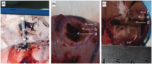
Although the experiment was conducted with no renal vascular pedicle clamping, no blood loss was found during the operation. The kidneys were removed enbloc, and the tissues of the ablation zone were dry and hard. Different colours of concentric circles surrounded the microwave needle: from the centre outwards was thick black, pale, and dark red (). The samples were sectioned along the needle tract, and the ablation shape was similar to a chestnut. With the needle tract as the centre, from the inner to the outer, the ablation lesions could be divided into three zones: the carbonisation zone (thick black, brittle and hard), the solidification zone (pale), and the inflammatory reaction zone (dark red). The pelvic tissues near the needle tract were not charred but were constricted and turned pale yellow, and the ablation areas near the renal pelvis were smaller than the contralateral ().
The microwave needle was inserted into the renal parenchyma by 2 cm or 3 cm, and the kidneys were ablated with different power (50–60 W) and time (300–600 s) combinations. Six pigs with 12 kidneys had 34 thermoablations were applied, 32 of which occurred with a single needle and the other two with a double needle. The maximum diameter parallel and perpendicular to the needle tract was measured in the ablation lesions. The data were analysed with statistical software to obtain the average ablation range ( and ).
Table 1. Maximum diameter of ablation zone with different power/time combinations, microwave needle depth was 2 cm (n = 4,  ).
).
Table 2. Maximum diameter of ablation zone with different power/time combinations, microwave needle depth was 3 cm (n = 4, ).
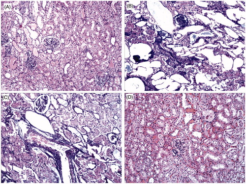
The pathological characteristics of the kidney ablation areas were observed under the microscope (H&E staining, 100×). The normal kidney tissue had a regular structure and the nuclei were intact (). The carbonisation zone was thoroughly necrotic with many broken cores, the glomerulus and tubules were distorted, the epithelium of the renal ubules shed off, and parts of them occluded, and the vascular endothelial cells underwent necrosis (). Coagulative necrosis was observed in the coagulation area and presented as swelling with obscured structural outlines. No ‘skipped’ areas were noted in the ablation zone (). The structure outlines of the inflammatory reaction zone were clear, the renal tubular epithelial cells were slightly swollen, and the interstitial small blood vessels were highly expanded and congested with infiltrating inflammatory cells ().
Figure 2. Pathological characteristics of kidney ablation areas (HE staining, 100x). (A) Normal kidney tissue. (B) Carbonization zone. (C) Coagulation zone. (D) Inflammatory reaction zone.
Discussion
With the current increases in the incidence and detection rate of kidney tumours, the choice of proper treatment for patients, including patients who are not suitable for surgical resection and who require nephron sparing with serious complications, renal dysfunction, hereditary kidney cancer, bilateral kidney cancers, recurrence after partial nephrectomy, solitary kidney, tumours in renal allografts and small renal tumours (<4 cm), has attracted more attention. Ablation technology is becoming popular in the field of minimally invasive treatment of tumours because of its security, minimal invasiveness and effectiveness [Citation39–41]. Related research has reported that energy ablation therapies can be considered for these patients [Citation42–45].
As a minimally invasive therapy, MWA had been applied in the clinical treatment of solid tumours such as liver cancer, lung cancer, uterine fibroids, bone neoplasms, thyroid cancer and breast cancer, and has shown good therapeutic effects [Citation46–50]. In 2007 MWA was applied in kidneys by Clark et al. [Citation51], who first reported a study of MWA of kidney tumours. After this report some relevant studies reported the efficacy of MWA of renal tumours, and most of the studies reported promising results [Citation52,Citation53]. Currently, basic research on MWA in terms of in vivo kidneys is rare. But the concept of minimally invasive treatment and renal preservation requires a higher level of consideration.
We have employed laparoscopic therapy combined with MWA. With the assistance of laparoscopy, clear surgical fields and a wide operating range can be provided for MWA, which makes this method safe and reliable. The trauma of laparoscopic surgery is relatively small, and the pneumoperitoneum has an impact on ablation due to its pressure on the renal vasculature, which reduces renal perfusion to some extent. This method is applicable for kidney tumours near the hilum, pelvis, ureter, large blood vessels and neighbouring organs.
Most kidney tumours are located in the surface or exhibit convex growth. In this study the depth of the microwave needle was set at 2 cm or 3 cm and the tissue was ablated with different power and time combinations. The ablation zone expanded with increasing power and time. When two needles were combined, the maximum diameter of the ablated lesions significantly increased. For example, for the two needles spaced at 1.0 cm or 1.5 cm and ablated with 50 W/480 s, the maximum diameters of the ablation were 4.1 cm and 4.8 cm, respectively. The temperature of the solidification and carbonisation zone was reliable was higher than 60 °C. According to the literature, when the tissue temperature is greater than 60 °C, proteins begin to degrade and coagulate and cells undergo necrosis. When the heating time is extended or the temperature rises, irreversible changes in the tissue become quicker [Citation54–56]. According to these data, we can use the tumour size and location to choose a single or double needle combined with different time/power and appropriate depth of the needle to thoroughly ablate the tumours.
During ablation we observed that the kidney tissues continued to carbonise and shrink, and therefore the final measurement may be smaller than the actual ablated area. In other words, we measured the ablation range to 3 cm in diameter, but the actual ablated tumour size should be greater than 3 cm. Thus, if we refer to the parameters above, not only can the tumours be ablated completely but also a part of the normal kidney tissues will be ablated as a security boundary to achieve optimal ablation results.
In addition, with the same frequency and time, the inflammatory reaction zone became wider when the power rose. The microwave heat conducted more broadly with the improvement of power, which increased heat damage to the normal renal tissues adjacent to the tumours. In practice, facing the same size of tumour, the time needed will be longer with lower power, but the damage to the surrounding normal kidney tissues will be smaller. For the risk of thermal damage of MWA to the surrounding tissues and organs, direct vision with the laparoscope may be avoided. Therefore, to ensure therapeutic efficacy, we need to consider the conditions of the patients and select the appropriate power and time to achieve a beneficial curative effect.
In this study we observed that when the microwave applicator was near the renal pelvis, the ablation size close to the pelvis was smaller compared with the contralateral side. This phenomenon suggested that the dense structure of the renal pelvis or the urine might reduce or absorb some of the heat and inhibit the effects of the microwaves. Therefore, in dealing with tumours adjacent to the pelvis,we need to extend the ablation time to improve the effectiveness of cancer treatment; however, this method may increase the incidence of post-operative urinary fistula.
The post-operative pathological examination revealed that the renal tissues of the carbonisation zone were thoroughly necrotic. Coagulative necrosis was observed in the coagulation area. No skipped areas were noted in the ablation zone. Therefore, on the basis of the pathological results, MWA is safe, effective and feasible; however, the post-operative outcomes of the ablated areas are not known, and there is no definite pathological evidence. If we can obtain tissue pathology 3–6 months after ablation we will be better able to evaluate the effects of MWA.
The present study has some limitations. In this study, the experimental model included normal porcine kidneys, but a kidney tumour model would be more reliable. Single needle data and only two double needle ablation datasets for laparoscopic MWA were obtained in this study. More double needle ablation data could be collected in future studies.
In conclusion, this study shows that laparoscopic MWA is safe and reliable, and provided excellent results in an in vivo porcine model. No blood was lost during the operation, with no renal vascular pedicle clamping. We have obtained relevant data about the laparoscopic MWA of kidneys and offered guidelines for the use of a 2450-MHz MWA system. We found that the ablation diameters were significantly dependent on the time/power interactions and the depth of the needle insertion. For patients with poor physical condition or small renal masses (<4 cm), we can refer to these data and select an appropriate combination. Further studies are needed to determine the role of MWA in the treatment of renal tumours. For example, the relationship between the renal parenchymal thickness and depth of puncture as well as the local or systemic changes of inflammatory factors after ablation and the impact on prognosis must be clarified.
Acknowledgements
Division of author responsibilities: data collection, management, analysis and manuscript writing, Baoan Hong; project development and manuscript editing, Ning Zhang; help with the pathological image analysis, Yuan Zhao; experiment instruction, Xiaodong Zhang and Yong Yang; help with data collection and management, Xin Du and Guowei Chen. We would also like to express our thanks to Kefei Qi, Jun Zhang, and Yi Wang from Nanjing KangYou company for their expertise in MWA instruments.
Declaration of interest
This study was supported by the 2014 Beijing Natural Science Foundation (grant no.7142059). The authors report no conflicts of interest. The authors alone are responsible for the content and writing of the paper.
References
- Altekruse SF, Dickie L, Wu XC, Hsieh MC, Wu M, Lee R, et al. Clinical and prognostic factors for renal parenchymal, pelvis, and ureter cancers in SEER registries: Collaborative stage data collection system, version 2. Cancer 2014;120(Suppl 23):3826–35
- Bretheau D, Lechevallier E, Eghazarian C, Grisoni V, Coulange C. Prognostic significance of incidental renal cell carcinoma. Eur Urol 1995;27:319–23
- Hegarty NJ, Gill IS, Desai MM, Remer EM, O’Malley CM, Kaouk JH. Probe-ablative nephron-sparing surgery: Cryoablation versus radiofrequency ablation. Urology 2006;68:7–13
- Chow WH, Devesa SS, Warren JL, Fraumeni JF Jr. Rising incidence of renal cell cancer in the United States. JAMA 1999;281:1628–31
- Stevens LA, Li S, Wang C, Huang C, Becker BN, Bomback AS, et al. Prevalence of CKD and comorbid illness in elderly patients in the United States: Results from the Kidney Early Evaluation Program (KEEP). Am J Kidney Dis 2010;55:S23–33
- Kouba E, Smith A, McRackan D, Wallen EM, Pruthi RS. Watchful waiting for solid renal masses: Insight into the natural history and results of delayed intervention. J Urol 2007;177:466–70; discussion 470
- Canter D, Kutikov A, Sirohi M, Street R, Viterbo R, Chen DY, et al. Prevalence of baseline chronic kidney disease in patients presenting with solid renal tumors. Urology 2011;77:781–5
- Ljungberg B, Cowan NC, Hanbury DC, Hora M, Kuczyk MA, Merseburger AS, et al. EAU guidelines on renal cell carcinoma: The 2010 update. Eur Urol 2010;58:398–406
- Campbell SC, Novick AC, Belldegrun A, Blute ML, Chow GK, Derweesh IH, et al. Guideline for management of the clinical T1 renal mass. J Urol 2009;182:1271–9
- Chung JS, Son NH, Lee SE, Hong SK, Lee SC, Kwak C, et al. Overall survival and renal function after partial and radical nephrectomy among older patients with localised renal cell carcinoma: A propensity-matched multicentre study. Eur J Cancer 2015;51:489–97
- Sandberg JM, Krane LS, Hemal AK. A nonrandomized prospective comparison of robotic-assisted partial nephrectomy in the elderly to a younger cohort: An analysis of 339 patients with intermediate-term follow-up. Urology 2014;84:838–43
- Lau WK, Blute ML, Weaver AL, Torres VE, Zincke H. Matched comparison of radical nephrectomy vs nephron-sparing surgery in patients with unilateral renal cell carcinoma and a normal contralateral kidney. Mayo Clin Proc 2000;75:1236–42
- Kim SP, Thompson RH, Boorjian SA, Weight CJ, Han LC, Murad MH, et al. Comparative effectiveness for survival and renal function of partial and radical nephrectomy for localized renal tumors: A systematic review and meta-analysis. J Urol 2012;188:51–7
- Lopez-Garibay LA, Cendejas-Gomez Jde J, Rodriguez-Covarrubias F, Gomez-Conzatti A, Gabilondo-Navarro F, Sotomayor-de-Zavaleta M. Long-term renal function in patients with renal-cell carcinoma treated surgically: Comparison between radical and partial nephrectomy. Rev Invest Clin 2013;65:7–11
- Tan HJ, Norton EC, Ye Z, Hafez KS, Gore JL, Miller DC. Long-term survival following partial vs radical nephrectomy among older patients with early-stage kidney cancer. JAMA 2012;307:1629–35
- Lesage K, Joniau S, Fransis K, Van Poppel H. Comparison between open partial and radical nephrectomy for renal tumours: Perioperative outcome and health-related quality of life. Eur Urol 2007;51:614–20
- Kyriazis I, Ozsoy M, Kallidonis P, Panagopoulos V, Vasilas M, Liatsikos E. Current evidence on lasers in laparoscopy: Partial nephrectomy. World J Urol 2014;33:589–94
- Miller AJ, Kurup AN, Schmit GD, Weisbrod AJ, Boorjian SA, Thompson RH, et al. Percutaneous clinical T renal mass ablation in the octogenarian and nonagenarian: Oncologic outcomes and morbidity. J Endourol 2015;29:671–6
- Ritchie RW, Leslie TA, Turner GD, Roberts IS, D’Urso L, Collura D, et al. Laparoscopic high-intensity focused ultrasound for renal tumours: A proof of concept study. BJU Int 2011;107:1290–6
- Sung HH, Park BK, Kim CK, Choi HY, Lee HM. Comparison of percutaneous radiofrequency ablation and open partial nephrectomy for the treatment of size- and location-matched renal masses. Int J Hyperthermia 2012;28:227–34
- Sommer CM, Lemm G, Hohenstein E, Stampfl U, Bellemann N, Teber D, et al. Bipolar versus multipolar radiofrequency (RF) ablation for the treatment of renal cell carcinoma: Differences in technical and clinical parameters. Int J Hyperthermia 2013;29:21–9
- Katsanos K, Mailli L, Krokidis M, McGrath A, Sabharwal T, Adam A. Systematic review and meta-analysis of thermal ablation versus surgical nephrectomy for small renal tumours. Cardiovasc Intervent Radiol 2014;37:427–37
- Lubner MG, Brace CL, Hinshaw JL, Lee FT Jr. Microwave tumor ablation: Mechanism of action, clinical results, and devices. J Vasc Interv Radiol 2010;21:S192–203
- Skinner MG, Iizuka MN, Kolios MC, Sherar MD. A theoretical comparison of energy sources – microwave, ultrasound and laser – for interstitial thermal therapy. Phys Med Biol 1998;43:3535–47
- Brace CL. Radiofrequency and microwave ablation of the liver, lung, kidney, and bone: What are the differences? Curr Probl Diagn Radiol 2009;38:135–43
- Brace CL, Hinshaw JL, Laeseke PF, Sampson LA, Lee FT Jr. Pulmonary thermal ablation: Comparison of radiofrequency and microwave devices by using gross pathologic and CT findings in a swine model. Radiology 2009;251:705–11
- Carrafiello G, Lagana D, Pellegrino C, Fontana F, Mangini M, Nicotera P, et al. Percutaneous imaging-guided ablation therapies in the treatment of symptomatic bone metastases: Preliminary experience. Radiol Med 2009;114:608–25
- Lu DS, Raman SS, Limanond P, Aziz D, Economou J, Busuttil R, et al. Influence of large peritumoral vessels on outcome of radiofrequency ablation of liver tumors. J Vasc Interv Radiol 2003;14:1267–74
- Wright AS, Sampson LA, Warner TF, Mahvi DM, Lee FT Jr. Radiofrequency versus microwave ablation in a hepatic porcine model. Radiology 2005;236:132–9
- Yu NC, Raman SS, Kim YJ, Lassman C, Chang X, Lu DS. Microwave liver ablation: Influence of hepatic vein size on heat-sink effect in a porcine model. J Vasc Interv Radiol 2008;19:1087–92
- Nan Q, Zheng W, Fan Z, Liu Y, Zeng Y. Analysis to a critical state of thermal field in microwave ablation of liver cancer influenced by large vessels. Int J Hyperthermia 2010;26:34–8
- Haemmerich D, Lee FT Jr. Multiple applicator approaches for radiofrequency and microwave ablation. Int J Hyperthermia 2005;21:93–106
- Farina L, Weiss N, Nissenbaum Y, Cavagnaro M, Lopresto V, Pinto R, et al. Characterisation of tissue shrinkage during microwave thermal ablation. Int J Hyperthermia 2014;30:419–28
- Lopresto V, Pinto R, Cavagnaro M. Experimental characterisation of the thermal lesion induced by microwave ablation. Int J Hyperthermia 2014;30:110–18
- Ai H, Wu S, Gao H, Zhao L, Yang C, Zeng Y. Temperature distribution analysis of tissue water vaporization during microwave ablation: Experiments and simulations. Int J Hyperthermia 2012;28:674–85
- Cavagnaro M, Amabile C, Cassarino S, Tosoratti N, Pinto R, Lopresto V. Influence of the target tissue size on the shape of ex vivo microwave ablation zones. Int J Hyperthermia 2015;31:48–57
- Hope WW, Schmelzer TM, Newcomb WL, Heath JJ, Lincourt AE, Norton HJ, et al. Guidelines for power and time variables for microwave ablation in an in vivo porcine kidney. J Surg Res 2009;153:263–7
- Bartoletti R, Cai T, Tosoratti N, Amabile C, Crisci A, Tinacci G, et al. In vivo microwave-induced porcine kidney thermoablation: Results and perspectives from a pilot study of a new probe. BJU Int 2010;106:1817–21
- Hinshaw JL, Lubner MG, Ziemlewicz TJ, Lee FT Jr, Brace CL. Percutaneous tumor ablation tools: Microwave, radiofrequency, or cryoablation–-what should you use and why? Radiographics 2014;34:1344–62
- Thompson RH, Atwell T, Schmit G, Lohse CM, Kurup AN, Weisbrod A, et al. Comparison of partial nephrectomy and percutaneous ablation for cT1 renal masses. Eur Urol 2015;67:252–9
- Park SY, Park BK, Kim CK. Thermal ablation in renal cell carcinoma: What affects renal function? Int J Hyperthermia 2012;28:729–34
- Ankem MK, Nakada SY. Needle-ablative nephron-sparing surgery. BJU Int 2005;95(Suppl2):46–51
- Baird AD, Woolfenden KA, Desmond AD, Fordham MV, Parsons KF. Outcome and survival with nonsurgical management of renal cell carcinoma. BJU Int 2003;91:600–2
- Bird VG, Carey RI, Ayyathurai R, Bird VY. Management of renal masses with laparoscopic-guided radiofrequency ablation versus laparoscopic partial nephrectomy. J Endourol 2009;23:81–8
- Kunkle DA, Uzzo RG. Cryoablation or radiofrequency ablation of the small renal mass: A meta-analysis. Cancer 2008;113:2671–80
- Acksteiner C, Steinke K. Percutaneous microwave ablation for early-stage non-small cell lung cancer (NSCLC) in the elderly: A promising outlook. J Med Imaging Radiat Oncol 2015;59:82–90
- Pusceddu C, Sotgia B, Fele RM, Melis L. Treatment of bone metastases with microwave thermal ablation. J Vasc Interv Radiol 2013;24:229–33
- Liu SR, Liang P, Yu XL, Cheng ZG, Han ZY, Yu J. Percutaneous microwave ablation for liver tumours adjacent to the marginal angle. Int J Hyperthermia 2014;30:306–11
- Yue W, Chen L, Wang S, Yu S. Locoregional control of recurrent papillary thyroid carcinoma by ultrasound-guided percutaneous microwave ablation: A prospective study. Int J Hyperthermia 2015;31:403–8
- Zhang J, Feng L, Zhang B, Ren J, Li Z, Hu D, et al. Ultrasound-guided percutaneous microwave ablation for symptomatic uterine fibroid treatment–-a clinical study. Int J Hyperthermia 2011;27:510–16
- Clark PE, Woodruff RD, Zagoria RJ, Hall MC. Microwave ablation of renal parenchymal tumors before nephrectomy: Phase I study. Am J Roentgenol 2007;188:1212–14
- Liang P, Wang Y, Zhang D, Yu X, Gao Y, Ni X. Ultrasound guided percutaneous microwave ablation for small renal cancer: Initial experience. J Urol 2008;180:844–8; discussion 848
- Moreland AJ, Ziemlewicz TJ, Best SL, Hinshaw JL, Lubner MG, Alexander ML, et al. High-powered microwave ablation of t1a renal cell carcinoma: Safety and initial clinical evaluation. J Endourol 2014;28:1046–52
- McGahan JP, Browning PD, Brock JM, Tesluk H. Hepatic ablation using radiofrequency electrocautery. Invest Radiol 1990;25:267–70
- Rossi S, Fornari F, Pathies C, Buscarini L. Thermal lesions induced by 480 KHz localized current field in guinea pig and pig liver. Tumori 1990;76:54–7
- Stauffer PR, Goldberg SN. Introduction: Thermal ablation therapy. Int J Hyperthermia 2004;20:671–7