
Key words:
Citation counting is a comparably new procedure that has become widely used to evaluate single papers but also to rank journals by the so-called impact factor (IF), often determined to the third decimal. This is a phenomenon of the information society and therefore only reliable for later years. However, we can go back to the 1960s. When doing so for Upsala Journal of Medical Sciences, there is one paper which has been cited much more than any other, and that is Lars Grimelius’s ‘A Silver Nitrate Stain for α2 Cells in Human Pancreatic Islets’, published in the journal in 1968 (Citation1). According to the Thomson Reuters database it has been cited 897 times to date. None of the journal’s other papers is even close to that record. The value would have been even higher if the method had not been incorporated in many laboratories as a routine staining for which a citation was regarded as unnecessary.
Silver impregnation (‘staining’) has been used in histology and histopathology for a long time and was initially particularly developed for studies of nervous structures. Like in photography, the methods utilize reduction of a silver salt to inorganic silver or silver oxide which is deposited on specific structures. Subsequently, it was found that silver techniques were able to visualize certain specific cells outside the nervous system. Several methods had been developed that turned out to be useful for the studies of the endocrine pancreas. One often-used technique was the Gross–Schultze method, which was extensively studied by Grimelius’s and my teacher Professor Gösta T. Hultquist (Citation2). The Gross–Schultze technique was later severely questioned, however, since it was found not to be completely specific and small changes in the staining protocol resulted in other impregnation patterns (Citation3).
When this story started, the most frequent pancreatic islet cells were basically defined and included β-cells, α1 (D), and α2 (A) cells. β-Cells were known to produce insulin, and α2 cells were supposed to produce glucagon, while the function of α1 was unknown. In addition to the Gross–Schultze technique there were a couple of other silver methods which were often employed for pancreatic studies. One was the silver proteinate method, originally developed by Bodian (Citation4) and further modified by several investigators including Lars Grimelius (Citation5). Another silver impregnation method was that of Davenport, modified by Hellerström and Hellman in Uppsala (Citation6). This last method was found to discriminate between two populations of non-β-cells shown to include α1 and α2 cells.
When Lars Grimelius () entered the scene the importance of islet composition in the pathogenesis of diabetes (type 1 and type 2 were not clearly defined at that time) was still somewhat unclear. A number of trials to determine the total mass of the different islet cells had produced controversial results (for review, see Warren et al. (Citation7)). The professor who recruited Lars Grimelius, the above-mentioned Gösta Hultquist, who preferred to be called Hqt, had a long history of studies of islets of Langerhans. Hqt was a brilliant pathologist, particularly in anatomical pathology, but his heart beat more strongly for experimental pathology. He used a technique to ligate the rat pancreatic duct in order to induce atrophy of the exocrine but not endocrine tissue. Such tissue was then transplanted into the anterior chamber of diabetic or control rats (Citation8). It was possible by such means to study effects of diabetes and of antidiabetic drugs (Citation9).
Figure 1. Lars Grimelius on a tour in Swedish Lapland together with the author in August 1969.

We should remember that when Lars Grimelius started his career at the Department of Pathology at Uppsala University, in the 1950s, there were no graduate student programmes, no graduate courses, and not necessarily any projects to start with. Rather, there was a demand for the students themselves to find a suitable problem to solve and also find useful methods for the task. At best there could be a suggestion of a broad problem to study—not so easy for an inexperienced student. Luckily, for a student in pathology there were a limited set of methods to use, and most of them were based on light microscopy. The time for writing a doctoral thesis was not restricted, and it could sometimes take a decade or more. It was suggested to Grimelius that he should study the effects of a new antidiabetic drug on the rat pancreas. I can already here unveil that this study was never accomplished.
Instead of taking up the suggested problem, Grimelius (a methodical person) started to evaluate different staining methods for islet cells. The mentioned silver proteinate method gave very nice results. However, the supply of the chemical at the Department was limited, and therefore Grimelius ordered the substance from 10 different suppliers. Only one of the received substances gave satisfactory results. Grimelius therefore ordered 1 kg of this specific preparation but got the response from the company that they had tried to produce more but results were unreliable. This sad fact led Grimelius to develop his so successful silver impregnation method. Like many advances in science, this one sprang from a failure.
Against this background Lars Grimelius decided at least to try to develop a method based only on simple, well-controlled chemicals. Since there was no firm biochemical knowledge on how silver impregnation methods worked or what exactly they demonstrated, development of a procedure was basically empiric. That meant a lot of experimental work. Lars Grimelius identified the following variables: Concentration of silver salt, concentration of reduction agent, pH, temperature, duration and fixation of tissue, and, with human material, autolysis. It is easy to understand that combinations of all these variables can create an almost endless number of possibilities. Grimelius, of course, was assisted in performing sections of all tissue materials needed, but he carried out the mixing of solutions, the incubations, mountings, and evaluations himself, without assistance. After years of work, a method finally saw the light of day. This was about the time when I came to the Department as a young and enthusiastic medical student with an intention to start with a research project. Grimelius was about to finish the laboratory work, and it was time to write the papers. Part of that time I came to share an office with him. The room was a somewhat historical one since it had been the office of probably the most famous pathologist in Uppsala, Robin Fåhraeus (1888–1968), and it had still some of his furniture and fixtures, including a large hibiscus and a green sofa on which Grimelius lay down and slowly wrote his papers with tiny handwriting with a short old-fashioned pencil. I also remember the frequent use of an eraser. Now and then he went out to the laboratory to do some late checking experiments. Above us both, a copy of a Greek (or Roman?) sculpture was looking down. I am looking back at this time through a nostalgic mist, but the situation in the laboratory was certainly very relaxed.
All this writing took its time for Grimelius. There was no real feeling of hurry since there was no fixed time for a thesis work, and today’s severe competition was unknown. Funding was safe through money directly allocated to the Department. We should also remember that there were no computers, so all text and the large number of tables had to be written and re-written on a typewriter. Finally, two meticulous papers describing and evaluating the Grimelius silver impregnation method were finished, and it was time for submission. The method manuscript was sent to the journal Stain Technology, which was the journal that most often published new methods. The journal, however, unexpectedly rejected the paper. This was, as it turned out, a big mistake by the editor. What to do then? Luckily, there was Acta Societatis Medicorum Upsaliensis, later renamed to Upsala Journal of Medical Sciences, a journal very often used for doctoral thesis papers. Lars Grimelius has told me how he submitted the papers there. He went to the editor, Gunnar Ågren, Professor of Medical Biochemistry, who inspected them and asked, ‘Has Professor Hultquist looked at them?’ The answer was yes, and then the papers were accepted.
The first paper, ‘A silver nitrate stain for α2 cells in human pancreatic islets’ (Citation1), which is the description of the method, rapidly became used and has been frequently cited, until now 897 times according to the Thomson Reuters database. It became a ‘citation classic’ in 1984. It is obvious that the rejection by Stain Technology (present impact factor 1.0) was a big mistake, as it became the most cited of any single papers in Upsala Journal of Medical Sciences (present impact factor 1.71). Grimelius later got his revenge when he was invited to contribute with a review article on silver staining methods for the journal Stain Technology, now renamed to Biotechnology and Histochemistry (Citation10). Lars Grimelius later got a second ‘citation classic’ with the paper that shows that pancreatic α1 cells (D cells) store somatostatin (Citation11). The second paper in Grimelius’s thesis (Citation12) contains all control experiments with a great number of variables. Included in the doctoral thesis, defended in the spring of 1969, was also an electron microscopic study of the distribution of silver particles in labelled cells. The electron microscope, a Zeiss EM9, had been bought a few years earlier, partially with money raised by the thoracic surgeon Viking Olov Björk, who wanted Hqt to study heart tissue electron microscopically, which he also did (Citation13). Grimelius showed that silver particles were localized to secretory granules and more specifically in the electron-lucent peripheral area (Citation14). This was an interesting observation, since while the electron-dense area contains glucagon, other granule components are present in the translucent periphery. Among these is chromogranin A, which is one of the substances that have been found to be responsible for the argyrophilic properties of endocrine cells (Citation15,16). Other components are certainly of importance as well (Citation15). I had much benefit both from the use of the silver method and from Grimelius himself for one of the papers in my own thesis (Citation17). Examples of the Grimelius silver impregnation are shown in .
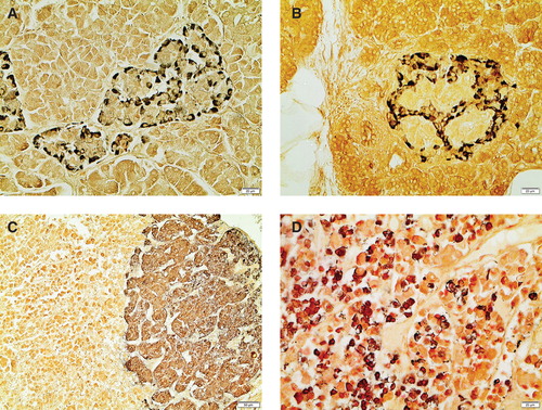
Figure 2. Examples of Grimelius stain, all performed in 1972. A: Normal human islet of Langerhans. B: Human islet of Langerhans with large deposits of amyloid. Most remaining cells are α2 (A) cells. (Patient with type 2 diabetes.) C: Part of adrenal gland. Cells in medulla are argyrophilic, while cortical cells are unlabelled. (Same case as A.) D: Section from anterior pituitary. One population of cells is argyrophilic. (Same case as A).
Lars Grimelius widened the use of his own as well as other silver staining methods to other areas in endocrine anatomy and pathology. Grimelius’s stain turned out to visualize a number of cells, e.g. in gastrointestinal tract and parathyroid and thyroid glands, and it played an important role in the mapping of such cells () (Citation18,19). The Grimelius silver impregnation method turned out to be very useful for discrimination of neuroendocrine tumours from other neoplasms.
Grimelius was wise to understand that he should widen his methodological arsenal, and in 1974–1975 he was guest scientist in the laboratory of Professor A. G. Everson Pearse at Hammersmith Hospital, London. Pearse was a pioneer in histochemistry, well-known to everyone working in histopathology. This started a long period of collaboration and friendship with Pearse and with Julia M. Polak, who succeeded as professor at the department. During his stay in London, Grimelius learnt immunohistochemistry, which was under development, and brought the technique back to Sweden. He then turned more and more towards endocrine tumour pathology, employing immunohistochemistry, and started a new career in clinical pathology. But that is another story.
Declaration of interest: The author reports no conflicts of interest. The author alone is responsible for the content and writing of the paper.
References
- Grimelius L. A silver nitrate stain for alpha2 cells in human pancreatic islets. Acta Soc Med Upsal. 1968;73:243–70.
- Hultquist GT, Dahlen M, Helander CG. Über die Technik Darstellung und Zählung der sogen. Silberzellen in den Langerhansschen Inseln. Schweiz Zeitschr Path Bakt. 1948;11:570–89.
- Creutzfeldt W, Theodossiou A. Die Relation der A- und B-zellen in den Pankreasinseln bei Nichtdiabetikern und Diabetikern. Beitr Pathol Anat. 1957;117:235–52.
- Bodian D. A new method for staining nerve fibres and nerve endings in mounted paraffin sections. Anat Rec. 1936;65:89–97.
- Grimelius L. A modified silver protein method for studying the argyrophil cells of the islets of Langerhans. In Brolin SE, Hellman B, Knutsson H, editors. The structure and metabolism of pancreatic islets. Oxford: Pergamon Press: 1964. p. 99–104.
- Hellerström C, Hellman B. Some aspects of silver impregnation of the islets of Langerhans in the rat. Acta Endocrinol (Copenh). 1960;35:518–32.
- Warren S, LeCompte PM, Legg MA. The pathology of diabetes mellitus. 4th ed. Philadelphia: Lea & Febiger; 1966.
- Hultquist GT. The ultrastructure of pancreatic tissue from duct-ligated rats implanted into anterior chamber of rat eyes. Ups J Med Sci. 1972;77:8–18.
- Grimelius L, Hultquist GT, Thorell J, Winbladh L. Studies on islet tissue transplants in the anterior chamber of the eye in rats. In Brolin SE, Hellman B, Knutsson H, editors. The structure and metabolism of the pancreatic islets. Oxford: Pergamon Press; 1964. p 173–8.
- Grimelius L. Silver stains demonstrating neuroendocrine cells. Biotech Histochem. 2004;79:37–44.
- Polak JM, Pearse AG, Grimelius L, Bloom SR. Growth-hormone release-inhibiting hormone in gastrointestinal and pancreatic D cells. Lancet. 1975;1:1220–2.
- Grimelius L. The argyrophil reaction in islet cells of adult human pancreas studies with a new silver nitrate procedure. Acta Soc Med Ups. 1968;73:271–94.
- Björk VO, Hultquist G. Ultrastructural, enzyme histochemical and light microscopic investigation of the myocardium in cases undergoing open-heart surgery. Scand J Thorac Cardiovasc Surg. 1967;1:27–41.
- Grimelius L. An electron microscopic study of silver stained adult human pancreatic islet cells, with reference to a new silver nitrate procedure. Ups J Med Sci. 1969;74:28–48.
- Lundqvist M, Arnberg H, Candell J, Malmgren M, Wilander E, Grimelius L, et al. Silver stains for identification of neuroendocrine cells. A study of the chemical background. Histochem J. 1990;22:615–23.
- Cetin Y. Chromogranin A immunoreactivity and Grimelius’ argyrophilia. A correlative study in mammalian endocrine cells. Anat Embryol (Berl). 1992;185:207–15.
- Westermark P, Grimelius L. The pancreatic islet cells in insular amyloidosis in human diabetic and non-diabetic adults. Acta Pathol Microbiol Scand A. 1973;81:291–300.
- Grimelius L, Wilander E. Silver stains in the study of endocrine cells of the gut and pancreas. Invest Cell Pathol. 1980;3:3–12.
- DeLellis RA, Balogh K. Histochemical characteristics of parafollicular cells and medullary thyroid carcinoma. Am J Pathol. 1973;72:119–28.