Abstract
Background. The influence of gender on the association between metabolic syndrome (MS) and subclinical organ damage (OD) has been poorly investigated. The aim of this study was to investigate whether the risk of developing left ventricular hypertrophy (LVH) and carotid atherosclerosis is different in men and women with MS. Methods. A total of 3752 untreated and treated hypertensive patients (mean age 53.3 ± 12.6, 52.7% men) were considered for this analysis. All patients underwent standard ultrasonographic investigations searching for LVH and carotid atherosclerosis. The MS was defined according to ATP III criteria. Results. LVH was more prevalent in women and men with the MS compared with their counterparts (58% vs 34% and 48% vs 33%, respectively, p < 0.001). This was also the case for carotid plaque prevalence (61% vs 42% and 57% vs 44%, p < 0.001). The prevalence of OD was not different between men and women with MS, after adjusting for confounders. In multivariate analysis, abdominal obesity was the most important MS component independently related to LVH in both genders, followed by blood pressure. As for carotid plaques, blood pressure, hyperglycemia and hypertriglyceridemia turned out to be independent correlates regardless of gender. Conclusions. Our data indicate that MS is associated with a higher risk of LVH and carotid atherosclerosis irrespective of gender; these findings do not support a gender influence in the association between MS and subclinical OD.
Key Words::
Introduction
The metabolic syndrome (MS) is a clinical entity characterized by the coexistence of multiple risk factors for cardiovascular disease (CVD) and diabetes mellitus (Citation1,Citation2), such as raised blood pressure (BP), hyperglycemia, elevated triglyceride levels, low high-density cholesterol levels and central obesity. The pathogenesis of MS is incompletely defined; increasing evidence indicates that both insulin resistance and activation of the renin–angiotensin–aldosterone system play a central role in this syndrome, which is associated with an adverse cardiovascular (CV) prognosis (Citation3,Citation4). Individuals with MS have a two- to three-fold higher risk of developing CVD within 10 years than their counterparts (Citation2); the increased CV risk may be related to the development of more pronounced subclinical alterations in cardiac and vascular structures in subjects with MS.
Echocardiographic studies in population-based samples or hypertensive cohorts have consistently shown that subjects with metabolic risk factors or with MS have increased left ventricular mass (LVM), higher prevalence of left ventricular hypertrophy (LVH) and diastolic dysfunction compared with their counterparts without MS (Citation5–8). Over the past decade, the impact of MS on large arterial vessels (i.e. carotid wall) has been analyzed by ultrasonographic studies either in hypertensive subjects as in the general population. In most of these studies, both carotid intima-media thickness (IMT) and plaque prevalence were found to be higher in subjects with MS and were significantly related with the number of MS components (Citation9–11). Despite a large body of data on the relationship of MS with cardiac and extra-cardiac organ damage (OD), only few studies focused on gender-related differences in such an association and their results provided non-univocal information (Citation12–16). Thus, the aim of the present study in a large cohort of subjects with essential hypertension was to investigate whether the likelihood of having LVH and carotid atherosclerosis is different between men and women with MS.
Methods
Study population
The analysis was performed on data from the Evaluation of Target Organ Damage in Hypertension (ETODH) study, a cross-sectional observational registry providing detailed information on hypertension-related OD in untreated and treated subjects with uncomplicated essential hypertension. Details of the study protocol have been previously reported (Citation12). Entry criteria included the absence of previous clinically overt CV disease, secondary causes of hypertension and life-threatening conditions. After their informed consent had been obtained during the initial visit, all patients underwent the following procedures, which were completed within 1–4 weeks: medical history and physical examination, clinic BP measurement, blood and urine sampling, standard 12-lead electrocardiogram, non-mydriatic retinography, cardiac, renal and carotid ultrasonography. The study protocol was approved by the Ethics Committee of one of the institutions involved.
Blood pressure measurement
Clinic BP was measured at two different visits in the outpatient clinic using a mercury sphygmomanometer and taking the first and fifth phases of Koroktoff sounds to identify systolic (SBP) and diastolic BP (DBP), respectively. Measurements started after the subjects had rested for 5 min in the sitting position. Three measurements were taken at 1-min interval in each occasion and the average of six values was used to define clinic SBP and DBP.
Echocardiography
Echocardiographic examinations were carried out according to standardized procedures. M-mode, two-dimensional, Doppler examinations were made with commercially available instruments. LVM was estimated from end-diastolic left ventricular internal diameter (LVIDd), interventricular septum (IVST) and posterior wall thickness (PWT) according to the Devereux's formula (Citation17) and normalized to BSA and height2.7 to obtain LVM indexes (LVMI). Relative wall thickness (RWT) was calculated as (2 × PWT)/LVIDd; left ventricular (LV) filling was assessed by mitral flow with standard pulsed Doppler technique, the following parameters were considered: early diastolic peak flow velocity (E), late diastolic peak flow velocity (A) and their ratio (E/A).
Carotid ultrasonography
Images of extra-cranial carotid walls (common, bifurcation and internal carotid arteries) were obtained in several projections by high resolution, linear array 7.5–10.0-MHz probes. Plaques were sought in the near and far walls of the entire extra-cranial tree, and intima-media thickness (IMT) was measured in the posterior wall of both common carotid arteries 5, 10, 15, 20 and 25 mm caudally to the bifurcation (Citation18). For statistical purposes, all 10 measurements were averaged to obtain the mean value of common carotid IMT. Details about reproducibility of LVM and IMT measurements in our laboratory have been previously reported (Citation19).
Definition of organ damage
OD was defined by the presence of ultrasonographic LVH and vascular alterations. In particular, LVH was defined as LVMI equal to or higher than 51 g/m2.7 in men and 47 g/m2.7 in women (Citation20,Citation21). The presence of at least one carotid atherosclerotic plaque or diffuse intima-media (IM) thickening was taken as evidence of vascular alterations.
A plaque was defined as a focal thickening greater than 1.3 mm in any segment of carotid arteries (Citation22). Diffuse IM thickening was diagnosed when the average common carotid wall thickness exceeded 0.9 mm (Citation23).
Definition of the metabolic syndrome
MS was diagnosed when three or more of the following criteria were present: abdominal obesity (waist circumference > 102 cm in men and > 88 cm in women), hypertriglyceridemia (> 150 mg/dl or 1.69 mmol/l or drug treatment for elevated triglycerides), reduced high-density lipoprotein (HDL) cholesterol (< 40 mg/dl or 1.04 mmol/l in men and < 50 mg or 1.29 mmol/l in women) or drug treatment for reduced HDL-cholesterol, high BP (SBP ≥ 130 mmHg or DBP ≥ 85 mmHg or drug treatment for hypertension) and high fasting blood glucose (≥ 100 mg/dl or ≥ 6.1 mmol/l) or drug treatment for elevated glucose (Citation1,Citation2).
Statistical analysis
Statistical analysis was performed by the SAS system (version 6.12; SAS Institute Inc., Cary, North Carolina, USA). Values were expressed as means± SD or as percentages. Continuous variables were compared by analysis of variance (ANOVA), using the Student's t-test for dual comparisons. Analysis of categorical data was carried out by the χ2 test or Fischer's exact test when appropriate. To investigate the association between individual components of MS and markers of OD such as LVH and carotid plaques, multiple logistic regression analyses were performed in both genders by calculating odd ratios and their 95% confidence limits. For all analyses, a p < 0.05 was considered statistically significant.
Results
The ETODH registry started in January 1999 and by the end of July 2006 had enrolled 3752 subjects (52.7% men) with untreated (29.5%) or treated essential hypertension. Mean age was 53.3 ± 12.6 (range 18–90) years. Mean SBP and DBP were 146 ± 18 and 92 ± 10.0 mmHg, respectively; 22.2% of the study sample were smokers, 5.2% had type 2 diabetes mellitus and 40.9% had the MS. Men were younger than women, had higher DBP, body mass index, fasting blood glucose, triglyceride and uric acid, but lower SBP, heart rate, total and HDL- cholesterol values. The prevalence of LVH tended to be higher in women (43.0% vs 39.0%) whereas that of carotid plaques showed an opposite trend (48.9% vs 50.0), respectively, without achieving statistical significance in both cases (p = 0.06 and p = 0.50).
In men, the most common component of the MS (other than high BP) was high fasting blood glucose (43.1%), followed by low HDL-cholesterol (40.5%), hypertriglyceridemia (35.7%) and abdominal obesity (25.8%). In women, low HDL-cholesterol (39.4%) was the most frequent feature of the MS followed by abdominal obesity (35.5%), hypertriglyceridemia (24.9%) and high fasting blood glucose (28.1%).
For the present analysis, patients were divided in four groups according to the gender and the presence of MS. shows that both men and women with MS were older, were more frequently treated with antihypertensive drugs, had higher SBP and uric acid values, and showed increased prevalence of type 2 diabetes mellitus than their gender counterparts, the differences being in all instances statistically significant.
Table I. Clinical characteristics of the study population divided by gender and the presence of metabolic syndrome (MS).
As reported in , prevalence of LVH was approximately 40% higher in men and 70% higher in women with MS compared with their gender counterparts (p < 0.01 for both). A similar trend was observed for carotid IMT and carotid plaque, this latter alteration being 30% and 45% more frequent in men and women with MS, respectively, compared with their gender counterparts (p < 0.01 for both). Of note, the prevalence of LVH and carotid plaque was not different between men and women with MS, after adjusting for age, clinic BP and anti-hypertensive treatment.
Table II. Ultrasonographic findings in the study population as a whole and divided by gender.
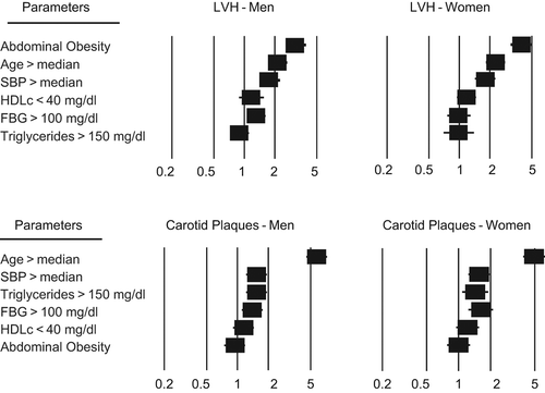
In order to analyze the factors involved in the pathogenesis of OD, the relation of LVH and carotid plaque with abdominal obesity, hyperglycemia, hypertriglyceridemia, low HDL-cholesterol, above median values of clinic SBP and age was analyzed by a logistic regression model in both genders. As shown in , in men, the strongest independent correlate of LVH was abdominal obesity, followed by age, SBP and fasting blood glucose; neither HDL-cholesterol nor triglyceride were related with this cardiac phenotype. In women, abdominal obesity, age and SBP but not fasting blood glucose were independently correlated with LVH. In both genders, age, SBP, fasting blood glucose and triglycerides were independently associated with carotid plaque.
Figure 1. Forrest plots showing odds ratios and 95% confidence intervals (CI) of independent variables associated with left ventricular hypertrophy (LVH, upper panel) and carotid plaques (lower panel) in hypertensive men and women. SBP, systolic blood pressure; FBG, fasting blood glucose.
Finally, prevalence rates of LVH and carotid plaque were significantly higher in patients with MS regardless of the absence (35%, 42%, vs 27%, 31%, p at least < 0.05) or the presence of antihypertensive treatment (59%, 62%, vs 38%, 51%, p at least < 0.001).
Discussion
This cross-sectional study compared the extent of subclinical cardiac and vascular OD in a large sample of untreated and treated hypertensive subjects categorized according to the MS defined by the Adult Treatment Panel III and gender. This study clearly indicates that MS is associated with a greater risk of LVH and carotid atherosclerosis both in men and women; our data, however, do not support a gender difference in the association between MS and subclinical cardiac and carotid OD. This conclusion is strengthen by the observation that: (i) LVMI, carotid IMT as well as LVH and carotid plaque prevalence were significantly higher in patients with MS compared with their non-MS counterparts irrespective of gender; (ii) the extent of cardiac and extra-cardiac OD was similar in men and women with MS after adjusting for confounders; (iii) in multivariate analyses abdominal obesity, age and SBP were the best independent correlates of LVH both in men and women; this was also the case for carotid plaque, being age and SBP the major determinants of this ultrasonographic phenotype in both genders. Thus, our data provide a new piece of information on the controversial interaction between sex and OD in the MS by showing that the correlates and extent of cardiac and extra-cardiac damage in patients with MS are not different between men and women.
Our results are not in keeping with previous reports indicating that the effects of MS on LV structure and early carotid atherosclerosis are prevalent in women. Among 618 non-diabetic, untreated hypertensive subjects, a greater prevalence of LVH was found by Schillaci et al. (Citation13) in women with MS compared with those without the syndrome (37% vs 14%, p < 0.001); such a difference was not present in men (39% vs 29%, p < 0.09). In a population-based survey of 1588 participants in the Salzburg Atherosclerosis Prevention program at High Individual Risk (SAPHIR) study, Iglseder et al. (Citation14) reported that the extent of carotid plaque and IMT were greater in subjects with MS; after adjusting for established risk factors, however, the difference in carotid plaque score remained significant only in women. The higher prevalence of MS in our study sample (41%) compared with the above-mentioned studies (27% and 16%, respectively) may partly explain this discrepancy. Thus, our results do not support the view that MS is a condition at less risk for subclinical OD in men than in women. Our conclusion is in agreement with the findings provided by two European population-based studies. In the Pressioni Arteriose Monitorate E Loro Associazioni (PAMELA) study, MS was associated with increased LV mass index in the whole sample (97.4 ± 23.8 vs 83.6 ± 19.9 g/m2), as well as in men and women without differences between genders, in younger and older subjects (Citation24); the differences remained significant after adjustment for age, gender, smoking, alcohol consumption, 24-h average SBP and history of CV events. Among 2228 individuals (2036 men and 2192 women) living in Stockholm County, Sweden (Citation16), the prevalence of LVH was 12.8% in men with the MS and 7.9% in men without the syndrome (p = 0.0003); the corresponding figures in women were 9.9% and 3.3%, respectively (p < 0.0001). In men, hypertension was the only component significantly associated with LVH after adjustments (OR = 3.40; 95% CI = 1.99–5.82); in women, waist circumference (OR = 1.63; 95% CI 1.03–2.57) and glycemia (OR = 1.77; 95% CI 1.13–2.79) were also independently associated with LVH, after hypertension (OR = 4.41; 95% CI 2.50–7.76). Sex-specific relations between LV mass, left atrial size and glucose metabolism, estimated by the homeostasis model assessment, have been investigated in a pioneering study in the Framingham population (Citation25), showing that a relation between insulin resistance and LV mass was present only in women; additional adjustments for BMI, however, rendered this gender- related difference non-significant.
Several mechanisms may explain the interaction of the MS with cardiac and vascular structures. Available evidence indicates that BP plays a pivotal role in the development of preclinical OD and that the impact of BP on the heart and vessels may be amplified by metabolic factors. The adverse cardiac effects of MS have been mostly related to high-normal or frankly elevated BP values and to central obesity, namely to a combination of pressure and volume overload (Citation26). Recently Guerra et al. demonstrated in 380 overweight/obese hypertensives that the effects of the MS on LV parameters are mainly driven by the degree of adiposity (Citation27). Metabolic components, such as hyperglycemia and dyslipidemia, probably contribute to the abnormal loading conditions by decreasing large artery compliance; hyperinsulinemia may also stimulate the growth of myocardial cells. Finally, the role of cytokines produced by the abdominal adipose tissue should be considered in the pathogenesis of LVH and vascular damage over and beyond hemodynamic factors.
The main finding of the present study is that among the MS components abdominal obesity and BP were the most important correlates of LVH without significant sex-related differences. In a clinical perspective, this observation suggests that obese hypertensive men and women should be screened for LVH by echocardiography and that prevention/treatment of obesity should reduce the likelihood of new-onset and persistent LVH.
Our data also point toward a synergic effect of raised BP levels and metabolic alterations such as hyperglycemia and hypertriglyceridemia in determining carotid atherosclerosis in either men and women. Thus carotid subclinical alterations should be systematically searched in hypertensive patients with concomitant metabolic risk factors, regardless the gender in order to optimize the clinical management.
Study limitations
Our study sample included only Caucasian subjects and hypertensive patients referred to a specialist centre; thus, our results should not be extended to different ethnic groups or to the general population. The inclusion of patients on antihypertensive treatment probably affected the relationship between MS and OD, as treatment-related BP decrease might have reduced the prevalence and degree of OD in our study. We found, however, that cardiac and extra-cardiac OD was significantly greater in patients with MS independently of the antihypertensive drugs; in particular, the difference in organ involvement was even more pronounced in treated than in untreated individuals. Lastly, our cross-sectional analysis does not permit any causal inference.
Conflict of interest:
The authors declare no conflict of interest. The authors alone are responsible for the content and the writing of the paper.
References
- Grundy SM, Cleeman JI, Daniels SR, Donato KA, Eckel RH, Franklin BA, et al. Diagnosis and management of the metabolic syndrome. An American Heart Association/National Heart, Lung, and Blood Institute Scientific Statement. Circulation. 2005;112:2735–2352.
- Alberti KGMM, Eckel RH, Grundy SM, Zimmet PZ, Cleeman JI, Donato KA, et al. Harmonizing the metabolic syndrome. A Joint Interim Statement of the International Diabetes Federation Task Force on Epidemiology and Prevention; National Heart, Lung, and Blood Institute; American Heart Association; World Federation; International Atherosclerosis Society; and International association for the Study of Obesity. Circulation. 2009;120:1640–1645.
- Essick EE, Sam F. Cardiac hypertrophy and fibrosis in the metabolic syndrome: A role for aldosterone and the mineralcorticoid receptor. Int J Hypertens. 2011;346985.
- Thethi T, Kamiyama M, Kobori H. The link between the renin–angiotensin–aldosterone system and renal injury in obesity and the metabolic syndrome. Curr Hypertens Rep.2012;14:160–169.
- Chinali M, Devereux RB, Howard BV, Roman MJ, Bella JN, Liu JE, et al. Comparison of cardiac structure and function in American Indians with and without the metabolic syndrome (The Strong Heart Study). Am J Cardiol. 2004;93: 40–44.
- Ferrara LA, Cardoni O, Mancini M, Zanchetti A. Metabolic syndrome and left ventricular hypertrophy in a general population. Results from the Gubbio Study. J Hum Hypertens. 2007;21:795–801.
- Cuspidi C, Meani S, Fusi V, Severgnini B, Valerio C, Catini E, et al. Metabolic syndrome and target organ damage in untreated essential hypertensives. J Hypertens. 2004;22: 1991–1998.
- Leoncini G, Ratto E, Viazzi F, Vaccaro V, Parodi D, Parodi A, et al. Metabolic syndrome is associated with early signs of organ damage in non-diabetic, hypertensive patients. J Intern Med. 2005;257:454–460.
- Mulè G, Nardi E, Cottone S, Cusimano P, Volpe V, Piazza G. et al.Influence of the metabolic syndrome on hypertension-related target organ damage. J Intern Med. 2005;257: 503–513.
- Tzou WS, Douglas PS, Srinivasan SR, Bond MG, Tang R, Chen W, et al. Increased subclinical atherosclerosis in young adults with metabolic syndrome. The Bogalusa Heart Study. J Am Coll Cardiol. 2005;46:457–463.
- Zanchetti A, Hennig M, Baurecht H, Tang R, Cuspidi C, Carugo S, Mancia G. Prevalence and incidence of the metabolic syndrome in the European Lacidipine Study on Atherosclerosis (ELSA) and its relation with carotid intima media thickness. J Hypertens. 2007;25:2463–2470.
- Cuspidi C, Meani S, Valerio C, Sala C, Fusi V, Zanchetti A, Mancia G. Age and target organ damage in essential hypertension: Role of the metabolic syndrome. Am J Hypertens. 2007;20:296–303.
- Schillaci G, Pirro M, Pucci G, Mannarino MR, Gemelli F, Siepi D, et al. Different impact of the metabolic syndrome on left ventricular structure and function in hypertensive men and women. Hypertension. 2006;47:881–886.
- Iglseder B, Cip P, Malaimare L, Ladurner G, Paulweber B. The metabolic syndrome is a stronger risk factor for early atherosclerosis in women than in men. Stroke. 2005;36: 1212–1217.
- Aijaz B, Ammar KA, Lopez-Jimenez S, Redfield MM, Jacobsen SJ, Redeheffer RJ. Abnormal cardiac structure and function in the metabolic syndrome: A population-based study. Mayo Clin Proc. 2008;83:1350–1357.
- Halldin M, Fahlstadius P, De Faire U, Vikstrom M, Hellenius ML. The metabolic syndrome and left ventricular hypertrophy, the influence of gender and physical activity. Blood Press. 2012;21:153–160.
- Devereux RB, Reickek N. Echocardiographic determination of left ventricular mass in man. Anatomic validation of the method. Circulation. 1977;55:613–618.
- Pignoli P, Tremoli E, Poli A, Paoletti R. Intimal plus media thickness of the arterial wall: A direct measurement with ultrasound imaging. Circulation. 1986;74:1399–1408.
- Cuspidi C, Lonati L, Macca G, Sampieri L, Fusi V, Michev I, et al. Prevalence of left ventricular hypertrophy and carotid thickening in a large selected population: Impact of different echocardiographic and ultrasonographic diagnostic criteria. Blood Press. 2001;10:142–149.
- Lang RM, Bierig M, Devereux RB, Flachskampf FA, Foster E, Pellikka PA, et al. Chamber Quantification Writing Group; American Society of Echocardiography's Guidelines and Standards Committee; European Association of Echocardiography. Recommendations for chamber quantification: A report from the American Society of Echocardiography's Guidelines and Chamber Quantification Writing Group, developed in conjunction with the European Association of Echocardiography, a branch of the European Society of Cardiology. J Am Soc Echocardiogr. 2005;18:1440–1463.
- Cuspidi C, Facchetti R, Sala C, Bombelli M, Negri F, Carugo S, et al. Normal values of left ventricular mass: Echocardiographic findings from the Pamela population. J Hypertens. 2012;30:997–1003.
- Zanchetti A, Bond MG, Hennig M, Neiss A, Mancia G, Dal Palù C, et al. Risk factors associated with alterations in carotid intima-media thickness in hypertension: Baseline data from the European Lacidipine Study on Atherosclerosis. J Hypertens. 1998;16:949–961.
- 2007 Guidelines for the Management of Arterial Hypertension. The Task Force for the Management of Arterial Hypertension of the European Society of Hypertension (ESH) and of the European Society of Cardiology (ESC). J Hypertens. 2007:25:1105–1187.
- Mancia G, Bombelli M, Corrao G, Facchetti R, Madotto F, Giannattasio C et al. Metabolic syndrome in the Pressioni Arteriose Monitorate E Loro Associazioni (PAMELA) study: Daily life blood pressure, cardiac damage and prognosis. Hypertension. 2007;49:40–47.
- Rutter MK, Parise H, Benjamin EJ, Levy D, Larson MG, Meigs JB, et al. Impact of glucose intolerance and insulin resistance on cardiac structure and function: Sex-related differences in the Framingham Heart Study. Circulation. 2003;107:448–454.
- de Simone G. State of the art in the metabolic syndrome. Nutr Metab Cardiovasc Dis. 2005;15:239–241.
- Guerra F, Mancinelli L, Angelini L, Fortunati M, Rappelli S, Dessì-Fulgheri P, Sarzani R. The association between left ventricular hypertrophy with metabolic syndrome is dependent on body mass index in overweight or obese hypertensives. PLoS ONE. 2011;6(1):e16630.