Abstract
Introduction: Positive pressure ventilation influences hemodynamics and impairs renal function. The effects of inverse ratio ventilation (IRV) on hormonal response and renal function need to be investigated because this represents a highly invasive form of positive pressure ventilation. Materials and methods: Ten male patients were treated postoperatively for 60 min using five different ventilation modes. At the end of these periods, hemodynamics, urine production, fractional excretion of sodium (FESo), as well as the hormones, atrial natriuretic peptide (ANP), renin, angiotensin II, aldosterone, and antidiuretic hormone (ADH), were measured in plasma. Results: Central venous pressure (CVP), ADH, and renin with a positive end-expiratory pressure (PEEP) of 15 cm H2O and an inspiration/expiration ratio (I:E) of 1:2 revealed significant increases from baseline, whereas CVP, renin, and FESo showed an increase with an PEEP of 15 cm H2O and an I:E ratio of 2:1. Urine production significantly decreased with an PEEP of 15 cm H2O and an I:E ratio of 2:1. Conclusions: IRV with PEEP induced clear effects on hemodynamics and hormonal responses (renin) as well as a decrease in urine production in patients with healthy renal and pulmonary systems after an operation. However, all parameters apart from FESo and renin remained within the normal range. Whether pathological values are also observed after longer periods of positive pressure ventilation shall be the subject of other studies.
INTRODUCTION
Positive pressure mechanical ventilation with positive end-expiratory pressure (PEEP) is often used in intensive care for patients with impaired lung function.
This positive effect on oxygenation and CO2 elimination, and thereby also the stabilization of lung function, entails disadvantages for other organ systems. These relate primarily to the influences on hemodynamics because of the increase in intrathoracic pressure and heart filling. As a result of these pressure and volume changes, and the hormonal responses associated with them, renal function is particularly affected by positive pressure ventilation.
Forms of ventilation using increased PEEP and altered inspiration/expiration ratios [I:E ratio, inverse ratio ventilation (IRV)], as used wherever lung function is greatly impaired, induce pronounced effects on hemodynamics and filling pressure in the circulation.
Positive pressure ventilation with PEEP and a normal I:E ratio have been the subject of earlier investigations.Citation1 However, other forms of ventilation still remain to be investigated. Results of other studies showed a decrease in perfusion pressure, an increase in central venous pressure (CVP), and a stimulation of the renin–angiotensin–aldosterone system (RAAS).Citation2–6 In this study, we intended to investigate the effects of more invasive forms of ventilation on hormonal regulation and renal function where different I:E ratios were applied using a controlled ventilation with an PEEP.
MATERIALS AND METHODS
After approval was sought from the ethics committee and written informed consent was acquired from the patients, 10 male patients from the surgery department of the Justus-Liebig University hospital of Giessen were recruited into the study immediately after they had undergone surgery.
Exclusion criteria included urological, thoracic, and cardiovascular surgery interventions, as well as cardiopulmonary disorders, kidney disease, contrast media application during the preceding 4 weeks, and the intake of potentially nephrotoxic drugs such as aminoglycoside antibiotics.
All patients were transferred from the out-patient (OP) area to the ICU in both an intubated and a ventilated state. During a 30-min equilibration phase, the CVP was set using Ringer solution to a value of 8 cm H2O before the baseline values for hemodynamics were recorded.
After the equilibration phase (PEEP = 0 cm H2O), the initial respirator settings for the positive pressure ventilation with PEEP were set. After this, the increase in extrinsic PEEP level was set to 5 cm H2O after 60 min, and with the constant PEEP level and the two different I:E settings (1:2 and 2:1), these were also applied for 60 min.
After each change of respirator settings, a 60-min equilibration phase elapsed before the parameters were measured.
Electrolytes were quantified using the autoanalyzer. The fractional excretion of sodium (FESo) was calculated from the sodium and creatinine concentrations in urine and plasma.
To study hormonal changes, EDTA plasma was taken after each study period and the parameters ADH, atrial natriuretic peptide (ANP), renin, angiotensin II, and aldosterone were measured by radio-immuno-assay (RIA) in our own laboratory.
Statistics
Descriptive statistics were first obtained for all the parameters. Mean values and standard errors of the means (SEMs) were determined. All parameters were tested for the normality of their distribution using Fisher's test.
The results at the end of the study period were compared with the baseline and values during the time course using a t-test. The significance level was set to p < 0.05.
RESULTS
Patients and treatment regimes
Over the entire course of the study, all patients could be studied, extubated, and transferred to a regular ward without the necessity to make any changes to the study protocol.
Hemodynamics, urea, and creatinine
The systolic and diastolic blood pressure values, as well as the mean pressures, stayed within the normal range both before and during the study periods (average pressures at the beginning of the study are listed in ). Because of the constancy of the values, we refrained from illustrating any blood pressure time courses.
TABLE 1. Patients' biometric data, hemodynamics, score values, operation, and transfusion data
The intraoperative PEEP level, the fluid balance, and the urine output were comparable ().
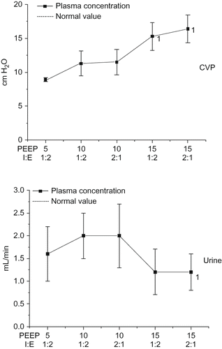
The CVP rose significantly with an PEEP of 15 cm H2O in comparison to baseline ().
FIGURE 1. Changes in central venous pressure and diuresis during the study period (1 = p < 0.05 difference to the begin of the study).
At the start of the study, creatinine, urea, sodium, and potassium in serum were all within normal ranges and remained so throughout the study. Even on the next day, no detectable changes could be found. The baseline values at the start of the study are shown in . We refrained from any other forms of illustration because no further changes could be observed over the entire course of the study.
The urinary excretion increased at an PEEP of 10 cm H2O and an I:E ratio of 1:2 and fell from this time point until a significant change was seen with an PEEP of 15 cm H2O and an I:E ratio of 2:1 ().
The FESo increased during the study periods and showed a significant difference at an PEEP of 15 cm H2O and an I:E ratio of 2:1 ().
FIGURE 2. Changes in fractional excretion of sodium and atrial natriuretic period during the study period (1 = p < 0.05 difference to the begin of the study).
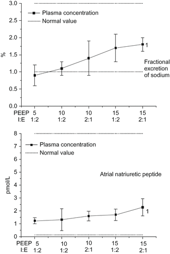
ANP during the first study period showed only a small increase, although this became significant with an PEEP of 15 cm H2O and an I:E ratio of 2:1 (). Over the entire study course, however, the values remained within the normal range.
Levels of antidiuretic hormones (ADH) also lay within the normal range (adiuretin, vasopressin). At an PEEP of 15 cm H2O and an I:E ratio of 1:2, however, they showed a significant increase compared to baseline ().
FIGURE 3. Changes in vasopressin and renin during the study period (1 = p < 0.05 difference to the begin of the study).
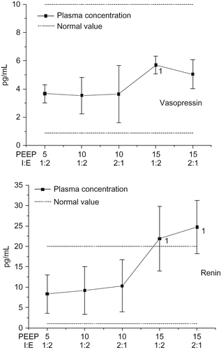
The renin concentration in serum showed a significant increase from an PEEP of 15 cm H2O as compared to baseline ().
The angiotensin II concentration in serum revealed a divergent course. After rising at an PEEP of 10 cm H2O, there was a decrease and then a further increase at an PEEP of 15 cm H2O and an I:E ratio of 2:1 (). The changes were not, however, statistically significant and were all within the normal range (angiotensin 2–500 pg/mL).
FIGURE 4. Changes in angiotensin II and aldosterone during the study period.
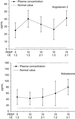
Concentrations of aldosterone were also within the normal range and showed no significant changes over the course of the study ().
DISCUSSION
Controlled ventilation with PEEP is necessary for many patients who have impaired lung function associated with differing underlying conditions.Citation7–9 For example, pulmonary diseases (e.g., pneumonia) or cardiac insufficiency with pulmonary edema are indications for invasive ventilation. In surgical medicine, postsurgical ventilation is often indicated after major surgery, after severe trauma, or if sepsis occurs. With such vital indications, side effects of ventilation therapy to other organ systems (e.g., hemodynamics, intrathoracic pressure) need to be taken into account.Citation10,Citation11 All 10 patients in this study suffered no hemodynamic or other problems over its entire course. After ending the study, they regained consciousness either on the evening or the next day and showed no pathological side effects.Citation12,Citation13
Urine
With regard to kidney function, reductions in urinary excretion, creatinine clearance, and sodium excretion were all observed. In both human and animal clinical studies, decrease in urine volume of 28–40%, creatinine clearance of 23%, and sodium excretion of 63% were observed after 20–60 min with PEEP values of 10 cm H2O.Citation14–16 In dogs, an immediate normalization of urine production was found after the termination of ventilation with PEEP.Citation14 Only with an PEEP of 15 cm H2O and an IRV (I:E of 2:1) was a significant reduction in urine excretion seen because of the increase in pressure in the thorax and the hormonal responses. The increase in vasopressin played a particularly important role here.Citation17,Citation18
CVP
The CVP increased during the course of the study. This observation can quite easily be attributed to the increased PEEP that causes a direct increase in pressure in the thorax, and subsequently also the capacity vessels.Citation3,Citation17
Fractional excretion of sodium
To distinguish a functional (prerenal) renal insufficiency from tubulointerstitial damage, assessment of sodium reabsorption in the tubule is important. Prerenal renal failure is accompanied by a reduced glomerular filtration rate (GFR) and a reduced filtered sodium content. Less than 1% of the filtered sodium is excreted through the urine. In tubulointerstitial dysfunction, the percentage of sodium excreted through the urine increases to over 3% of the sodium filtered by the glomeruli.Citation16
In this study, we have to assume that the GFR was altered because of prerenal influences, that is, especially by the changes in atrial pressure, and the changes occurring in the RAAS.
ANP
Up until now three natriuretic peptides have been described in human plasma, namely, ANP, brain natriuretic peptide (BNP), and c-type natriuretic peptide (PNP). ANP and BNP are cardiac hormones with natriuretic, vasodilatory, and aldosterone inhibiting properties. Both hormones are secreted more in disorders that lead to increased cardiac output or an increase in intravascular pressure. Confirmed stimuli for ANP secretion include an increase in transmural pressure and an increase in heart rate. One can assume that an increase in atrial pressure of 1 mmHg will lead to an increase in plasma ANP levels of 10–14 pmol/L. This effect could not be observed to the same extent in our study ( and ).Citation19
Increased ANP secretion leads to a decrease in renin and aldosterone concentration in the blood, where in part the central effects of angiotensin II, such as increased ADH secretion, aggravated thirst and increased blood pressure, are also inhibited.Citation18 This effect contradicts with our own findings, where increases in renin and angiotensin II were found ( and ). Earlier studies showed that positive-pressure ventilation leads to an increase in right and left atrial pressure, a decrease in ANP, and an increase in CVP.Citation4 This decrease in ANP concentration could not be found in our own results, and instead we even saw a rise during the last period. Our literature-inconsistent findings may have been related to the study design. Although a period of 60 min leads to a steady state regarding hemodynamic parameters, different results were apparent regarding the hormonal changes.
The positive correlation between an elevated CVP and ANP/FESo described elsewhere was also clear in our own results.Citation19 A more rapid equilibration seems to have taken place.
ANP causes an increase in sodium excretion, an increase in water excretion, and a reduction in plasma volume. This was also clear from our own results where there was an increase in FESo and diuresis during the first study period ( and ).
Vasopressin
Vasopressin is a nonapeptide hormone that is formed in the hypothalamus and stored in the posterior lobe of the pituitary gland. Vasopressin secretion is promoted by increasing the effective osmotic pressure, reducing the extracellular fluid volume, thirst, medications, and emotional factors. Conversely, it is reduced by lowering the effective osmotic pressure, increasing the extracellular fluid volume, and consuming alcohol. Using a secondary messenger as an intermediary, vasopressin causes water retention as well as urinary concentration through its effects particularly on the distal tubules and collecting ducts where permeability is increased.
Peripherally, ADH is a thirst hormone. When water is lacking in the body, the blood serum becomes hypertonic. This is detected by osmoreceptors found in the hypothalamus, which in turn induce the release of vasopressin from the posterior pituitary gland. Vasopressin secretion can also be stimulated if there is a reduced volume within the arterial system, as registered by baroreceptors in the right atrium of the heart and the aortal arch. This mechanism should also contribute to the increase of vasopressin seen with an PEEP of 15 cm H2O, after the diuresis was increased during the first study period.Citation20
Renin
At the beginning of the RAAS cascade, the enzyme renin is released from a specialized region within the kidney tissue, that is, the so-called juxtaglomerular apparatus. This consists of cells within the blood vessel that supplies the glomerulus with blood. The following factors lead to an increased release of renin. In the blood renin cleaves the N-terminal end of angiotensinogen, that is, angiotensin I, which is then converted by angiotensin-converting enzyme to angiotensin II which raises the blood pressure. In ventilated patients, increased plasma renin and aldosterone activity were measured in plasma after a 20-min PEEP ventilation.Citation4,Citation20
A redistribution of blood flow occurs in favor of the inner cortex, even though the total renal perfusion remains constant.Citation15 Other studies have shown that under PEEP a reduction of blood flow to the kidney occurs that is proportional to the reduced cardiac output.Citation5,Citation14 The observed stimulation of renin secretion was probably caused by a reduced renal perfusion as a result of pressure changes in the major vessels. The early inhibition of renin release in the first study period was primarily due to angiotensin II and the aldosterone released by it.
Angiotensin II
Angiotensin II promotes the aldosterone increase, the sodium chloride increase, and the increase in water retention. In the adrenal cortex, angiotensin II induces the release of the hormone aldosterone. This promotes the resorption of renal sodium and water from the urine into the blood, making the salt content of the blood and the blood volume to increase. The angiotensin II formed interactions with angiotensin receptors. By activating these receptors, the following effects can be brought about: an overall contraction in the blood vessels, a decreased glomerular filtration with a simultaneously reduced sodium excretion in the kidneys, a stimulation of aldosterone and adrenaline release in the adrenals, and a release of vasopressin from the pituitary. A sensation of thirst can also be attributed to an acute stimulation of the receptors in the hypothalamus. Here a feedback mechanism seems to have been in effect, which initially inhibited and then stimulated renin release over the course of the study.
Aldosterone
The most important mineral corticoid of the adrenal cortex regulates the electrolyte and water balance (through the RAAS) and is as such influenced by blood volume and blood pressure. Aldosterone increases sodium resorption, especially in the renal tubules, and promotes potassium excretion. Aldosterone increases the resorption of sodium ions in the connecting tubuli and collecting ducts of the kidney (which also increases the extracellular volume), as a result of the increased incorporation of sodium channels within the apical plasma membrane. A direct influence on the FESo as well as renin and angiotensin II levels could not be demonstrated in our own results.
Controlled ventilation with PEEP has effects on numerous organ systems.Citation21–23 The extent to which this form of ventilation should be carried out, or measures for preventing kidney damage are realizable, has been debated many times over the years.Citation24–26
Some of the investigated parameters such as the CVP increase, the decrease in diuresis, and the increase in FESo and ANP can be observed in various forms of positive pressure ventilation. The activation of the RAAS was only observed in part of this study. Only a renin increase was demonstrated, while angiotensin II and aldosterone were not significantly changed. In summary, it can be stated that provided there is adequate filling of the heart, invasive ventilation only results in clear hormonal and functional renal effects from an PEEP of 15 cm H2O and in the presence of an IRV.
Declaration Of Interest: The authors report no conflicts of interest. The authors alone are responsible for the content and writing of the paper.
REFERENCES
- Brienza N, Dalfino L, Cinnella G, Diele C, Bruno F, Fiore T. Jaundice in critical illness: Promoting factors of a concealed reality. Intensive Care Med. 2006;32(2):267–274.
- Neumann P, Berglund JE, Andersson LG, Maripu E, Magnusson A, Hedenstierna G. Effects of inverse ratio ventilation and positive end-expiratory pressure in oleic acid-induced lung injury. Am J Respir Crit Care Med. 2000;161(5):1537–1545.
- Lessard MR, Guerot E, Lorino H, Lemaire F, Brochard L. Effects of pressure-controlled with different I:E ratios versus volume-controlled ventilation on respiratory mechanics, gas exchange, and hemodynamics in patients with adult respiratory distress syndrome. Anesthesiology. 1994;80:983–991.
- Andrivet P, Adnot S, Sanker S, Hormonal interactions and renal function during mechanical ventilation and ANF infusion in humans. J Appl Physiol. 1991;70(1):287–292.
- Beyer J. Regional blood flow and tissue oxygenation in positive end expiratory pressure ventilation. Fortschr Med. 1983;17(7):287–290.
- Duke GJ. Cardiovascular effects of mechanical ventilation. Crit Care Resusc. 1999;1(4):388–399.
- Shanholtz C, Brower R. Should inverse ratio ventilation be used in adult respiratory distress syndrome? Am J Respir Crit Care Med. 1994;149:(5)1354–1358.
- Botha J, Mudholkar P, Le Blanc V. The effect of changing from pressure support ventilation to volume control ventilation on renal function. Crit Care Resusc. 2005;7:303–309.
- Amato MB, Barbas CS, Medeiros DM, Effect of a protective-ventilation strategy on mortality in the acute respiratory distress syndrome. N Engl J Med. 1998;5:347–354.
- Pannu N, Mehta RL. Effect of mechanical ventilation on the kidney. Best Pract Res Clin Anaesthesiol. 2004;18:189–203.
- Luecke T, Pelosi P. Clinical review: Positive end-expiratory pressure and cardiac output. Crit Care. 2005;9(6):607–621.
- Sural S, Sharma RK, Singhal M, Etiology, prognosis, and outcome of post-operative acute renal failure. Ren Fail. 2000;1:87–97.
- Vieira JM Jr, Castro I, Curvello-Neto A, Effect of acute kidney injury on weaning from mechanical ventilation in critically ill patients. Crit Care Med. 2007;35(1):184–191.
- Gammanpila S, Bevan DR, Bhudu R. Effect of positive and negative expiratory pressure on renal function. Br J Anaesth. 1977;(3):199–205.
- Hall SV, Johnson EE, Hedley-Whyte J. Renal hemodynamics and function with continuous positive-pressure ventilation in dogs. Anesthesiology. 1974;41(5):452–461.
- Jarnberg PO, de Villota ED, Eklund J, Granberg PO. Effects of positive end-expiratory pressure on renal function. Acta Anaesthesiol Scand. 1978;22(5):508–514.
- Payen DM, Farge D, Beloucif S, No involvement of antidiuretic hormone in acute antidiuresis during PEEP ventilation in humans. Anesthesiology. 1987;1:17–23.
- Annat G, Viale JP, Bui Xuan B, Effect of PEEP ventilation on renal function, plasma renin, aldosterone, neurophysins and urinary ADH, and prostaglandins. Anesthesiology. 1983;2:136–141.
- Raufhake C, Walter M, Weber T, Van Aken H, Berendes E. Serum concentrations of atrial and brain natriuretic peptide during and after cardiopulmonary bypass. Anesth Analg. 2000;90:84–92.
- Kaczmarczyk G, Vogel S, Krebs M, Bünger H, Scholz A. Vasopressin and renin-angiotensin maintain arterial pressure during PEEP in non-expanded, conscious dogs. Am J Physiol Regul Integr Comp Physiol. 1996;271:R1396–R1402.
- Lefrant J-Y, Juan J-M, Bruelle P, Regional blood flows are affected differently by PEEP when the abdomen is open or closed: An experimental rabbit model. Can J Anaesth. 2002;49:302–308.
- Popovich M. Moon phases and ocean tides: The relationship between the inspiratory-expiratory phases of mechanical ventilation and right ventricular function. Crit Care Med. 1999;27:864–865.
- Theres H, Binkau J, Laule M, Phase related changes in right ventricular cardiac output under volume-controlled mechanical ventilation with positive end-expiratory pressure. Crit Care Med. 1999;27:953–958.
- Byrick RJ, Rose DK. Pathophysiology and prevention of acute renal failure: The role of the anaesthesist. Can J Anaesth. 1990;37:457–467.
- Wilkes BM, Mailloux LU. Acute renal failure. Pathogenesis and prevention. Am J Med. 1986;80:1129–1136.
- Uchino S, Kellum JA, Bellomo R, Beginning and ending supportive therapy for the kidney (BEST kidney) investigators. Acute renal failure in critically ill patients: A multinational, multicenter study. JAMA. 2005;294:813–818.