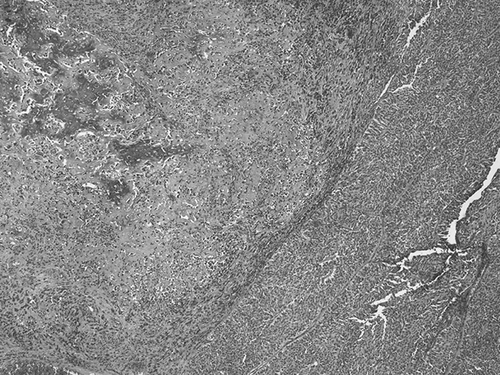
We have recently observed a sarcomatoid variant of urothelial carcinoma in the right renal pelvis of a 69-year-old man. Heterologous elements (chondrosarcoma and osteosarcoma) were found (). The nephrectomy specimen showed in the renal pelvis a polypoid tumor measuring 4 × 3 cm. The cut surface was gray white in color and hard in consistency. Histologically the tumor was composed by grade 2/3 transitional cell carcinoma (). The heterologous elements were prevalent (60% of the lesion) and invaded beyond muscularis propria into peripelvic fat (pT3). Immunohistochemically, epithelial elements were characterized by strong staining with keratins (AE1/AE3). The surrounding renal parenchyma was free of disease. We read with great interest Chiu KC and colleagues’ article “Renal carcinosarcoma: case report and review of literature” in Renal Failure (2008;30:1034–1039).Citation1 These authors present a “histologically proven case of renal carcinosarcoma extending from renal pelvis.” The first abdominal CT scan “revealed a well-defined 10 × 10 × 12 cm3 heterogeneous mass at the right kidney. Right arteriography was performed, which showed an avascular mass with a stretching of the renal arteries. A small area of renal parenchymal blush was found without definite hypervascular tumor stain.” The renal pelvis is not described. Another abdominal CT was performed previous to right radical nephrectomy and “revealed a well-defined 10 × 10 × 12 cm3 heterogeneous mass at the right kidney. Right arteriography was performed, which showed an avascular mass with a stretching of the renal arteries. A small area of renal parenchymal blush was found without definite hypervascular tumor stain.” The renal pelvis is not reported. On bisection, the renal parenchyma revealed extensive hemorrhage and necrosis with focal cystic formation. The calyces and pelvis were hardly identified. The ureter measured 4.0 cm in length and 0.5 cm in diameter. No hilar lymph node was grossly identified. Multiple sections of the kidney revealed extensive hemorrhage, necrosis, and focal cystic degeneration. The residual tumoral tissue showed an admixture of epithelial and mesenchymal elements. A few bizarre giant cells with one or two large nucleoli were present. The nuclei of the tumor cells had a coarsely clumped, chromatin pattern, and prominent nucleoli; a high mitotic rate was also noted. The carcinomatous component showed a tubular structure lined by a single layer of epithelial cells, which varied from low cuboidal to columnar, nuclear enlargement, and hyperchromasia. Immunohistochemical studies demonstrated positive staining for vimentin and negative staining against cytokeratin, desmin, and smooth muscle actin. The tubular cell areas were positive for cytokeratin. The renal pelvis is not described and is not recognizable in Figure 4 of the article. In conclusion, these authors have not documented the renal pelvis origin of the carcinosarcoma. In the title of the article, the authors reported “review of the literature.” This review of the literature is inaccurate. In the “References,” Chiu et al. cited only nine papers regarding carcinosarcoma of the renal pelvis.Citation2–10 Other reports of carcinosarcoma of the renal pelvis have not been reviewed by Chiu et al.Citation11–14 According to Kobayashi et al.’s review,Citation15 a total of 19 cases of carcinosarcoma or sarcomatoid carcinoma of the renal pelvis have been reported up until 1994. Eleven cases were reported as “true” carcinosarcoma and eight as sarcomatoid carcinoma. The carcinomatous component was transitional cell carcinoma in 14 cases, while the sarcomatous component in “true” carcinosarcomas was documented as rhabdomyosarcoma, osteosarcoma, and chondrosarcoma. In conclusion, the origin of carcinosarcoma from renal pelvis is not documented in Chiu et al.’s report and the authors performed an incomplete review of the literature regarding this neoplasm.
Figure 1. Heterologous elements (chondrosarcoma and osteosarcoma) were evident (H&E).
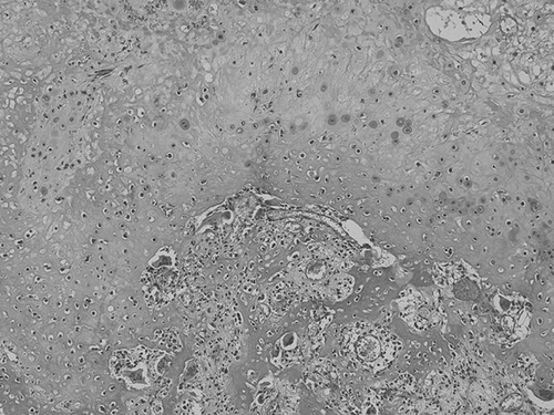
Figure 2. Mixture of heterologous elements and transitional cell carcinoma was evident (H&E).
Declaration of interest: The authors report no conflicts of interest. The authors alone are responsible for the content and writing of the paper.
Teresa Pusiol
Doriana Morichetti
Maria Grazia Zorzi
Francesco Piscioli
Institute of Anatomic Pathology, Rovereto Hospital, P.le S.Maria, 7, 38068 Rovereto (TN), Italy
E-mail: [email protected]
References
- Chiu KC, Lin MC, Liang YC, Chen CY. Renal carcinosarcoma: Case report and review of literature. Ren Fail. 2008;30:1034–1039.
- Fauci Jr. PA, Therhag HG, Davis JE. Carcinosarcoma of the renal pelvis. J Urol. 1961;85:897–902.
- Fisher ER, Davis ER. Carcinosarcoma of kidney. J Urol. 1962;87:109–118.
- Hou LT, Willis RA. Renal carcinosarcoma: True and false. J Pathol Bacteriol. 1963;85:139–144.
- Farrow GM, Harrison EG, Utz DC. Sarcomatous and sarcomatoid and mixed malignant tumors of the kidney in adults. Cancer. 1968;22:556–563.
- Ellioitt JT, Pontius EE, McCallum DC. Carcinosarcoma of kidney. Urology. 1973;1:151–153.
- Gallagher JC, Winslow DJ, Grossman A. Coexistent chondrosarcoma and transitional cell carcinoma in kidney. Urology. 1974;3:473–477.
- Ridolfi RL, Eggleston JC. Carcinosarcoma of the renal pelvis. J Urol. 1978;119:569–572.
- Tarry WF, Morabito RA, Belis JA. Carcinosarcoma of the renal pelvis with extension into the renal vein and inferior vena cava. J Urol. 1982;128:582–585.
- Chen KTK, Workman RD, Flam MS, DeKlotz RJ. Carcinosarcoma of renal pelvis. Urology. 1983;22:429–431.
- Dimitriou RJ, Gattuso P, Coogan CL. Carcinosarcoma of the renal pelvis. Urology. 2000;11(56):508.
- Vermeulen P, Hoekx L, Colpaert C, Wyndaele JJ, Van Marck E. Biphasic sarcomatoid carcinoma (carcinosarcoma) of the renal pelvis with heterologous chondrogenic differentiation. Virchows Arch. 2000;437(2):194–197.
- Lema Grille J, Blanco Parra M, Suárez Peñaranda JM, Durana Tonder C. Carcinosarcoma of the renal pelvis: Report of a case and review of the literature. Actas Urol Esp. 2002;26(7):509–512.
- Yilmaz E, Birlik B, Arican Z, Guney S. Carcinosarcoma of the renal pelvis and urinary bladder: A case report. Korean J Radiol. 2003;4(4):255–259.
- Kobayashi M, Hashimoto S, Hara Y, . Squamous carcinoma with pseudosarcomatous stroma of the renal pelvis and ureter: A case report. Hinyokika Kiyo. 1994;40:55–59 (in Japanese with an English abstract).