Abstract
Purpose: To report a case of posterior scleritis associated with angle closure glaucoma and transient myopia.
Methods: A previously healthy 29-year-old male was referred for YAG laser iridotomy to treat angle closure glaucoma. He was suffering from severe pain and vision loss in his right eye. Findings on examination were hyperemia, proptosis, and myopia measuring approximately 6 diopters. Diagnosis of posterior scleritis was made due to presence of choroidal thickening, sub-Tenon effusion and the classical “T” sign observed on orbital ultrasonography.
Results: Treatment was initiated using oral prednisolone. After 8 days, the patient’s symptoms regressed and visual acuity returned to 20/20.
Conclusions: Posterior scleritis is an often misdiagnosed eye disease. Pain is the main symptom and may be accompanied by decrease in visual acuity. Early diagnosis and treatment is important to avoid permanent visual loss.
Posterior scleritis is an unusual granulomatous inflammatory disease, which affects the posterior segment of the eye causing changes in the retina, choroid, and optic nerve. The clinical differential diagnosis of posterior scleritis includes angle closure glaucoma, choroidal folds, optic disc edema, intraorbital mass, choroidal detachment, and exudative retinal detachment. Ultrasonography (USG) and computerized tomography (CT) are usually effective in demonstrating thickened sclera with fluid in the sub-Tenon’s space.Citation1
CASE REPORT
A 29-year-old male patient who had been diagnosed with acute angle closure glaucoma was referred to our clinic for YAG laser iridotomy. His complaints were severe right eye pain for 1 week, loss of vision of the same eye, and a gradually increasing same-sided eyelid swelling. He had used dorzolamide HCl 2% drops bid, asetazolamide tablets qid, and pilocarpine HCl 2% drops qid for the past 3 days.
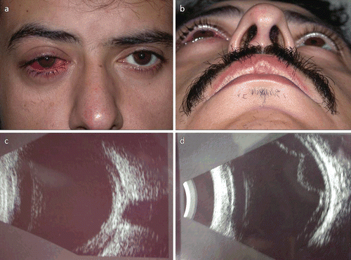
His uncorrected visual acuity was measured 20/400, which improved to 20/20 with –6.0 dioptres spherical correction. The visual acuity of the left eye was 20/20 without correction. Anterior segment examination of the right eye showed conjunctival hyperemia, shallow anterior chamber, angle closure, and bulging of the lens anteriorly. The retina and the optic nerve head were normal. Proptosis was present in the right eye. Hertel exophthalmometer was 20 mm in the right eye and 16 mm the left eye (lateral interorbital width measurement was 106 mm) (,). The orbital USG identified subchoroidal effusion and choroidal thickening in the right eye ().
Magnetic resonance imaging (MRI) performed the following day failed to show any mass or vascular abnormality. A repeat orbital USG, however, detected choroidal effusion and exudative retinal detachment (). At this point, posterior scleritis was considered to be the cause of the angle closure glaucoma. Systemic laboratory tests were positive for antinuclear antibody (ANA).
The patient was treated with 1 mg/kg/day prednisolone acetate. On the third day of treatment, decreased congestion and resolving proptosis were noted (Hertel exophthalmometry readings were 17 mm for right eye and 16 mm for left). The USG also showed a decrease in subchoroidal effusion and choroidal thickening. By the eighth day of treatment, the right visual acuity was 20/20 without correction and proptosis had completely resolved.
FIGURE 1 (a)The right eye is injected, and with marked proptosis (4 mm). (b) The proptosis in the right eye is more significant in the supine position. (c) B-scan USG showing a sub-Tenon’s effusion at the junction of the optic nerve and globe (T-sign). (d) B-scan USG showing choroidal effusion and exudative retinal detachment.
DISCUSSION
Posterior scleritis is usually seen unilaterally between the ages of 40–60 years. Although other symptoms, such loss of vision and the narrowing of the visual fields, are observed, the main symptom is pain in the affected eye. Exudative retinal detachment, cystoid macular edema, retinal pigment epithelial detachment, and choroidal folds may also be observed as in Vogt Kayanagi Harada syndrome.Citation2 Fundus examination may be suspicious for presence of posterior segment mass. This appearance may be confused with malignant melanoma, uveal metastatic carcinoma, choroidal hemangioma, or lymphoid hyperplasia.Citation3 It can be difficult to diagnose posterior scleritis and diagnosis usually depends on the observed symptoms. USG, fundus fluorescein angiography, and CT may be required for differential diagnosis.Citation4 The USG and CT will show the sub-Tenon effusion and any thickening of the posterior sclera, contributing to a definitive diagnosis. The basic sign in the USG is the “T” sign due to retrobulbar edema.Citation5 The “T” sign was clearly observed in our case. Symptoms and signs usually depend on the characteristics of inflammation, including location, and severity of involved adnexal choroid, retinal pigment epithelium, and extraocular muscles.
In cases of posterior scleritis, intraocular pressure is increased by 12–46%, and a number of mechanisms contribute to the increase in pressure. The mechanisms include inflammation of the outflow pathways, increasing of intraocular fluid viscosity, occlusion of trabecular meshwork by inflammatory cells and debris, peripheral anterior synechia, neovascularization, and increasing episcleral venous pressure.Citation6 Secondary angle closure glaucoma is usually seen in posterior episcleritis. The pathophysiology beneath the secondary angle closure glaucoma may be vascular congestion and inflammation of the sclera, ciliary body, and choroid. The inflammation of sclera may cause fluid collection posterior to the ciliary body and choroid. Supraciliary, choroidal effusion and ciliary body edema cause rotation of the ciliary body and iris base leading to displacement of the lens–iris diaphragm anteriorly and causing angle closure glaucoma.Citation6 The shallow anterior chamber and angle closure glaucoma in our case supports this thesis.
Myopia of approximately 6.00 diopters is another significant pathologic condition in our case. We assume a decrease in radius of the ciliary circle secondary to fluid collection behind the ciliary body. This would then cause a decrease in the tightness of the zonular fibers and would increase refractiveness of the lens. Another mechanism leading to myopia is caused by the anterior displacement of the lens–iris diaphragm, which increases the distance between the fovea and the lens. The alterations in lens refractiveness and axial length changes are the two possible mechanisms leading to myopia.
Inflammation of the macula and the optic disc may cause optic disc edema, retinal folds, macular exudations, and the detachment of retinal pigment epithelium, all of which may be responsible for loss of visual acuity.Citation1 However, in our case, the choroidal thickening and subchoroidal effusion led to proptosis, pushing from the anterior due to the mass effect.
Nonsteroidal anti-inflammatory drugs, corticosteroids, and immunosuppressive drugs may be effective in the treatment of posterior scleritis.Citation7 After treatment, inflammation and effusion usually subside and the anterior chamber depth is regained. Cycloplegic drugs, β–adrenergic antagonists, alpha 2 agonists, and carbonic anhydrase inhibitors may be useful in the treatment of posterior scleritis. Cycloplegic drugs help by pulling the lens–iris diaphragm posteriorly and by enabling tight zonules and open angle. Posterior scleritis cases are usually accompanied by anterior scleritis and anterior uveitis. Therefore, preventing synechia is an important aim of treatment.Citation2 Our case was successfully treated with systemic corticosteroids.
Posterior scleritis has a tendency to reoccur and if not treated may lead to sight-threatening complications. Posterior scleritis should be considered in the differential diagnosis of subretinal lesions with mass-like behavior. Early diagnosis is crucial because prompt treatment may have excellent outcome.
Declaration of interest: The authors report no conflicts of interest. The authors alone are responsible for the content and writing of the paper.
REFERENCES
- Dodds EM, Lowder CY, Barnhorst DA, et al. Posterior scleritis with annular ciliochoroidal detachment. Am J Ophthalmol. 1995; 120(5):677–679.
- Benson WE, Shields JA, Tasman W, et al. Posterior scleritis: a cause of diagnostic confusion. Arch Ophthalmol. 1979; 97(8):1482–1486.
- Stanga PE, Lim JI, Hamilton P. Indocyanine green angiography in chorioretinal diseases: indications and interpretation: an evidence-based update. Ophthalmology. 2003; 110(1):15–21.
- Maggioni F, Ruffatti S, Viaro F, et al. A case of posterior scleritis: differential diagnosis of ocular pain. J Headache Pain. 2007; 8(2):123–126.
- Biswas J, Mittal S, Ganesh SK, et al. Posterior scleritis: clinical profile and imaging characteristics. Indian J Ophthalmol. 1998; 46(4):195–202.
- Jain SS, Rao P, Kothari K, et al. Posterior scleritis presenting as unilateral secondary angle-closure glaucoma. Indian J Ophthalmol. 2004; 52(3):241–244.
- Jabs DA, Mudun A, Dunn JP, et al. Episcleritis and scleritis: clinical features and treatment results. Am J Ophthalmol. 2000; 130(4):469–476.