
Abstract
Stress-induced changes in plasma corticosterone and central monoamine levels were examined in mouse strains that differ in fear-related behaviors. Two DxH recombinant inbred mouse strains with a DBA/2J background, which were originally bred for a high (H-FSS) and low fear-sensitized acoustic startle reflex (L-FSS), were used. Levels of noradrenaline, dopamine, and serotonin and their metabolites 3,4-dihydroxyphenyacetic acid (DOPAC), homovanillic acid (HVA), and 5-hydroxyindoleacetic acid (5-HIAA) were studied in the amygdala, hippocampus, medial prefrontal cortex, striatum, hypothalamus and brainstem. H-FSS mice exhibited increased fear levels and a deficit in fear extinction (within-session) in the auditory fear-conditioning test, and depressive-like behavior in the acute forced swim stress test. They had higher tissue noradrenaline and serotonin levels and lower dopamine and serotonin turnover under basal conditions, although they were largely insensitive to stress-induced changes in neurotransmitter metabolism. In contrast, acute swim stress increased monoamine levels but decreased turnover in the less fearful L-FSS mice. L-FSS mice also showed a trend toward higher basal and stress-induced corticosterone levels and an increase in noradrenaline and serotonin in the hypothalamus and brainstem 30 min after stress compared to H-FSS mice. Moreover, the dopaminergic system was activated differentially in the medial prefrontal cortex and striatum of the two strains by acute stress. Thus, H-FSS mice showed increased basal noradrenaline tissue levels compatible with a fear phenotype or chronic stressed condition. Low corticosterone levels and the poor monoamine response to stress in H-FSS mice may point to mechanisms similar to those found in principal fear disorders or post-traumatic stress disorder.
Introduction
Aberrant fear conditioning is a primary component in the pathology of anxiety disorders (Lissek, Citation2012). Pavlovian fear conditioning is based on pairing a neutral stimulus with an unconditioned stimulus (US), such as an aversive footshock that transforms the neutral stimulus to a conditioned stimulus (CS). Fear extinction is the gradual weakening of this learned fear response upon repetitive presentations of the CS in the absence of the US (Davis et al., Citation2003; Sotres-Bayon et al., Citation2004). Footshock delivery both induces fear conditioning and provokes acute stress responses in rodents (Hajos-Korcsok et al., Citation2003), similar to stress responses induced by forced swimming (e.g. Reyes et al., Citation2012; Vranjkovic et al., Citation2012). Such stress responses, as well as the acquisition, expression and extinction of conditioned fear responses, are under the control of prefrontal-limbic networks that include the amygdala, hippocampus and medial prefrontal cortex (Cisler et al., Citation2010; Hitchcock et al., Citation1989; Maren & Hobin, Citation2007; Quinn et al., Citation2005; Sotres-Bayon et al., Citation2004; Vertes, Citation2004; Yilmazer-Hanke, Citation2008). Moreover, acute swim stress and footshocks presented during fear-conditioning training both (i) activate the hypothalamic-pituitary-adrenal (HPA) axis, resulting in corticosteroid release (Kioukia-Fougia et al., Citation2002; Yang le et al., Citation2012), and (ii) alter monoaminergic function in prefrontal-limbic networks, including ventral basal ganglia and downstream brain areas in the hypothalamus and midbrain (Bennett, Citation2011; Hajos-Korcsok et al., Citation2003; Shishkina et al., Citation2012; Stanford, Citation1996).
Screening for impaired fear extinction is a useful translational tool, which can facilitate the development of novel therapeutics for treating anxiety-related disorders (Holmes & Singewald, Citation2013). Inbred rodent strains are widely used to generate new bidirectionally selected, congenic, or recombinant inbred strains with specific behavioral characteristics and genetic backgrounds (Bailey, Citation1971; Bignami, Citation1965; Hitzemann et al., Citation1995; Steimer & Driscoll, Citation2005; Vadasz et al., Citation1982; Yilmazer-Hanke et al., Citation2004). The genealogy of inbred mouse strains show that the C3H/HeJHd and DBA/2JHd strains originate from a common ancestor (Beck et al., Citation2000). The two mouse strains exhibit a similar anxiety phenotype in the elevated plus maze, but they differ in their fear-sensitized acoustic startle response (FSS), which correlates with the density of serotonin and kainate binding sites in the amygdala (Yilmazer-Hanke et al., Citation2003). Thus, they are ideal candidates for generating new recombinant inbred strains that differ in fear- and stress-related behaviors (Yilmazer-Hanke, Citation2008).
In the present study, we utilized two newly generated DxH recombinant inbred mouse strains that were bidirectionally selected for a high FSS and low FSS, but share a common DBA/2J background. Previously, we investigated baseline monoamine levels in the brain of the two mouse strains (Browne et al., Citation2013), and now focus on stress-related changes. Based on the differences in fear-/stress-related behaviors and the amygdalar serotoninergic system of the parental C3H/HeJHd and DBA/2JHd strains (Yilmazer-Hanke et al., Citation2003), we hypothesized that a higher stress responsivity in the novel recombinant inbred strains is related to altered corticosterone release and/or monoaminergic transmission in prefrontal-limbic networks and associated brain regions. Therefore, we confirmed the fear phenotype in the Pavlovian fear-conditioning paradigm by measuring fear recall and extinction, and analyzed basal and stress-induced corticosterone and monoamine levels/turnover in prefrontal limbic regions, and downstream brain areas in the high-FSS and low-FSS strains.
Materials and methods
Animals
The two DxH recombinant inbred strains were generated in the Neuroanatomy Section, Institute of Anatomy, University of Magdeburg, Germany. First, offspring of C3H/HeJHd mice with a high FSS were backcrossed onto DBA/2JHd mice with a low FSS (Yilmazer-Hanke et al., Citation2003). During backcrossing, mice with a high FSS were monitored for their microsatellite loci (altogether 94 loci on chromosomes 1–19 and X chromosome at a distance of 10–30 centimorgan). At the B7th-B9th backcrossing generation, mice with a high FSS differed only in five microsatellite loci from DBA/2JHd mice. However, these backcrossed mice remained heterozygous, because they carried one C3H/HeJHd and one DBA/2JHd allele. Starting from the B7th-B9th generation, two recombinant inbred mouse strains with opposing FSS behaviors, the high (H)-FSS and low (L)-FSS strains, were obtained. Bidirectional selection pressure was continued during inbreeding for another 6–7 generations. This allowed random distribution of the non-FSS-related alleles between the H-FSS and L-FSS strains, whereas alleles associated with a high or low FSS were subject to selection pressure (for review: Yilmazer-Hanke, Citation2008). During the inbreeding process, mice with a high FSS exhibited C3H/HeJHd-type microsatellite markers. Therefore, monitoring of the five microsatellite markers helped to identify optimum breeders for accelerating the inbreeding process as generally recommended (JAX®-Genome-science-services, Citation2013). One microsatellite locus marking a high FSS was lost during inbreeding. Thus, the H-FSS strain is a C3H-like recombinant inbred strain with a DBA/2J background, which is homozygous for C3H/HeJHd alleles linked to the microsatellite markers D13Mit147, D13Mit291, D14Mit154 and D14Mit161 located on chromosomes 13 and 14. The L-FSS recombinant inbred strain was obtained during the inbreeding process from mice that showed a low FSS and carried only microsatellite alleles of DBA/2JHd mice (Hanke et al., Citation2005; Walsh et al., Citation2008). Therefore, the two mouse strains studied here are congenic-like recombinant inbred strains with a DBA/2JHd background, which differ in chromosomal segments originating from the C3H/2JHd strain.
The mice were maintained under a 12 h light/dark cycle at a room temperature of 21 °C ± 1 °C. Food and water was provided ad libitum. The H-FSS and L-FSS mice were obtained from colonies maintained by brother-and-sister-mating. Experimental mice were weaned at 3–4 weeks of age and housed in groups of 2–5 mice. Behavioral testing and collection of samples (blood and brain) occurred during the light phase. All procedures were carried out in accordance with the European Communities Council Directive of 24 November 1986 (86/609EEC) and NIH guidelines approved by the Animal Experimentation Ethics Committee of University College Cork (UCC) and the IACUC at Creighton University (CU). Age-matched separate cohorts of 6–8 weeks old male H-FSS and L-FSS mice were used for different experiments. Individual experiments were carried out by the same investigator. The cohorts were used to measure conditioned fear (H-FSS: n = 12; L-FSS: n = 9, UCC), immobility behavior during the acute forced swim test variant (H-FSS: n = 14; L-FSS: n = 14, CU), corticosterone levels following acute swim stress (H-FSS stress: n = 5; H-FSS control: n = 5; L-FSS stress: n = 8; L-FSS control: n = 8, UCC), and stress-induced alterations in brain monoamines (H-FSS stress: n = 11--12; H-FSS control: n = 9; L-FSS stress: n = 8; L-FSS control: n = 8--9, UCC).
Fear recall and extinction
Mice were transported in their home cages to the neighboring behavioral study room and habituated for 30 min to the new room before experiments were initiated. Fear conditioning was performed in a sound-attenuating isolation cabinet (San Diego Instruments, CA) illuminated by a light-emitting diode (LED). Mice were habituated for 30 min to the behavioral laboratory and placed in a transparent acrylic cylindrical animal enclosure that contained footshock grids (12.7 cm × 3.8 cm). After an initial 120 s acclimation period, they received 3 pairings (90–120 s variable inter-pairing interval) of a conditioned (CS; 30 s, 80 dB, white noise) and unconditioned stimulus (US; 2 s, 0.5 mA scrambled footshock), the latter co-terminated with the CS. The last US-CS pairing was followed by a 120 s no-stimulus consolidation period, and the mice were returned to their home cages. Within-session CS-recall and extinction learning was assessed 24 h later in a novel context (checkered cardboard walls, 25.5 cm × 12.7 cm enclosure with smooth bottom, lemon essence). After 120 s acclimation, the mice received 50 presentations of the CS (30 s, 5 s interstimulus intervals). Freezing time was scored by a trained observer using custom-made software (Yilmazer-Hanke et al., Citation2003). Fear behavior during acclimation and fear recall were calculated as the percentage of freezing time. Fear extinction was calculated as the ratio of the freezing time during the first (x) and second halves (y) of acoustic stimulation, respectively (100 – y/x*100).
Acute forced swim test
Exposure to swimming for 15 min is an effective physical and psychological stressor in rodents (Browne et al., Citation2012). For inducing acute swim stress and testing immobility, a shortened variant of the forced swim test (Bogdanova et al., Citation2013), called the forced swim test variant (FSTv), was applied. Thirty minutes before the experiment, mice were transported in their home cages to the neighboring behavioral study room. They were placed in a cylinder (internal diameter of 24 cm × 21 cm) filled with water to a depth of 15 cm, which was maintained at a temperature of approximately 24–25 °C. After stress exposure for 15 min, animals were taken out of the water, dried with a paper towel, and returned to their home cages. The water was renewed for each mouse with clean water. Behavior was scored by a trained observer using custom-made software measuring the sequence of behavior (Dipl. Ing. C.Kurtz, Magdeburg, Germany; see Yilmazer-Hanke et al., Citation2003). Immobility was assessed in intervals of 5 min, as well as in the whole test period consisting of 15 min. Immobility (i.e. floating) was defined as the lack of movements except those that will aid in keeping the mouse’s head above water.
Brain dissection
Mouse brains were dissected to study monoamines 30 min after exposure to acute stress and in controls. Mice were exposed to acute swim stress in the behavioral study room near the animal holding room and were transported in their home cages to the accessory surgery room. Thus, contrary to our prior study on baseline monoamine levels (Browne et al., Citation2013), the mice stayed in the same facility. Mice were sacrificed by cervical dislocation and their brains were removed. Brain regions of interest were dissected out on an ice-cooled Petri dish using sharp tweezers according to the atlas of Franklin & Paxinos (Citation2008). The samples were snap frozen with liquid nitrogen and kept at−80° C until further use. At the ventral surface of the brain, the hypothalamus was peeled out from the thalamus starting directly behind the optic chiasm (approximately between Bregma −0.58 and −3.40). After removing the olfactory bulbs, the brains were placed back on their ventral surface and the medial prefrontal cortex was dissected out bilaterally (approximately between Bregma 2.68 and 1.34); hence the tissue blocks included the anterior cingulate, prelimbic and infralimbic divisions of the medial prefrontal cortex, and maybe some portions of the medial orbital cortex and motor area M2. After that, a coronal block was removed from the brain (approximately between Bregma 1.34 and 0.02), and the striata (including nucleus accumbens) were dissected from the subcortical white matter laterally and septum medially. In the mid-hemispheric block from approximately Bregma −0.94 to −2.18, first the hippocampi were dissected out from the white matter beneath the neocortex and stem of the adjacent thalamus and midbrain. Then, the amygdalae were separated from posteriorly from the optic tract and removed with ventral portions of the surrounding cortex. The brainstem block (including the midbrain and pons) was dissected by removing the cerebellum from the ventral surface of the brain (approximately from Bregma −3.38 to −4.44).
Corticosterone enzyme-linked immunosorbent assay
Corticosterone was measured in trunk blood that was collected upon decapitation in the accessory procedure room located next to the behavioral study and animal holding rooms. The blood was obtained 30 min after exposure to swim stress to allow the corticosterone response to reach its peak (Connor et al., Citation1997), and also from control mice. A commercially available enzyme-linked immunosorbent assay (ELISA) kit (Assay Designs Correlate-EIATM Corticosterone 96 well kit catalog No. 900-097) was used to quantify the levels of the stress hormone corticosterone in the plasma. Briefly, trunk blood was collected in heparin-coated vacutainer tubes (BD Vacutainer 5 ml sterile, Becton, Dickinson and Company, Oxford, England). These tubes were centrifuged (Hettich Mikro 22R centrifuge, Tuttlingen, Germany) at 6000 rpm at 4 °C for 15 min. Supernatant plasma was then collected and stored at −80 °C until analysis. The assay was carried out as per manufacturer instructions. In order to adequately analyze the levels of corticosterone, samples were diluted 1/100, enabling detection of all samples to fall within the linear part of the standard curve. A Biochrom ASYS Atlantis Plate washer was used during the step. On completion of the assay, plates were read at 405 nm and corrected at 580 nm using a Synergy HT BioTek® plate reader (BioTek, Vermont, VT). The coefficient of variation (CV) for all samples was determined using the same standards and controls across all plates to limit intra-plate variability, and only samples with a CV <10% were included in analysis. Results were calculated using Gen5 BioTek® Microplate Data Collection and Analysis software (BioTek, Vermont, VT). Data were then exported to Excel spreadsheets; concentrations were corrected and analyzed using appropriate statistical tests.
High-performance liquid chromatography
The concentrations of noradrenaline, dopamine, and serotonin and their respective metabolites were determined using reverse-phase high-performance liquid chromatography (HPLC) (Browne et al., Citation2011, Citation2013). Immediately prior to analysis, the samples were placed in an Eppendorf tube containing 1 mL of homogenization buffer [10mM PBS buffer with protease inhibitors (Sigmafast® protease inhibitor tablets, Sigma-Aldrich, UK)], which was spiked with the internal standard 4 ng/20 μl of N-methyl-serotonin (Sigma Chemical Co., UK). Homogenization was achieved by 2–4 s of sonication (duration dependent on sample size) using a Bandelin Sonoplus sonicator. The samples were centrifuged (Hettich Mikro 22 R centrifuge) at 14,000 rpm at 4 °C for 15 min. The mobile phase contained 0.1 M citric acid, 0.1 M sodium dihydrogen phosphate, 0.01 mM EDTA (Alkem/Reagecon, Cork), 5.6 mM octane-1-sulphonic acid (Sigma), and 9% (v/v) methanol (Alkem/Reagecon, Cork), and was adjusted to pH 2.8 using 4 N sodium hydroxide (Alkem/Reagecon, Cork). Forty microliters of sample supernatant was injected onto the HPLC system, which consisted of SCL 10-Avp system controller, LC-10AS pump, SIL-10A autoinjector (with sample cooler maintained at 4 °C), CTO-10A oven, LECD 6A electrochemical detector (Shimadzu), and an online Gastorr Degasser (ISS, UK). A new reverse-phase column (Kinetex 2.6u C18 100 A 100 × 4.6 mm, Phenomenex) maintained at 30 °C was employed in the separation (flow rate 0.9 mL/min); the glassy carbon working electrode combined with an Ag/Ag CL reference electrode (Shimadzu Europa GmbH, Duisburg, Germany) was operated at 0.8 V. Each neurotransmitter was identified by its characteristic retention time, as determined by standard injections that were run at regular intervals during the sampling period. Internal standard peak height ratios were measured and compared with standard injections. Results were expressed as nanograms of neurotransmitter per grams of fresh weight of the tissue. This method is based on that of Seyfried et al. (Citation1986) for detection of neurotransmitters and was modified to facilitate analysis using Class-VP 5 software (Shimadzu Europa GmbH, Duisburg, Germany) to generate the chromatograms.
Data analysis
Statistical analyses were performed with the aid of the IBM SPSS software package Version 19.0 (IBM, Armonk, NY). Univariate comparisons of two groups were performed using a t-test (Welch test with Satterthwaite’s approximation to compute the degree of freedom (df) where appropriate). Analyses of multiple groups were carried out by computing two-way analyses of variance (ANOVA) for the main factors strain and stress, followed by univariate posthoc t-tests. A two-tailed p value < 0.05 was deemed to be significant. Data were expressed as mean ± SEM.
Results
Fear recall and extinction
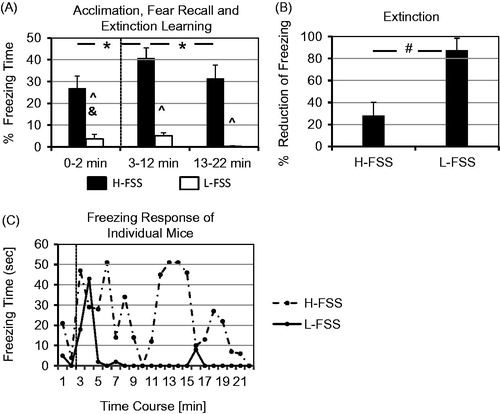
An ANOVA (main factor strain x repeated measures on fear recall) showed a significant strain difference (strain: F1,19 = 26.895, p < 0.001) and an increase in freezing duration upon stimulus presentation compared to the acclimation period (strain: F1,19 = 8.843, p < 0.01) with a strain x fear recall interaction (strain: F1,19 = 5.827, p < 0.05). During recall of auditory cue-conditioned fear, H-FSS mice showed an approximately four times longer freezing duration than L-FSS mice upon presentation of the CS (). A significant strain difference in freezing (strain: F1,19 = 30.138, p < 0.001) and a significant extinction learning in both strains (extinction learning: F1,19 = 9.489, p < 0.01) was found without an interaction between the two factors, as indicated by an ANOVA (main factor strain x repeated measures on the factor extinction learning). However, despite their ability to acquire some extinction, H-FSS mice displayed a severe deficit in extinction as compared to L-FSS mice (, t = − 3.491, df = 19, p < 0.01). Moreover, individual L-FSS mice displayed initial freezing responses of short duration upon CS presentation, which showed rapid extinction, whereas H-FSS mice specifically lacked this rapid extinction ().
Figure 1. The DxH recombinant inbred mouse strains both have a DBA/2J background, but the high-fear sensitized startle (H-FSS) strain has an insertion of four chromosomal C3H/2JHd segments into the DBA/2JHd background compared to the low (L)-FSS strain. (A) Increase in freezing duration following acclimation (0–2 min), and extinction learning during the first versus last 10 min of presentation of conditioned acoustic stimuli (CS). *p < 0.05 for fear recall (acclimation versus 3–12 min) and extinction learning (3–12 min versus 13–22 min), ^p < 0.05 for strain differences, &p < 0.05 strain x fear recall interaction. (B) H-FSS mice also show a severe extinction deficit in their freezing response. #p < 0.05 for strain differences. (C) Brief freezing in individual L-FSS mice upon CS presentation that declines rapidly, whereas H-FSS mice show generalized fear responses during acclimation and a deficit in extinction learning.
Acute forced swim test variant and corticosterone response
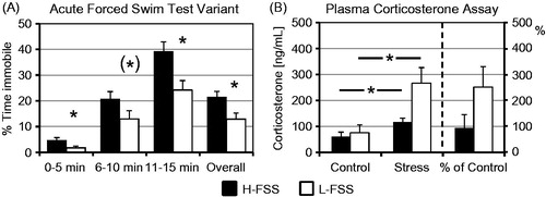
The H-FSS mice spent a significantly longer time immobile in the acute FSTv compared to L-FSS mice during the first (t = 2.320, df = 26, p < 0.05) and last 5-min block of the acute swim stress (t = 2.901, df = 26, p < 0.01), as well as during the whole test (t = − 2.790, df = 26, p = 0.01). In addition, a trend toward significance was found in the second block of the test (t = 1.832, df = 26, p = 0.078; ). A two-way ANOVA (strain x stress) revealed that acute swim stress induced by exposure to the FSTv significantly increased plasma corticosterone levels in both H-FSS and L-FSS mice (; stress effect: F1,22 = 8.745, p < 0.01). In addition, there was a trend towards a strain difference, the L-FSS mice showing higher basal and stress-induced corticosterone levels than H-FSS mice (strain effect: F1,22 = 3.862, p = 0.062). No strain x stress interaction was observed in plasma corticosterone levels. The average stress-induced increase in plasma corticosterone corresponded to 94% of control levels in H-FSS mice and 252% of control levels in L-FSS mice (), although this difference did not reach significance level (t = −1.446, df = 11, p = 0.176).
Figure 2. Immobility in the acute-15 min variant of the forced swim test (FSTv) and endocrine changes 30 min after exposure to acute swim stress (15 min duration). (A) The more fearful H-FSS mice spent a longer time immobile (i.e. floating) than the less fearful L-FSS mice in the acute FSTv. (B) There were no significant differences between basal plasma corticosterone concentrations of H-FSS and L-FSS mice, but acute swim stress induced by exposure to the FSTv resulted in a significant increase in corticosterone levels in both strains. L-FSS mice showed a higher stress-induced increase in plasma corticosterone than H-FSS mice (% of control), but this difference was not significant at the time point investigated after exposure to acute swim stress. *p < 0.05.
Monoamine levels
Noradrenaline
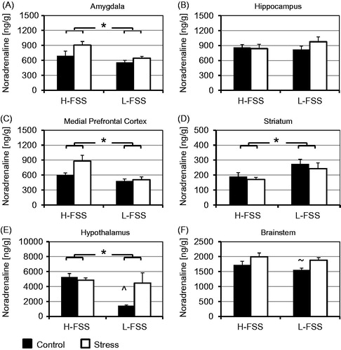
Stress induced by exposure to the acute FSTv significantly altered noradrenaline levels in the brainstem (), as indicated by the two-way ANOVA (stress x strain) (F1,34 = 7.188, p < 0.05). In addition, there was a significant interaction between the factors stress and strain in the hypothalamus for this monoamine (F1,34 = 5.562, p < 0.05). Post hoc analyses verified an increase of noradrenaline in the hypothalamus (p < 0.05) and brainstem of L-FSS mice (p < 0.05) but not H-FSS mice. The two-way ANOVA also showed strain differences. In H-FSS mice, basal and stress-induced noradrenaline levels were higher in the amygdala (F1,34 = 7.840, p < 0.01), medial prefrontal cortex (F1,34 = 8,377, p < 0.01), and hypothalamus (F1,34 = 8.439, p < 0.01), but were lower in the striatum compared to L-FSS mice (F1,34 = 8.133, p < 0.01).
Figure 3. Basal noradrenaline levels and changes in noradrenaline 30 min after exposure to acute swim stress in the FSTv. The H-FSS mice display higher noradrenaline concentrations in the amygdala (A), medial prefrontal cortex (C), and hypothalamus (E), and lower noradrenaline concentrations in the striatum (D) than L-FSS mice, but the two mouse strains do not differ in hippocampal noradrenaline levels (B). A stress x strain interaction is seen in the hypothalamus (E), and a stress effect in the brainstem (F).∼p < 0.05 for stress effect, *p < 0.05 for strain effect and ^p < 0.05 for stress x strain interaction in the two-way ANOVA.
Dopamine
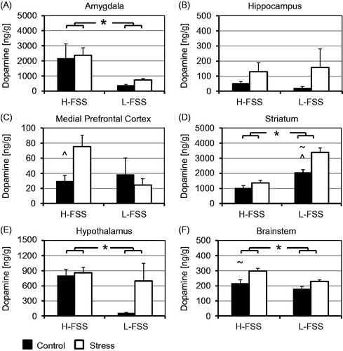
A two-way ANOVA (stress x strain) revealed a stress-induced increase in dopamine in the striatum (F1,34 = 15.291, p < 0.001) and brainstem (F1,34 = 12.969, p < 0.001) following exposure to the acute FSTv (). Moreover, a significant stress-strain interaction was seen in the medial prefrontal cortex (F1,34 = 4.181, p < 0.05) and striatum (F1,34 = 5.423, p < 0.05). Post hoc analyses showed a stress-induced dopamine increase in the striatum of L-FSS mice (p < 0.01), and medial prefrontal cortex (p < 0.05) and brainstem of H-FSS mice (p < 0.05). As with noradrenaline, the two-way ANOVA showed strain differences for dopamine levels in the amygdala and hypothalamus. Overall, dopamine concentrations were significantly higher in the amygdala (F1,34 = 9.196, p < 0.01), hypothalamus (F1,34 = 5.639, p < 0.05), and brainstem (F1,34 = 8.798, p < 0.01), but lower in the striatum of H-FSS mice compared to L-FSS mice (F1,34 = 51.819, p < 0.001). No strain differences were observed in the hippocampus.
Figure 4. Basal dopamine levels and changes in dopamine 30 min after exposure to acute swim stress in the FSTv. Dopamine levels are higher in the amygdala (A), hypothalamus (E), and brainstem (F) and lower in the striatum (D) of H-FSS than L-FSS mice. No differences are observed in the hippocampus (B). The stress-induced increase in dopamine levels is significant in the striatum (D) and brainstem (F), and an additional stress x strain interaction is seen in the medial prefrontal cortex (C) and striatum (D). ∼p < 0.05 for stress effect, *p < 0.05 for strain effect, and ^p < 0.05 for stress x strain interaction in the two-way ANOVA.
Serotonin
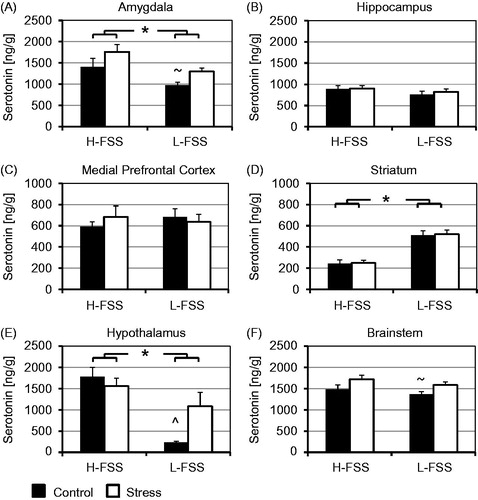
The two-way ANOVA (stress x strain) indicated that acute swim stress induced in the FSTv significantly increases serotonin levels in the amygdala (F1,34 = 4.683, p < 0.05) and brainstem (F1,34 = 6.005, p < 0.05). There was also a stress x strain interaction in the hypothalamus (F1,34 = 5.786, p < 0.05) (). Post hoc analyses confirmed an increase in serotonin levels in the amygdala (p < 0.05), hypothalamus (p < 0.05), and brainstem (p < 0.05) of L-FSS mice but not H-FSS mice. In addition to alterations in monoamines induced by stress, the two-way ANOVA also revealed strain differences. Both basal and stress-induced serotonin levels were significantly higher in the amygdala (F1,34 = 8.013, p < 0.01) and hypothalamus (F1,34 = 20.430, p < 0.001), and significantly lower in the striatum of H-FSS mice compared to L-FSS mice (F1,34 = 58.474, p < 0.001).
Figure 5. Basal serotonin levels and changes in serotonin 30 min after exposure to acute swim stress in the FSTv. Serotonin levels are higher in the amygdala (A) and hypothalamus (F) and lower in the striatum (D) of H-FSS than L-FSS mice. As with dopamine, no differences are observed in the hippocampus (B) and medial prefrontal cortex (C). The stress-induced increase in serotonin levels is significant in the amygdala (A) and brainstem (F), and a stress x strain interaction is seen in the hypothalamus (E). ∼p < 0.05 for stress effect, *p < 0.05 for strain effect, and ^p < 0.05 for stress x strain interaction in the two-way ANOVA.
Metabolites of monoamines
A two-way ANOVA (stress x strain) () revealed that acute swim stress induced in the FSTv significantly altered tissue concentrations of the dopamine metabolites 3,4-dihydroxyphenylacetic acid (DOPAC) in the brainstem (F1,34 = 6.928, p < 0.05), and of homovanillic acid (HVA) in the hippocampus (F1,34 = 4.199, p < 0.05) and brainstem (F1,34 = 14.887, p < 0.001). Significant stress-strain interactions were seen for DOPAC in the medial prefrontal cortex (F1,34 = 7.396, p = 0.01), striatum (F1,34 = 12.687, p = 0.001), and brainstem (F1,34 = 20.319, p < 0.001), and for HVA in the medial prefrontal cortex (F1,34 = 13.088, p = 0.001) and brainstem (F1,34 = 9.686, p < 0.01). In post hoc analyses, there was an increase of DOPAC in the medial prefrontal cortex of H-FSS mice (p < 0.05) and brainstem of both mouse strains (H-FSS p < 0.001, L-FSS p < 0.05). In addition, stress caused an increase of HVA in the brainstem of H-FSS mice (p < 0.001), whereas stress reduced DOPAC in the striatum (p < 0.01), and HVA in the medial prefrontal cortex of L-FSS mice (p < 0.05). The two-way ANOVA (stress x strain) further indicated a stress-strain interaction for the serotonin metabolite 5-hydroxyindoleacetic acid (5-HIAA) in the amygdala (F1,34 = 13.408, p = 0.001) and medial prefrontal cortex (F1,34 = 4.989, p < 0.05). Post hoc analyses showed an increase of 5-HIAA in the amygdala of H-FSS mice (p < 0.05) and a decrease in the medial prefrontal cortex of L-FSS mice (p < 0.05).
Table 1. Measurements of the dopamine metabolites 3,4-dihydroxyphenylacetic acid (DOPAC) and homovanillic acid (HVA) as well as the serotonin metabolite 5-hydroxyindoleacetic acid (5-HIAA) show significant differences in several brain regions in the two-way ANOVA.
The two-way ANOVA (stress x strain) also revealed a number of significant strain effects on the metabolites. DOPAC levels were higher in the amygdala (F1,34 = 4.418, p < 0.05), medial prefrontal cortex (F1,34 = 16.693, p < 0.001), and brainstem (F1,34 = 83.172, p < 0.001), and HVA levels were higher in the amygdala (F1,34 = 8.373, p < 0.01) of H-FSS mice than L-FSS mice. However, the levels of all three metabolites were reduced in the striatum of H-FSS mice compared to L-FSS mice (DOPAC: F1,34 = 19.914, p < 0.001; HVA: F1,34 = 24.054, p < 0.001; 5-HIAA: F1,34 = 67.698, p < 0.001). Levels of 5-HIAA were also lower in the brainstem of H-FSS mice than L-FSS mice (5-HIAA: F1,34 = 4.571, p < 0.05).
Monoamine turnover
The two-way ANOVA (stress x strain) () revealed significant stress-induced changes in dopamine turnover in the striatum (F1,34 = 10.029, p < 0.01) and hypothalamus (F1,34 = 20.874, p < 0.001) after exposure to the acute FSTv, as indicated by the DOPAC/dopamine ratios. Likewise, the HVA/dopamine ratio was significantly altered in the striatum (F1,34 = 5.259, p < 0.05) and hypothalamus (F1,34 = 8.723, p < 0.01), as shown by the two-way ANOVA (stress x strain). A stress–strain interaction was found for the DOPAC/dopamine ratio in the amygdala (F1,34 = 6.376, p < 0.05), striatum (F1,34 = 16.458, p < 0.001), hypothalamus (F1,34 = 18.578, p < 0.001), and brainstem (F1,34 = 10.231, p < 0.01). In addition, a stress-strain interaction was observed for the HVA/dopamine ratio in three of the four regions, i.e. in the striatum (F1,34 = 16.940, p < 0.001), hypothalamus (F1,34 = 7.932, p < 0.01), and brainstem (F1,34 = 12.769, p = 0.001). Post hoc analyses showed a significant reduction in the DOPAC/dopamine ratio in the amygdala (p < 0.001), hypothalamus (p < 0.001), and brainstem (p < 0.001) and HVA/dopamine ratio in the amygdala (p < 0.01), hypothalamus (p < 0.05), and brainstem (p < 0.05) of L-FSS mice. In contrast, stress significantly increased the HVA/dopamine ratio in the brainstem of H-FSS mice (p < 0.05). A two-way ANOVA (stress x strain) further showed that stress significantly altered serotonin turnover (5-HIAA/serotonin ratio) in the amygdala (F1,34 = 19.766, p < 0.001) and hypothalamus (F1,34 = 8.406, p < 0.001). Furthermore, a strain-stress interaction was found for serotonin turnover in the amygdala (F1,34 = 31.841, p < 0.001), hypothalamus (F1,34 = 16.409, p < 0.001), and brainstem (F1,34 = 7.669, p < 0.01). Post hoc analyses indicated a decrease in serotonin turnover in response to stress in the amygdala (p < 0.001), hypothalamus (p < 0.001), and brainstem (p < 0.05) of L-FSS mice, whereas no differences were seen in H-FSS mice.
Table 2. Dopamine turnover calculated as the ratio of the metabolites DOPAC and HVA to dopamine (DA) and serotonin turnover as the ratio of 5-HIAA to serotonin (5-HT). Strain differences and stress significantly influence dopamine and serotonin turnover in several brain regions as indicated by the two-way ANOVA.
The two-way ANOVA (stress x strain) further indicated significant strain differences in dopamine and serotonin turnover. The DOPAC/dopamine ratio was lower in the hippocampus (F1,34 = 11.776, p < 0.01) and hypothalamus (F1,34 = 68.726, p < 0.001), and the HVA/dopamine ratio was lower in the hippocampus (F1,34 = 11.062, p < 0.01), hypothalamus (F1,34 = 35.617, p < 0.001), and brainstem (F1,34 = 21.092, p < 0.001) of H-FSS mice compared to L-FSS mice. Likewise, the 5-HIAA/serotonin ratio was lower in the amygdala (F1,34 = 26.839, p < 0.001), hypothalamus (F1,34 = 42.748, p < 0.001), and brainstem (F1,34 = 12.562, p = 0.001) of H-FSS mice, although the 5-HIAA/serotonin ratio was slightly increased in the medial prefrontal cortex (F1,34 = 7.185, p < 0.05) in H-FSS mice. In addition, H-FSS mice had a higher DOPAC/dopamine ratio in the medial prefrontal cortex (F1,34 = 6.725, p < 0.05) and striatum (F1,34 = 6.031, p < 0.05) than L-FSS mice, although their HVA/dopamine ratio was lower in the medial prefrontal cortex (F1,34 = 6.592, p < 0.05).
Discussion
The H-FSS mice, which were originally bred for a high FSS, exhibited enhanced levels of cue-conditioned fear, a deficit in fear extinction, a higher immobility in the acute FSTv, and higher basal monoamine levels in most prefrontal-limbic brain regions compared to the less fearful L-FSS mice. Acute stress significantly elevated dopamine levels in the medial prefrontal cortex of H-FSS and striatum of L-FSS mice. Stress also augmented monoamine levels in the hypothalamus and brainstem of L-FSS mice, but H-FSS mice were mostly insensitive to stress-induced changes in monoamines, and also showed a trend toward lower basal and stress-induced corticosterone levels. Furthermore, basal dopamine/serotonin turnover was lower in many brain regions of H-FSS mice and showed few changes following stress exposure, whereas stress- reduced monoamine turnover in L-FSS mice.
Stable fear- and stress-related behaviors in H-FSS mice compared to L-FSS mice
H-FSS and L-FSS mice display stable differences in fear-related behaviors in two different paradigms. In the FSS test used for behavioral selection during generation of the strains, footshocks were delivered in an interval between acoustic stimuli (Yilmazer-Hanke et al., Citation2003) known to enhance the acoustic startle response through amygdala-dependent rapid contextual fear conditioning (Hitchcock et al., Citation1989). In the auditory cue-conditioning paradigm studied here, H-FSS mice again showed a more prominent fear response than L-FSS mice and a severe fear extinction deficit. Furthermore, the fearful H-FSS mice exhibited a depressive-like phenotype in the acute FSTv, as indicated by a higher immobility. Although direct comparisons of our results with human disorders must be regarded with caution, patients with depressive symptoms also show a high comorbidity of principal fear disorders, such as social phobia and panic disorder or post-traumatic stress disorder (PTSD), which has been associated with exaggerated fear learning and a deficit in extinction (Fusar-Poli et al., Citation2014; Hirschfeld, Citation2001; VanElzakker et al., Citation2014; Waters et al., Citation2014). H-FSS mice were specifically selected for fear- and stress-related traits during backcrossing and have a congenic-like background compared to the control L-FSS strain, but they may still possess alleles not related to fear-/stress-related behaviors, which could confound the neurochemical measures studied here. Therefore, analyses of behavioral data of a cross between H-FSS and L-FSS mice may provide mechanistic information on changes in the endocrine and central monoaminergic systems of our strains.
H-FSS mice lack an enhanced corticosterone surge when compared to L-FSS mice
Next, we tested whether the higher immobility of H-FSS mice evoked by swim stress is related to a higher corticosterone release compared to L-FSS mice. Contrary to our expectation, here we only found a trend towards low corticosterone levels and a blunted corticosterone response in H-FSS mice compared to higher basal corticosterone levels and a clear stress-induced elevation in corticosterone in L-FSS mice following the acute FSTv. Nevertheless, changes in corticosterone levels can last up to 90 min following stress exposure (Connor et al., Citation1997), and we measured the corticosterone response only at a single time point. In another genetically derived model of emotionality, Maudsley rats did not differ in their peripheral corticosterone response to acute restraint stress, although the more reactive strain exhibited a stronger adrenocorticotropic hormone (ACTH) response than the less reactive strain. The blunted corticosterone response to ACTH in the more reactive strain resulted from an adaptation of the adrenal cortex (Kosti et al., Citation2006). In high (HAB) and low anxiety behavior (LAB) rats bidirectionally selected in the elevated plus maze paradigm, the reactivity of the HPA axis/corticosterone response to various stressors correlated with a polymorphism in the arginin–vasopressin promoter, resulting in a potentiated vasopressin release. However, like our H-FSS/L-FSS mice, the HAB/LAB rats did not differ in their endocrine response after exposure to the forced swim test (Keck et al., Citation2003; Landgraf et al., Citation2007).
Mismatch between basal monoamine levels and indicators of emotional behavior
Basal concentrations of all three monoamines were elevated in the amygdala and hypothalamus of the more fearful H-FSS mice compared to L-FSS mice. H-FSS mice also had higher levels of dopamine in the brainstem and noradrenaline in the medial prefrontal cortex, the latter compatible with a trend we found previously. Yet, baseline serotonin levels were now elevated in the amygdala instead of the medial prefrontal cortex of H-FSS mice, although the exclusion of more anterior amygdalar/temporal regions in the current amygdala samples may have contributed to these differences (Browne et al., Citation2013). Additionally, our H-FSS mice displayed higher basal serotonin levels compared to the parental C3H/HeJ strain (Popova et al., Citation2001). The elevation in basal serotonin levels in H-FSS mice is in stark contrast to reduced serotonin tissue levels found in WKY rats, which also show depressive-like behavior like our H-FSS strain (Scholl et al., Citation2010).
While elevation of central serotonin/noradrenaline levels through treatment with reuptake inhibitors reduces anxiety (Dell'Osso et al., Citation2010; Nutt, Citation2005), a dramatic reduction of central serotonin levels in tryptophan hydroxylase 1 and 2 double knockout mice does not lead to an anxiety phenotype (Savelieva et al., Citation2008). Furthermore, treatment with an irreversible, monoaminoxidase (MAO) inhibitor from gestational day 12 to postnatal day 21 reduced both anxiety levels (Blazevic et al., Citation2012) and the serotonin concentration in the frontal cortex and midbrain raphe region of adult Wistar rats (Hranilovic et al., Citation2011). Consistent with the latter two observations, we found lower tissue concentrations of serotonin in the brainstem of the less fearful L-FSS strain. Nevertheless, microdialysis studies in rodents with monoamine transporter deficiency indicate that tissue monoamine concentrations can be decreased despite an increase in extracellular monoamine levels (Haenisch & Bonisch, Citation2011).
Acute and chronic stress can induce opposed changes in tissue monoamine levels
Acute stress led to an increase in hypothalamic noradrenaline concentrations in Wistar rats after footshock delivery (Shinba et al., Citation2010). Moreover, acute swim stress following the FSTv elevated hypothalamic noradrenaline and dopamine levels in L-FSS but not H-FSS mice, compatible with the trend towards a higher stress-induced elevation of corticosterone in L-FSS mice. Hypothalamic noradrenaline and dopamine have a stimulatory effect on the HPA axis (Belda & Armario, Citation2009; Daftary et al., Citation1998, Citation2000; Flak et al., Citation2009). In addition, monoaminergic changes in prefrontal-limbic brain areas (Herman et al., 2005; Radley et al., 2008) may contribute to stress-related dysfunction in the HPA axis of H-FSS mice. Nevertheless, effects of acute stress on noradrenaline levels are highly dependent on the rodent strain and possibly also on the stressor type used. For example, contrary to the stress-induced hypothalamic increase in noradrenaline in L-FSS mice and Wistar rats by acute swim stress in the acute FSTv or by footshock, restraint reduced noradrenaline in the hypothalamus and brainstem of C57BL/6J mice (Browne et al., Citation2011). Inescapable shock or restraint also decreased noradrenaline in the hypothalamus of CD-1 mice (Irwin et al., Citation1986) or prefrontal cortex/striatum of Sprague–Dawley rats (Ahmad et al., Citation2012). This clearly differs from the increase in hypothalamic noradrenaline levels seen in chronic stress paradigms (Irwin et al., Citation1986). Also, mouse strains showing increased fear-like behaviors, like the BALB/cJ strain (Browne et al., Citation2011) or our H-FSS mice, often lack stress-induced changes in noradrenaline levels in hypothalamic and brainstem regions consistent with a blunted noradrenaline response to stress.
In contrast, acute stress mostly enhanced and chronic stress decreased central dopamine and serotonin levels in a region-specific manner (Ahmad et al., Citation2010; Rasheed et al., Citation2010). In the prefrontal cortex, dopamine was increased by stressors like acute swim stress (H-FSS mice) or by acute restraint stress (Ahmad et al., Citation2012). Dopamine levels were also increased in the striatum of L-FSS mice, although in Sprague–Dawley rats, striatal dopamine was unchanged or reduced by acute stress (Abbas et al., Citation2011; Ahmad et al., Citation2012). In addition, acute swim stress induced by the FSTv increased serotonin levels in the amygdala, hypothalamus and brainstem of the less fearful L-FSS mice, similar to the elevation seen in the prefrontal cortex, striatum, and/or hippocampus of rats following acute swim stress or restraint (Abbas et al., Citation2011; Ahmad et al., Citation2012). Furthermore, adult rats exposed postnatally to maternal separation showed a blunted dopamine and serotonin response to acute restraint stress in the striatum and brainstem, like our fearful H-FSS mice, but their basal brain monoamine levels were unchanged or slightly reduced (Jahng et al., Citation2010).
Region-specific monoamine turnover is strain and stressor dependent
Acute restraint stress increased dopamine turnover mainly in prefrontal-striatal areas (Browne et al., Citation2011), although dopamine turnover was not altered in these areas in our fearful H-FSS or the fearful BALB/c strain (Browne et al., Citation2011). Instead, acute swim stress induced by the FSTv increased dopamine turnover in the brainstem of H-FSS mice but decreased it in the hypothalamus and brainstem of L-FSS mice. This also contrasts findings in F344 rats, in which the application of a milder stressor, like acute novelty stress, did not affect dopamine turnover in the brainstem (Browne et al., Citation2011). Chronic restraint or unpredictable stress further increased dopamine turnover in the prefrontal cortex of C57BL/6J mice and Sprague–Dawley rats, and the hippocampus and hypothalamus of BALB/c mice (Ahmad et al., Citation2010; Browne et al., Citation2011).
In general, stressors like acute restraint, acute novelty, and repeated swim stress enhanced serotonin turnover in prefrontal-limbic and associated areas of various rat and mouse strains (Ara & Bano, Citation2012; Browne et al., Citation2011; Drossopoulou et al., Citation2004; Miura et al., Citation2002). Following acute swim stress in the FSTv, serotonin turnover was elevated in the hypothalamus of H-FSS mice (this study) and in the amygdala and prefrontal cortex, but not hypothalamus, of Sprague–Dawley rats (Connor et al., Citation1997). Interestingly, stress decreased the serotonin turnover mostly in the less fearful strains; i.e. in the amygdala, hypothalamus, and brainstem of our L-FSS mice and hippocampus of C57BL/6J mice (Browne et al., Citation2011). Nevertheless, serotonin turnover was not altered 24 h after stress (Shishkina et al., Citation2012) or by chronic unpredictable stress (Ahmad et al., Citation2010), although social separation increased serotonin turnover in the hippocampus and median raphe (dos Santos et al., Citation2010). Furthermore, serotonin and dopamine levels/turnover were simultaneously increased or decreased within individual brain areas of our H-FSS mice. This could result from a reduced MAO-A activity in the amygdala and hypothalamus and an increased MAO-A activity in the striatum, because MAO-A knockout or blockade of MAO-A activity with l-deprenyl increased central serotonin and/or dopamine concentrations (Boix et al., Citation1998; Popova et al., Citation2001).
Similarities with PTSD and panic disorder and conclusions
The more fearful H-FSS mice had higher tissue noradrenaline and serotonin levels and lower dopamine and serotonin turnover under basal conditions, but they were largely insensitive to stress-induced changes in neurotransmitter metabolism. High noradrenaline levels in the amygdala of H-FSS mice may be associated with a chronic stressed condition (Irwin et al., Citation1986) and the high cue-associated fear-learning in these mice, because amygdalar noradrenaline release contributes to memory enhancement of emotionally charged events and consolidation of fear memory (Ferry & McGaugh, Citation2000; LaLumiere et al., Citation2003; Tully et al., Citation2007). Consistent with the increased noradrenaline levels found in H-FSS mice, PTSD patients also have elevated noradrenaline levels in the cerebrospinal fluid (Zoladz & Diamond, Citation2013). Moreover, higher noradrenaline levels in the medial prefrontal cortex of H-FSS mice may deactivate this region, which controls the output of the amygdala (Fitzgerald, Citation2011). Our findings may also indicate a compensatory upregulation of basal serotonin levels, which may dampen noradrenaline-induced exaggerated stress- and fear-related or depressive-like behaviors. Nonetheless, enhanced baseline monoamine levels were not detectable in H-FSS mice compared to L-FSS mice in our previous study (Browne et al., Citation2013), which may be due to a higher overall variation in basal monoamine levels (e.g. due to effects of transport stress) or higher residual genetic variation in earlier generations of our strains.
Chronic stress conditions can also be associated with transient (Kant et al., Citation1987) or long-term (Katz et al., Citation1981; Uresin et al., Citation2004; Vogel & Jensh, Citation1988) increases in baseline plasma corticosterone concentrations, which was not observed in our H-FSS mice. However, lower corticosterone levels and a blunted corticosterone response to stress are typical hallmarks of PTSD (Chester et al., Citation2014; Zoladz & Diamond, Citation2013), which more closely resembles the endocrine findings in our H-FSS mice. The hypothesis that H-FSS mice suffer from a condition resembling PTSD or a principal fear disorder can be tested with a thorough analysis of the HPA axis, e.g. by presenting various stressors and measuring hormone levels at different time points following acute stress exposure and/or delivery of chronic stressors known to provoke PTSD symptoms in rodent models. Altogether, the enhanced fear levels and depressive-like behaviors linked to increased central noradrenaline concentrations and blunted monoamine, and possibly also blunted glucocorticoid responses, may be compatible with a chronic stressed or PTSD-like traumatic condition in H-FSS mice.
Declaration of interest
The mice used in the present study were generated and maintained through funding by the SFB 426-B5 to H.S./D.Y.-H., Neuroanatomy, Med. School, Univ. of Magdeburg, Germany, to H.S.; start-up funds/allocations in Dept Anatomy, University College Cork, Ireland to D.Y.-H.; the MeroPharm AG to D.Y.-H.; State of Nebraska LB692 to D.Y.-H.; and NIH–NIGMS 8P20GM103471-09 (Subaward 34-5507-2020-109). The MSc in Biotechnology Programme in 2007 (Cork, Ireland) funded I.W./D.Y.-H., and J.F.C. was funded by the European Community's FP7/2007--2013 (Grant Agreement 201714).
The authors have no conflicts of interests to disclose.
Acknowledgements
The authors acknowledge C. Kurtz for programming the custom-made software based on C++ (Anatomy, Univ. of Magdeburg, Germany), A. Kröber and P. Wendler (Anatomy, Univ. of Magdeburg, Germany) and J. Manning (Dept Anatomy, University College Cork, Ireland) for their assistance, and B. Bittner (Creighton University) for editing the text. We further thank Prof. Peter Wieacker and PD Dr Ilse Wieland (Human Genetics, Univ. of Magdeburg, Germany) for their support and valuable comments during generation of the strains.
Related Research Data
References
- Abbas G, Naqvi S, Mehmood S, Kabir N, Dar A. (2011). Forced swimming stress does not affect monoamine levels and neurodegeneration in rats. Neurosci Bull 27:319–24
- Ahmad A, Rasheed N, Ashraf GM, Kumar R, Banu N, Khan F, Al-Sheeha M, Palit G. (2012). Brain region specific monoamine and oxidative changes during restraint stress. Can J Neurol Sci (Le Journal canadien des sciences neurologiques) 39:311–18
- Ahmad A, Rasheed N, Banu N, Palit G. (2010). Alterations in monoamine levels and oxidative systems in frontal cortex, striatum, and hippocampus of the rat brain during chronic unpredictable stress. Stress 13:355–64
- Ara I, Bano S. (2012). Citalopram decreases tryptophan 2,3-dioxygenase activity and brain 5-HT turnover in swim stressed rats. Pharmacol Rep: PR 64:558–66
- Bailey DW. (1971). Recombinant-inbred strains. An aid to finding identity, linkage, and function of histocompatibility and other genes. Transplantation 11:325–7
- Beck JA, Lloyd S, Hafezparast M, Lennon-Pierce M, Eppig JT, Festing MF, Fisher EM. (2000). Genealogies of mouse inbred strains. Nat Genet 24:23–5
- Belda X, Armario A. (2009). Dopamine D1 and D2 dopamine receptors regulate immobilization stress-induced activation of the hypothalamus-pituitary-adrenal axis. Psychopharmacology 206:355–65
- Bennett MR. (2011). The prefrontal-limbic network in depression: modulation by hypothalamus, basal ganglia and midbrain. Prog Neurobiol 93:468–87
- Bignami G. (1965). Selection for high rates and low rates of avoidance conditioning in the rat. Animal Behav 13:221–7
- Blazevic S, Colic L, Culig L, Hranilovic D. (2012). Anxiety-like behavior and cognitive flexibility in adult rats perinatally exposed to increased serotonin concentrations. Behav Brain Res 230:175–81
- Bogdanova OV, Kanekar S, D'Anci KE, Renshaw PF. (2013). Factors influencing behavior in the forced swim test. Physiol Behav 118:227–39
- Boix F, Qiao SW, Kolpus T, Sagvolden T. (1998). Chronic l-deprenyl treatment alters brain monoamine levels and reduces impulsiveness in an animal model of Attention-Deficit/Hyperactivity Disorder. Behav Brain Res 94:153–62
- Browne CA, Clarke G, Dinan TG, Cryan JF. (2011). Differential stress-induced alterations in tryptophan hydroxylase activity and serotonin turnover in two inbred mouse strains. Neuropharmacology 60:683–91
- Browne CA, Clarke G, Hanke J, Dinan TG, Schwegler H, Yilmazer-Hanke DM, Cryan JF. (2013). Alterations in prefrontal cortical serotonin and antidepressant-like behavior in a novel C3H/HeJxDBA/2J recombinant inbred mouse strain. Behav Brain Res 236:283–8
- Browne CA, O'Brien FE, Connor TJ, Dinan TG, Cryan JF. (2012). Differential lipopolysaccharide-induced immune alterations in the hippocampus of two mouse strains: effects of stress. Neuroscience 225:237–48
- Chester JA, Kirchhoff AM, Barrenha GD. (2014). Relation between corticosterone and fear-related behavior in mice selectively bred for high or low alcohol preference. Addict Biol 19:663--75
- Cisler JM, Olatunji BO, Feldner MT, Forsyth JP. (2010). Emotion regulation and the anxiety disorders: an integrative review. J Psychopathol Behav Assess 32:68–82
- Connor TJ, Kelly JP, Leonard BE. (1997). Forced swim test-induced neurochemical endocrine, and immune changes in the rat. Pharmacol Biochem Behav 58:961–7
- Daftary SS, Boudaba C, Szabo K, Tasker JG. (1998). Noradrenergic excitation of magnocellular neurons in the rat hypothalamic paraventricular nucleus via intranuclear glutamatergic circuits. J Neurosci: Official J Soc Neurosci 18:10619–28
- Daftary SS, Boudaba C, Tasker JG. (2000). Noradrenergic regulation of parvocellular neurons in the rat hypothalamic paraventricular nucleus. Neuroscience 96:743–51
- Davis M, Walker DL, Myers KM. (2003). Role of the amygdala in fear extinction measured with potentiated startle. Ann N Y Acad Sci 985:218–32
- Dell'Osso B, Buoli M, Baldwin DS, Altamura AC. (2010). Serotonin norepinephrine reuptake inhibitors (SNRIs) in anxiety disorders: a comprehensive review of their clinical efficacy. Hum Psychopharmacol 25:17–29
- dos Santos L, de Andrade TG, Graeff FG. (2010). Social separation and diazepam withdrawal increase anxiety in the elevated plus-maze and serotonin turnover in the median raphe and hippocampus. J Psychopharmacol 24:725–31
- Drossopoulou G, Antoniou K, Kitraki E, Papathanasiou G, Papalexi E, Dalla C, Papadopoulou-Daifoti Z. (2004). Sex differences in behavioral, neurochemical and neuroendocrine effects induced by the forced swim test in rats. Neuroscience 126:849–57
- Ferry B, McGaugh JL. (2000). Role of amygdala norepinephrine in mediating stress hormone regulation of memory storage. Acta Pharmacol Sin 21:481–93
- Fitzgerald PJ. (2011). A neurochemical yin and yang: does serotonin activate and norepinephrine deactivate the prefrontal cortex? Psychopharmacology 213:171–82
- Flak JN, Ostrander MM, Tasker JG, Herman JP. (2009). Chronic stress-induced neurotransmitter plasticity in the PVN. J Comparative Neurol 517:156–65
- Franklin KBJ, Paxinos G. (2008). The mouse brain in stereotaxic coordinates. 3rd ed. Amsterdam: Academic Press
- Fusar-Poli P, Nelson B, Valmaggia L, Yung AR, McGuire PK. (2014). Comorbid depressive and anxiety disorders in 509 individuals with an at-risk mental state: impact on psychopathology and transition to psychosis. Schizophr Bull 40:120--31
- Genome Scanning Service (2013). Available at <http://jaxservices.jax.org/genome/scanningFAQ.html> (accessed 29 July 2013)
- Haenisch B, Bonisch H. (2011). Depression and antidepressants: insights from knockout of dopamine, serotonin or noradrenaline re-uptake transporters. Pharmacol Ther 129:352–68
- Hajos-Korcsok E, Robinson DD, Yu JH, Fitch CS, Walker E, Merchant KM. (2003). Rapid habituation of hippocampal serotonin and norepinephrine release and anxiety-related behaviors, but not plasma corticosterone levels, to repeated footshock stress in rats. Pharmacol Biochem Behav 74:609–16
- Hanke J, Rose C, Wieland I, Jakubizka S, Wieacker P, Schwegler H, Yilmazer-Hanke DM. (2005). The genetic basis of the fear-sensitized acoustic startle response in two closely related mouse strains. Ann Anat Suppl 187:107
- Herman JP, Ostrander MM, Mueller NK, Figueiredo H. (2005). Limbic system mechanisms of stress regulation: hypothalamo-pituitary-adrenocortical axis. Prog Neuropsychopharmacol Biol Psychiatry 29:1201--13
- Hirschfeld RM. (2001). The comorbidity of major depression and anxiety disorders: recognition and management in primary care. Primary Care Companion J Clin Psychiatr 3:244–54
- Hitchcock JM, Sananes CB, Davis M. (1989). Sensitization of the startle reflex by footshock: blockade by lesions of the central nucleus of the amygdala or its efferent pathway to the brainstem. Behav Neurosci 103:509–18
- Hitzemann R, Qian Y, Kanes S, Dains K, Hitzemann B. (1995). Genetics and the organization of the basal ganglia. Int Rev Neurobiol 38:43–94
- Holmes A, Singewald N. (2013). Individual differences in recovery from traumatic fear. Trends Neurosci 36:23–31
- Hranilovic D, Blazevic S, Ivica N, Cicin-Sain L, Oreskovic D. (2011). The effects of the perinatal treatment with 5-hydroxytryptophan or tranylcypromine on the peripheral and central serotonin homeostasis in adult rats. Neurochem Int 59:202–7
- Irwin J, Ahluwalia P, Anisman H. (1986). Sensitization of norepinephrine activity following acute and chronic footshock. Brain Res 379:98–103
- Jahng JW, Ryu V, Yoo SB, Noh SJ, Kim JY, Lee JH. (2010). Mesolimbic dopaminergic activity responding to acute stress is blunted in adolescent rats that experienced neonatal maternal separation. Neuroscience 171:144–52
- Kant GJ, Leu JR, Anderson SM, Mougey EH. (1987). Effects of chronic stress on plasma corticosterone, ACTH and prolactin. Physiol Behav 40:775–9
- Katz RJ, Roth KA, Carroll BJ. (1981). Acute and chronic stress effects on open field activity in the rat: implications for a model of depression. Neurosci Biobehav Rev 5:247–51
- Keck ME, Welt T, Muller MB, Uhr M, Ohl F, Wigger A, Toschi N, et al. (2003). Reduction of hypothalamic vasopressinergic hyperdrive contributes to clinically relevant behavioral and neuroendocrine effects of chronic paroxetine treatment in a psychopathological rat model. Neuropsychopharmacology: Official Publication Am College Neuropsychopharmacol 28:235–43
- Kioukia-Fougia N, Antoniou K, Bekris S, Liapi C, Christofidis I, Papadopoulou-Daifoti Z. (2002). The effects of stress exposure on the hypothalamic-pituitary-adrenal axis, thymus, thyroid hormones and glucose levels. Progress Neuro-psychopharmacol Biol Psychiatr 26:823–30
- Kosti O, Raven PW, Renshaw D, Hinson JP. (2006). Intra-adrenal mechanisms in the response to chronic stress: investigation in a rat model of emotionality. J Endocrinol 189:211–18
- LaLumiere RT, Buen TV, McGaugh JL. (2003). Post-training intra-basolateral amygdala infusions of norepinephrine enhance consolidation of memory for contextual fear conditioning. J Neurosci: The Official J Soc Neurosci 23:6754–8
- Landgraf R, Kessler MS, Bunck M, Murgatroyd C, Spengler D, Zimbelmann M, Nussbaumer M, et al. (2007). Candidate genes of anxiety-related behavior in HAB/LAB rats and mice: focus on vasopressin and glyoxalase-I. Neurosci Biobehav Rev 31:89–102
- Lissek S. (2012). Toward an account of clinical anxiety predicated on basic, neurally mapped mechanisms of Pavlovian fear-learning: the case for conditioned overgeneralization. Depress Anxiety 29:257–63
- Maren S, Hobin JA. (2007). Hippocampal regulation of context-dependent neuronal activity in the lateral amygdala. Learn Mem 14:318–24
- Miura H, Qiao H, Ohta T. (2002). Influence of aging and social isolation on changes in brain monoamine turnover and biosynthesis of rats elicited by novelty stress. Synapse 46:116–24
- Nutt DJ. (2005). Overview of diagnosis and drug treatments of anxiety disorders. CNS Spectr 10:49–56
- Popova NK, Gilinsky MA, Amstislavskaya TG, Morosova EA, Seif I, De Maeyer E. (2001). Regional serotonin metabolism in the brain of transgenic mice lacking monoamine oxidase A. J Neurosci Res 66:423–7
- Quinn JJ, Loya F, Ma QD, Fanselow MS. (2005). Dorsal hippocampus NMDA receptors differentially mediate trace and contextual fear conditioning. Hippocampus 15:665–74
- Radley JJ, Williams B, Sawchenko PE. (2008). Noradrenergic innervation of the dorsal medial prefrontal cortex modulates hypothalamo-pituitary-adrenal responses to acute emotional stress. J Neurosci 28:5806--16
- Rasheed N, Ahmad A, Pandey CP, Chaturvedi RK, Lohani M, Palit G. (2010). Differential response of central dopaminergic system in acute and chronic unpredictable stress models in rats. Neurochem Res 35:22–32
- Reyes BA, Szot P, Sikkema C, Cathel AM, Kirby LG, Van Bockstaele EJ. (2012). Stress-induced sensitization of cortical adrenergic receptors following a history of cannabinoid exposure. Exp Neurol 236:327–35
- Savelieva KV, Zhao S, Pogorelov VM, Rajan I, Yang Q, Cullinan E, Lanthorn TH. (2008). Genetic disruption of both tryptophan hydroxylase genes dramatically reduces serotonin and affects behavior in models sensitive to antidepressants. PLoS One 3:e3301
- Scholl JL, Renner KJ, Forster GL, Tejani-Butt S. (2010). Central monoamine levels differ between rat strains used in studies of depressive behavior. Brain Res 1355:41–51
- Seyfried CA, Adam G, Greve T. (1986). An automated direct-injection HPLC-method for the electrochemical/fluorimetric quantitation of monoamines and related compounds optimized for the screening of large numbers of animals. Biomed Chromatogr: BMC 1:78–88
- Shinba T, Ozawa N, Yoshii M, Yamamoto K. (2010). Delayed increase of brain noradrenaline after acute footshock stress in rats. Neurochem Res 35:412–17
- Shishkina GT, Kalinina TS, Dygalo NN. (2012). Effects of swim stress and fluoxetine on 5-HT1A receptor gene expression and monoamine metabolism in the rat brain regions. Cell Mol Neurobiol 32:787–94
- Sotres-Bayon F, Bush DE, LeDoux JE. (2004). Emotional perseveration: an update on prefrontal-amygdala interactions in fear extinction. Learn Mem 11:525–35
- Stanford SC. (1996). Stress: a major variable in the psychopharmacologic response. Pharmacol Biochem Behav 54:211–17
- Steimer T, Driscoll P. (2005). Inter-individual vs line/strain differences in psychogenetically selected Roman High-(RHA) and Low-(RLA) Avoidance rats: neuroendocrine and behavioural aspects. Neurosci Biobehav Rev 29:99–112
- Tully K, Li Y, Tsvetkov E, Bolshakov VY. (2007). Norepinephrine enables the induction of associative long-term potentiation at thalamo-amygdala synapses. Proc Natl Acad Sci USA 104:14146–50
- Uresin Y, Erbas B, Ozek M, Ozkok E, Gurol AO. (2004). Losartan may prevent the elevation of plasma glucose, corticosterone and catecholamine levels induced by chronic stress. J Renin-Angiotensin-Aldosterone Sys: JRAAS 5:93–6
- Vadasz C, Kobor G, Lajtha A. (1982). Neurobehavioral genetic analysis in recombinant inbred strains. In: Lieblich I, editor. Genetics of the brain. Amsterdam: Elsevier Biomedical Press. p 127–54
- VanElzakker MB, Kathryn Dahlgren M, Caroline Davis F, Dubois S, Shin LM. (2014). From Pavlov to PTSD: the extinction of conditioned fear in rodents, humans, and anxiety disorders. Neurobiol Learn Memory 113:3--18
- Vertes RP. (2004). Differential projections of the infralimbic and prelimbic cortex in the rat. Synapse 51:32–58
- Vogel WH, Jensh R. (1988). Chronic stress and plasma catecholamine and corticosterone levels in male rats. Neurosci Lett 87:183–8
- Vranjkovic O, Hang S, Baker DA, Mantsch JR. (2012). Beta-adrenergic receptor mediation of stress-induced reinstatement of extinguished cocaine-induced conditioned place preference in mice: roles for beta1 and beta2 adrenergic receptors. J Pharmacol Experiment Ther 342:541–51
- Walsh I, Hanke J, Yilmazer-Hanke DM. (2008). Effects of early postnatal amphetamine treatment on a DxH recombinant inbred strain with a deficit in the extinction of conditioned fear. Behav Pharmacol 19:A12
- Waters AM, Bradley BP, Mogg K. (2014). Biased attention to threat in paediatric anxiety disorders (generalized anxiety disorder, social phobia, specific phobia, separation anxiety disorder) as a function of ‘distress' versus ‘fear' diagnostic categorization. Psychol Med 44:607–16
- Yang le J, Liu X, Liu de X, Jiang H, Mao XQ, Wang C, Pan F. (2012). Effects of different adrenergic blockades on the stress resistance of Wistar rats. Neurosci Lett 511:95–100
- Yilmazer-Hanke DM, Roskoden T, Zilles K, Schwegler H. (2003). Anxiety-related behavior and densities of glutamate, GABAA, acetylcholine and serotonin receptors in the amygdala of seven inbred mouse strains. Behav Brain Res 145:145–59
- Yilmazer-Hanke DM, Wigger A, Linke R, Landgraf R, Schwegler H. (2004). Two Wistar rat lines selectively bred for anxiety-related behavior show opposite reactions in elevated plus maze and fear-sensitized acoustic startle tests. Behav Genet 34:309–18
- Yilmazer-Hanke DM. (2008). Morphological correlates of emotional and cognitive behaviour: insights from studies on inbred and outbred rodent strains and their crosses. Behav Pharmacol 19:403–34
- Zoladz PR, Diamond DM. (2013). Current status on behavioral and biological markers of PTSD: a search for clarity in a conflicting literature. Neurosci Biobehav Rev 37:860–95