Abstract
In minimally invasive surgery, a small field of view is often required to achieve a large magnification factor during micro-scale tasks such as coronary anastomosis. However, constantly changing the orientation and focal length of the laparoscope camera is cumbersome, and can impose extra visual and cognitive load on the operating surgeon in terms of realigning the visual pathways and anatomical landmarks. The purpose of this paper is to investigate the use of fixational movements in robotic-assisted minimally invasive surgery, such that the perceived resolution of the foveal field of view is greater than the intrinsic resolution of the laparoscope camera. The proposed technique is based on super resolution imaging using projection onto convex sets for monochrome images, and a maximum a posteriori method with a novel YIQ space-based prior for color images. Validation with both phantom and in vivo data from totally endoscopic coronary artery bypass surgery is provided.
Introduction
The use of computer-assisted technology in surgical applications has increased dramatically in recent years, contributing to a range of new methods for training, education and diagnosis. In surgery, advances in medical image computing have enabled detailed preoperative planning and intraoperative surgical guidance. One of the most promising advances in surgical technology in recent years has been the introduction of robotic-assisted Minimally Invasive Surgery (MIS) Citation[1]. This is increasingly being used to perform procedures that are otherwise prohibited by the confines of the operating environment. The technique offers a unique opportunity for deploying sophisticated surgical tools that can greatly enhance the manual dexterities of the operating surgeon Citation[2], Citation[3]. The future of robotic surgery lies in the intelligent use of preoperative data for more complex procedures such as beating-heart surgery, where there is large-scale tissue deformation.
Whilst the benefits of MIS with regard to patient recovery and surgical outcome are well established, its deployment is associated with complexity of instrument controls, restricted vision and mobility, difficult hand-eye coordination, and the lack of tactile perception, which together require a high degree of operator dexterity Citation[4]. For robotic-assisted MIS, dexterity is enhanced with microprocessor-controlled mechanical wrists, which allow motion scaling to reduce gross hand movements and enable performance of micro-scale tasks that are otherwise not possible. So far, however, there has been only limited progress towards resolving the issue of restricted vision. This is largely due to the restriction on the number of trocar ports used during MIS and the small field of view (FOV) required to achieve a large magnification factor for performing micro-scale tasks such as coronary anastomosis. The restricted vision significantly affects the visual-spatial orientation of the surgeon and his awareness of peripheral sites. During MIS, constantly changing the orientation and focal length of the laparoscope camera is cumbersome. This can impose extra visual and cognitive load on the operating surgeon in terms of realigning the visual pathways and anatomical landmarks. In certain cases, this can also contribute to surgical errors, as view rotation is known to affect 3D orientation and thus surgical performance. In MIS, the limited FOV has also made intraoperative surgical guidance difficult due to the paucity of unique surface features for aligning pre- or intraoperative 3D image data.
Alteration of an angle of vision during laparosopic surgery was shown by Ames et al. Citation[5] to lead to deterioration of performance in novice surgeons. The need to change the position of the laparoscopic camera can be avoided by the use of wide-angle endoscopes which cover a large FOV. Cao et al. Citation[6] showed that the use of a wide-FOV monitor does improve performance in terms of surgical maneuverability. They found that the wide-FOV monitor provided additional information to the participant, allowing more efficient performance at high magnifications without the need for zooming. Using super resolution, the additional trocar port insertion needed for a supplemental wide-FOV endoscope Citation[7], Citation[8] can be avoided.
A recent study has demonstrated the benefits of having simultaneous close-up and wide-angle views during MIS Citation[9]. Visual control in the da Vinci surgical systems was improved by adding a large, panoramic FOV visualization of the surgical field. In the study, a stereo endoscope was upgraded with a third optical channel, providing additional 2D wide-angle visualization which could be activated during surgery. Its application to totally endoscopic coronary artery bypass grafting resulted in marked improvement in the surgical work-flow. In this regard, super resolution can be used to simultaneously provide both a large FOV and a large magnification view without the need for the endoscope upgrade.
Recently, gaze-contingent robotic control has attracted significant research interest due to its unique capability in coupling human visual perception with machine vision Citation[10], Citation[11]. The research is based on the fact that, in terms of visual acuity, human eyes do not have a uniform visual response; in fact, the best visual acuity is only within a visual angle of 1-2 degrees. This is called foveal vision, and to see those areas that we do not direct our eyes towards when observing a scene we must rely on a cruder representation of the objects offered by non-foveal vision, the visual acuity of which drops off dramatically away from the center of focus. The limited extent of the fovea demands that the eyes be highly mobile and able to sweep across a large visual angle. As a result, the motion of the eyes is extremely varied in both the amplitude and frequency spectrum. During periods of visual fixation, small eye movements continuously move the projection of the image on the retina Citation[12]. These fixational eye movements include small saccades, slow drifts, and physiological nystagmus. Existing research has shown that micro-motions, including both micro-saccades of the eyes Citation[13] and subpixel (sub-sampling) movements of the visual scene Citation[14], can enhance visual resolution, although the mechanisms of the human visual system are unknown.
The purpose of this paper is to investigate the use of fixational eye movements in robotic-assisted MIS, such that the perceived resolution of the foveal FOV is greater than the intrinsic resolution of the laparoscope camera. In practice, this permits the use of a relatively large FOV for microsurgical tasks, such that the aforementioned drawbacks associated with the existing MIS setup can be avoided. With the proposed method, the position of the surgeon's gaze can be located by using eye tracking. Super resolution can then be used to provide a high-resolution image to the foveal FOV. The proposed technique for monochrome images is based on super resolution imaging using projection onto convex sets (POCS) Citation[15], Citation[16]. Extension to color images employs the maximum a posteriori (MAP) super resolution method Citation[17] with a novel YIQ prior. The YIQ model defines a color space in terms of one luminance (Y) and two chrominance components (I and Q). Validation with both phantom and in vivo data from Totally Endoscopic Coronary Artery Bypass (TECAB) surgery Citation[18] is provided.
Methods
Super resolution is a method for deriving a high-resolution image from a set of low-resolution images that have sub-pixel shifts Citation[14], Citation[19], Citation[20]. The additional information required to calculate the high-resolution image is provided by different “views” given by sub-pixel shifts of low-resolution images. It therefore provides a way of pushing the image resolution beyond the intrinsic limitation of the image sensors.
If one considers high-resolution (HR) image x to be the true representation of the scene, then a low-resolution (LR) image yi is equal towhere D is a down-sampling operator, M is a warp operator which accounts for translation, rotation and other transformations, H incorporates the point spread function (PSF) of the sensor such as motion blur, and vi describes the additive noise. The recovery of the HR image x from a set of sub-pixel shifted LR images yi is the goal of all super resolution algorithms. Existing approaches to the problem include non-uniform interpolation (which consists of sub-pixel LR image registration, interpolation to the HR grid and image restoration) Citation[21], projection onto convex sets (POCS) Citation[15], Citation[16], constrained least squares (CLS) Citation[22], and MAP Citation[17], Citation[23], Citation[24].
The number of non-redundant LR images K needed to achieve a super resolution ratio of L/M is equal towhere L × L and M × M denote the sizes of HR and LR images, respectively. In the underdetermined super resolution cases where K < [L/M]2, a regularization term is needed for efficient interpolation of missing data. In an overdetermined case where K > [L/M]2, additional information can lead to better noise reduction. For real-time applications where fast convergence is required, it is desirable to choose the number of LR images K to be larger than [L/M]2.
Projection onto convex sets
The super resolution reconstruction algorithm used in this work for monochrome images is based on the method of projection onto convex sets Citation[15], Citation[16]. The POCS method is an iterative method which incorporates prior knowledge by restricting the solution to the intersection of closed convex sets. In each iteration, a solution xn+1 is derived such thatwhere Pi are projection operators which project the arbitrary solution xn onto m closed convex sets. Each convex set represents a constraint on the solution such as amplitude, energy, or a reference-image constraint Citation[16]. In other words, the projection operators impose prior knowledge about the HR image on the solution.
Tekalp et al. Citation[15] introduced the following closed convex set constraint for each pixel of each low-resolution frame yi:where δ0 defines a bound that represents the statistical confidence in observed LR frames and is usually estimated from the noise present in the LR images. The
in a sense represents the difference between the HR image x convolved with a PSF and the ith LR image yi:
where hi is a PSF of the sensor for the ith frame, given by the ratio of the area where LR pixel (m, n) yi;m,n and HR pixel (k, l) xk,l overlap divided by the total area of the LR pixel. The projection of an HR image x onto convex set Ci defined in Equation (4) is given by Citation[15]:
In this study, the reconstruction of the HR images involves the use of the projection operator presented in Equation (6) together with amplitude and energy constraints. Amplitude constraint
restricts the image intensities to be within the lower and upper bounds, α and β respectively. Similarly, the energy constraint imposes the maximum permissible energy on the reconstructed image.
In practice, parameter δ0 can be estimated from the noise in low-resolution images automatically. The amplitude constraint bounds are usually set to be the minimum and maximum values of the intensities in the original image and do not require tuning from the user.
Maximum a posteriori super resolution algorithm
For color super resolution reconstruction, a regularization approach based on Bayesian estimation may also be used. As an example, MAP Citation[17], Citation[23], Citation[24] is used to estimate an unobserved quantity on the basis of observed data. The MAP estimate of the HR image is given by
where Pr(x|yi) is the likelihood function describing “the likelihood” of the HR image x given the LR observed images yi. By applying Bayes’ rule this becomes
This is tantamount to minimizing the negative log of the numerator since the denominator is independent of x:
The conditional density in the first term, which represents the data fidelity, is equal to
where Wi incorporates down-sampling, warp and blur operators for each of the K LR images, σ is the error variance, and N is the size of the LR images. For the prior term Pr(x) in Equation (10), it is common to choose a Gaussian Citation[25] or Huber-Markov Random Field (MRF) prior Citation[26].
YIQ color super resolution
The super resolution of color images is often performed by using the super resolution on the luminosity channel, then adding the interpolated color channels. In order to use the full available information to improve the resolution of color images, it is necessary to deal with separate color channels. Recently, super resolution has been combined with demosaicing, where an additional term that accounts for dependencies between the color channels has been used Citation[27].
The YIQ model defines a color space in terms of one luminance (Y) and two chrominance components (I and Q). YIQ color is the closest model to the color perception of the human eyes and has been used to derive the following MAP log-likelihood function:for each of the Y, I and Q channels. The coefficients of the coefficient vector dc for a clique c are chosen to impose smoothness on the HR images, so often first- or second-order derivative discrete approximations are used as a measure of smoothness. The “temperature” of the Gibbs density function Citation[28] used to model the HR image is given by parameter λ, which represents a smoothing parameter to provide a tradeoff between the smoothness of the image estimate and data fidelity. Cross validation Citation[29] can be used to estimate the value λ for a given set of images automatically.
The solution to Equation (12) represents the super resolution reconstructed image, which can be found by an optimization algorithm. In this study, the following algorithm based on gradient descent is used, with which the (n + 1)th iteration of the image is updated according towhere α is the optimization step size. The gradient is calculated separately for each channel. For example, the Y-channel gradient is equal to
Similar expressions can be derived for the I and Q chrominance channels. By combining the three separate super resolved images, true color super resolution can be achieved.
Image registration
For super resolution imaging, accurate image registration is crucial to the subsequent reconstruction result. The precision of the registration has to be accurate to a fraction of a pixel. While there are many methods that address registration to the pixel level, sub-pixel registration is more difficult and requires careful consideration. Phase correlation methods Citation[30] are based on the fact that in the frequency domain two shifted images differ only by a phase shift, and therefore correlation of the Fourier transforms of the images is used to calculate the relative shift between images. While accurate, phase domain methods are not practical for real-time applications due to the large computational load. Spatial domain methods are based on direct manipulation of the image data. Keren et al. Citation[31] developed an iterative motion estimation algorithm based on a Taylor series expansion. A pyramidal scheme is used to increase the precision of image registration for large motion parameters. This method proved to be fast and robust and is used for this work. Block Matching methods Citation[32], which treat small blocks of pixels in the image separately, are useful in situations where non-uniform shifts within the images are present and full-frame registration is not possible.
Experimental setups
To validate the proposed super resolution framework for MIS, a Stäubli RX60 robotic arm was used to control the sub-pixel movement of the camera. The system has six degrees of freedom (DOF) and a repeatability accuracy of ± 0.02 mm at high speed and acceleration. A phantom heart model was created by using thixotropic silicone mold rubber and pre-vulcanized natural rubber latex with rubber mask grease paint to achieve a specular appearance and high visual fidelity. A video sequence was captured using a camera mounted on the described robotic arm. The camera was moved in a zigzag pattern with the total movement spanning, on average, 2-4 pixels. From the video sequence, 40 images were extracted and each image was separately down-sampled to one fourth of the original resolution to ensure that the algorithm remained “blind” to individual frame shifts. To assess the in vivo applicability of the technique, two video sequences from a totally endoscopic coronary artery bypass (TECAB) surgery were used. The natural camera motion during the operation was sufficient to provide a set of non-redundant shifted images, as the shifts between the images enhanced by super resolution only need to differ by sub-pixel motion. There is no need to track the motion of the camera, as image registration methods can provide image shifts with the desired sub-pixel accuracy.
Results
shows one example image of the phantom heart experiment: a shows a video snapshot of the “true”, actual scene (original image); b is the corresponding LR image obtained by down-sampling; and d illustrates the result of the described POCS super resolution algorithm as applied to the set of 40 registered LR images. The insert in each panel shows a magnified view of the FOV indicated by the arrow, demonstrating the high-resolution details that have been recovered by the proposed POCS algorithm.
Figure 1. (a) A video snapshot of the actual scene (original image) of the phantom experiment, and (b) the corresponding LR images obtained by down-sampling. (c) For comparison, the LR image in (b) was re-sampled using cubic interpolation. (d) The resulting image after application of the described POCS super resolution algorithm; the insert demonstrates the high-resolution details recovered by the proposed algorithm.
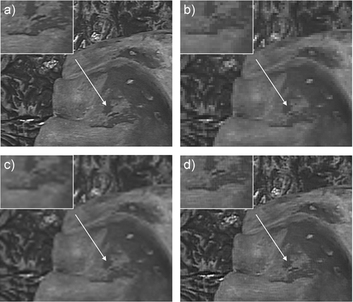
To quantify the level of detail reconstructed in the HR image, an entropy measure Citation[33] that represents the amount of information present in the images is calculated. Larger entropy values in general are indicative of the high-resolution details recovered. For the phantom study, the entropy values of the original, LR, interpolated, and POCS-reconstructed HR images are 6.74, 6.57, 6.56 and 6.72, respectively, which suggests the information recovered by the HR image is close to 99.7%, based on the entropy measure. It should be noted, however, that this is a global statistical measure, and may not represent the actual visual fidelity recovered. Nevertheless, it does provide a quantitative index when combined with visual feature assessment.
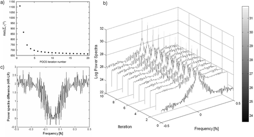
To examine the convergence behavior of the algorithm, a illustrates the RMS error in relation to the iteration steps. The algorithm in general has a rapid convergence rate, which makes it suitable for real-time implementations. In c, the power spectra of the LR and HR images are also provided, demonstrating the amount of high-frequency detail recovered.
Figure 2. (a) The convergence of the POCS algorithm as demonstrated by the RMS error defined in Equation (4). (b) The power spectra of the LR image (at the zero position) and HR images for the first 10 iterations (where fs denotes the spatial sampling frequency). (c) Plot showing the difference in power spectra between the LR and HR images for the 10th iteration.
For in vivo data, illustrates an FOV corresponding to the foveal region of the fixation and the high-resolution details recovered by the POCS algorithm, with the corresponding images before resolution enhancement provided for reference. This figure also indicates the position of the FOV in relation to the operating view. To provide a more accurate assessment of the level of detail recovered, illustrates the intensity profiles as marked in for each of the images presented. The corresponding entropy measures for these images are summarized in .
Figure 3. (a) Original video image frames from a TECAB sequence. (b) The corresponding magnified time frames from the region marked by the white square in (a). (c) HR reconstructions calculated using the described POCS method. White lines in b and c indicate rows for which intensity profiles are shown in .
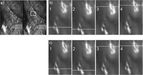
Figure 4. Pixel intensity profiles across image features on both HR (solid lines) and LR (dashed lines) images, showing the improved resolution of the reconstructed image for the time series shown in . Pixels used for line profiles are marked by white lines in .
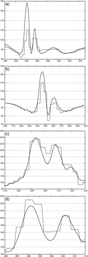
Table I. Entropy values for the TECAB images shown in .
The second in vivo experiment, as illustrated in , was used to demonstrate the benefits of the proposed color super resolution method. The corresponding results obtained by applying the proposed algorithm are provided in . It is evident that the HR image provides enhanced details compared to those of the LR image without suffering from color artefacts. To provide a more quantitative assessment of the image quality achieved, illustrates separate RGB line profiles of the LR and HR images shown in .
Figure 5. (a) Representative image frames from the 2nd in vivo experiment demonstrating the benefits of the proposed color super resolution method. (b) The relative shifts between the image frames. (c) One of the video frames with the region of interest marked; the super resolution details of this region are shown in . [Color version available online.]
![Figure 5. (a) Representative image frames from the 2nd in vivo experiment demonstrating the benefits of the proposed color super resolution method. (b) The relative shifts between the image frames. (c) One of the video frames with the region of interest marked; the super resolution details of this region are shown in Figure 6. [Color version available online.]](/cms/asset/d06137a7-d40e-4e02-85e6-5f8b14c0010e/icsu_a_272694_f0005_b.gif)
Figure 6. a) The LR image from the marked region in c. b) The corresponding SR image reconstructed from the MAP-YIQ method. [Color version available online.]
![Figure 6. a) The LR image from the marked region in Figure 5c. b) The corresponding SR image reconstructed from the MAP-YIQ method. [Color version available online.]](/cms/asset/e419409f-ffe2-40ce-bf7f-1a7a467c0077/icsu_a_272694_f0006_b.gif)
Figure 7. Color profiles of LR and HR images along the white lines shown in . [Color version available online.]
![Figure 7. Color profiles of LR and HR images along the white lines shown in Figure 6. [Color version available online.]](/cms/asset/c5618c14-dda0-4433-9484-35d48e38f151/icsu_a_272694_f0007_b.gif)
Discussion and conclusions
In this paper, we have demonstrated the use of super resolution for robotic-assisted MIS. The basic motivation of the technique is to investigate the use of fixational movements in robotic-assisted MIS such that the perceived resolution of the foveal FOV is greater than the intrinsic resolution of the laparoscope camera. This will allow the use of a relatively large FOV for microsurgical tasks such that super resolution is applied to fixations in response to real-time eye tracking. Experiments with both phantom data in a controlled environment and video sequences from a TECAB procedure have been used. The results derived by using both POCS and MAP–YIQ reconstructions demonstrate the potential value of the proposed technique in improving the apparent spatial resolution of the images. It is expected that, in combination with eye tracking, the described method can provide the operator with gaze-contingent super resolution foveal vision, thus minimizing the need for manual adjustment of the laparoscope camera during surgery. Specifically, reconstructed images can be used to replace the images available to the operating surgeon or, alternatively, they can be displayed on a separate screen to simultaneously show wide-FOV and zoomed-in views of the observed scene.
References
- Ballantyne GH. Robotic surgery, telerobotic surgery, telepresence, and telementoring. Surgical Endoscopy 2002; 16(10)1389–1402
- Byrn JC, Schluender S, Divino CM, Conrad J, Gurland B, Shlasko E, Szold A. Three-dimensional imaging improves surgical performance for both novice and experienced operators using the da Vinci Robot System. Am J Surg 2007; 193(4)519–522
- Yohannes P, Rotariu P, Pinto P, Smith AD, Lee BR. Comparison of robotic versus laparoscopic skills: is there a difference in the learning curve?. Urology 2002; 60(1)39–45
- Tendick F, Jennings R, Tharp G, Stark L. Sensing and manipulation problems in endoscopic surgery: experiment, analysis, and observation. Presence 1993; 2(1)66–81
- Ames C, Frisella AJ, Yan Y, Shulam P, Landman J. Evaluation of laparoscopic performance with alteration in angle of vision. J Endourol 2006; 20(4)281–283
- Cao A, Ellis RD, Composto A, Pandya AK, Klein MD. Supplemental wide field-of-view monitor improves performance in surgical telerobotic movement time. Int J Med Robotics Comput Assist Surg 2006; 2(4)364–369
- Kim K, Kim D, Matsumiya K, Kobayashi E, Dohi T. Wide FOV wedge prism endoscope. Proceedings of the 27th Annual International Conference of the IEEE Engineering in Medicine and Biology Society (IEEE-EMBS 2005), ShanghaiChina, September, 2005, 5758–5761
- Kobayashi E, Sakuma I, Konishi K, Hashizume M, Dohi T. A robotic wide-angle view endoscope using wedge prisms. Surgical Endoscopy 2004; 18(9)1396–1398
- Dogan S, Aybek T, Risteski P, Mierdl S, Stein H, Herzog C, Khan MF, Dzemali O, Moritz A, Wimmer-Greinecker G. Totally endoscopic coronary artery bypass graft: initial experience with an additional instrument arm and an advanced camera system. Surgical Endoscopy 2004; 18(11)1587–1591
- Mylonas GP, Darzi A, Yang G-Z (2004) Gaze contingent depth recovery and motion stabilisation for minimally invasive robotic surgery. Proceedings of Second International Workshop on Medical Imaging and Augmented Reality (MIAR 2004), BeijingChina, August, 2004, G-Z Yang, T Jiang. Springer, Berlin, 311–319
- Mylonas GP, Stoyanov D, Deligianni F, Darzi A, Yang G-Z (2005) Gaze-contingent soft tissue deformation tracking for minimally invasive robotic surgery. Proceedings of the 8th International Conference on Medical Image Computing and Computer-Assisted Intervention (MICCAI 2005), Palm Springs, CA, October, 2005, JS Duncan, G Gerig. Springer, Berlin, 843–850, Part I. Lecture Notes in Computer Science 3749
- Yang G-Z, Dempere-Marco L, Hu X, Rowe A. Visual Search: psychophysical models and practical applications. Image and Vision Computing Journal 2002; 20(4)291–305
- Martinez-Conde S, Macknik SL, Hubel DH. The role of fixational eye movements in visual perception. Nature Reviews in Neuroscience 2004; 5(3)229–240
- Park SC, Park MK, Kang MG. Super-resolution image reconstruction: a technical overview. IEEE Signal Processing Magazine 2003; 20(3)21–36
- Tekalp AM, Ozkan MK, Sezan MI. High-resolution image reconstruction from lower-resolution image sequences and space-varying image restoration. Proceedings of IEEE International Conference on Acoustics, Speech, and Signal Processing (ICASSP-92), San Francisco, CA, March, 1992, 169–172
- Stark H, Oskoui P. High-resolution image recovery from image-plane arrays, using convex projections. J Opt Soc Am A 1989; 6(11)1715–1726
- Schultz RR, Stevenson RL. Extraction of high-resolution frames from video sequences. IEEE Trans Image Processing 1996; 5(6)996–1011
- Falk V, Diegeler A, Walther T, Banusch J, Brucerius J, Raumans J, Autschbach R, Mohr FW. Total endoscopic computer enhanced coronary artery bypass grafting. Eur J Cardio-Thoracic Surg 2000; 17(1)38–45
- Chaudhuri S. Super-Resolution Imaging. Kluwer Academic Press, Boston 2001
- Farsiu S, Robinson D, Elad M, Milanfar P. Advances and challenges in super-resolution. Int J Imaging Systems Technol 2004; 14(2)47–57
- Ur H, Gross D. Improved resolution from subpixel shifted pictures. CVGIP: Graphical Model and Image Processing 1992; 54(2)181–186
- Hong M-C, Kang MG, Katsaggelos AK. An iterative weighted regularized algorithm for improving the resolution of video sequences. Proceedings of the 1997 IEEE International Conference on Image Processing 1997 (ICIP-97), Santa Barbara, CA, October, 1997, 474–477
- Hardie RC, Barnard KJ, Armstrong EE. Joint MAP registration and high-resolution image estimation using a sequence of undersampled images. IEEE Trans Image Processing 1997; 6(12)1621–1633
- Schultz RR, Stevenson RL. A Bayesian approach to image expansion for improved definition. IEEE Trans Image Processing 1994; 3(3)233–242
- Bouman C, Sauer K. A generalized Gaussian image model for edge-preserving MAP estimation. IEEE Trans Image Processing 1993; 2(3)296–310
- Li SZ. Markov Random Field Modeling in Image Analysis. Computer Science Workbench Series. Vol. 19. Springer-Verlag New York, Secaucus, NJ 2001
- Farsiu S, Elad M, Milanfar P. Multiframe demosaicing and super-resolution of color images. IEEE Trans Image Processing 2006; 15(1)141–159
- Geman S, Geman D. Stochastic relaxation, Gibbs distribution and the Bayesian restoration in images. IEEE Trans Pattern Anal Machine Intell 1984; 6(6)721–741
- Thompson AM, Brown JC, Kay JW, Titterington DM. A study of methods of choosing the smoothing parameter in image restoration by regularization. IEEE Trans Pattern Anal Machine Intell 1991; 13(4)326–339
- Kuglin C, Hines D. The phase correlation image alignment method. Proceedings of the IEEE 1975 International Conference on Cybernetics and Society, New York, NY, September, 1975, 163–165
- Keren D, Peleg S, Brada R. Image sequence enhancement using sub-pixel displacements. Proceedings of the Computer Society Conference on Computer Vision and Pattern Recognition (CVPR ’88), Ann Arbor, MI, June, 1988, 742–746
- Jain J, Jain A. Displacement measurement and its application in interframe image coding. IEEE Trans Communications 1981; 29(12)1799–1808
- Gonzalez RC, Woods RE. Digital Image Processing. Prentice-Hall, Inc., New Jersey 2002