Abstract
Several studies have shown that computer-navigated TKA reduces the rate of outliers. Thirty-one consecutive patients were operated on by the same surgeon using a computer assisted navigation system. Data collected by the system included the final mechanical axis of the extremity (HKA angle) and the coronal angle of the tibial and femoral implants. These same values were measured using CAD software on full weight-bearing long X-rays taken 6 weeks post-surgery. Deviations were observed when X-ray measurements were compared to intra-operative data collected from the navigation system. A statistically significant difference was found in the tibial cut (1.29° ± 1.35°; p < 0.0001) and in the HKA (1.59° ± 2.36°; p = 0.0007). Outliers of more than 3° were observed in the coronal plane of the tibial implant in 9.6% of patients, in the coronal plane of the femoral implant in 6.4% of patients, and in the HKA angle of 29% of patients. Our results indicate that the use of navigation alone is insufficient to prevent outliers beyond an acceptable range of 3°.
Introduction
The success of a Total Knee Arthroplasty (TKA) is dependent on multiple factors, including patient characteristics, implant selection, operative technique, component positioning, and limb alignment. It has been shown that there is a statistically significant positive correlation between a good clinical result and a well-positioned prosthesis Citation[1], Citation[2]. Proper coronal alignment has been correlated with good clinical outcomes, whereas misalignments of more than 3° of varus or valgus result in a higher failure rate Citation[3].
Computer Assisted Orthopaedic Surgery (CAOS) for TKA is gaining popularity among orthopaedic surgeons. The objective of CAOS in TKA is to improve the accuracy of implant positioning and extremity alignment.
There are three main categories of navigation systems for CAOS Citation[4]:
Intra-operative, image-free (no CT or radiograph) navigation systems;
Pre-operative, image-based (CT-based) navigation systems; and
Intra-operative, image-based (radiograph, no CT) systems.
Many studies comparing navigated TKA to conventional TKA have demonstrated that CAOS yields better limb alignment and improved implant positioning in the coronal plane Citation[5–11]. These studies showed that navigated TKAs have a lower number of outliers (results with a misalignment greater than 3°) in the femoral and tibial components, as well as in the lower limb mechanical axis. However, despite the use of navigation systems, alignment and implant positioning errors were still observed. In our own department, we use the ORTHOsoft Knee 2.1 Universal navigation system (ORTHOsoft, Montreal, Canada) for TKA, but have recently become aware of several cases of post-operative misalignment in tibial implants and limb mechanical axes despite the use of this system.
Very few studies have investigated the accuracy of computer-navigated TKA systems by comparing the navigation system data to actual measurements Citation[12–14]. In this study, we evaluate the accuracy of the ORTHOsoft Knee system by comparing key data obtained intra-operatively by the navigation system with the equivalent measurements obtained post-operatively from follow-up X-rays.
Methods
Patients
Between November 2007 and February 2008, 31 consecutive patients (11 males and 20 females; mean age 70.12 ± 10.66 years) suffering from knee arthrosis underwent TKA procedures performed by the same surgeon (D.J.Z.). All patients signed an informed consent form.
Computer Assisted Orthopaedic Surgery
All operations were performed with the assistance of ORTHOsoft Knee 2.1 Universal (ORTHOsoft, Montreal, Canada).
Operative technique
Under spinal anesthesia, a midline skin incision was performed followed by a medial parapatellar arthrotomy. The ACL was sectioned and the menisci were excised. Once exposure of the knee joint was completed, two wires were drilled into the distal medial femoral condyle and two more wires into the proximal anteromedial tibia. The corresponding passive optical trackers (ORTHOsoft Navitrack ER) were then secured onto the wires and the landmarks digitized as instructed by the ORTHOsoft software. The digitizer tool has a sharp point for landmark selection and has a tracker (ORTHOsoft Navitrack ER) attached to it. An infra-red camera connected to the navigation workstation locates the trackers to provide real-time positional information. With the assistance of the navigation system, the tibia and femur were cut and prepared according to the implant guidelines. The patella was evaluated in each case and was either electrocauterized circumferentially and resurfaced or only electrocauterized. Once the trial components were placed, a fitted spacer was placed between them and the range of motion (ROM) was evaluated using the navigation system. Capsule and soft tissue releasing was performed accordingly. The trial components were then removed and the real implants cemented in place. After a final reduction, the ROM and the mechanical axis of the extremity were evaluated. Closure of the retinaculum, subcutaneous tissue and skin followed. Lastly, a compressive dressing and a Jones-type bandage were applied to the extremity.
Implants
The Depuy P.F.C. Sigma Fixed Bearing Knee System (Cruciate–Substituting) was used in 29 patients and the Depuy P.F.C. Sigma RP Rotating Platform Knee System was used in two patients.
Data collection in the operating room
Data was collected by the navigation system throughout each surgery and the following information was used for research purposes: the final mechanical axis of the operated extremity (HKA–Hip-Knee-Ankle); the coronal angle of the tibial implant; and the coronal angle of the femoral implant.
Data collection at follow-up clinic
For all patients, a full weight-bearing long-axis X-ray of the lower extremities was obtained at 6 weeks post-surgery. Standing radiographs were obtained with the patient standing in a bipedal stance in front of the long film cassette. The radiography tube was positioned at a distance of 305 cm. The film included the hips, knees and ankles, with the beam centered on the knee joint. Data derived from the X-rays were calculated using techniques and reference points analogous to the algorithm used by the ORTHOsoft navigation software. To avoid bias, measurements on each X-ray were performed by an orthopaedic surgeon who did not participate in the surgery (Y.S.B.). Measured values were obtained using CAD software (AutoCAD 2008, Autodesk, Inc., San Rafael, CA). The CAD software was used to draw lines representing the axes as described below, and the software's dimensioning tool was then used to compute the angles.
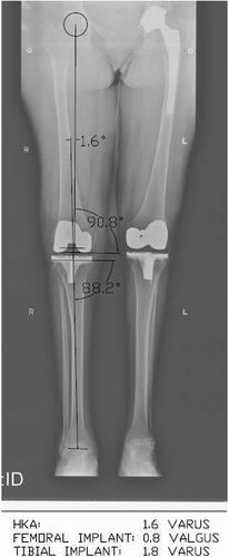
The following measurements were made from the X-rays ():
The mechanical axis of the operated extremity (HKA). The mechanical axis of the femur was represented by a line from the center of the femoral head to the center of the distal femur, equivalent to the center of the intercondylar femoral notch. The mechanical axis of the tibia was represented by a line from the center of the tibial cut, equivalent to the middle of the intercondylar eminence, to the center of the talus. The HKA angle was recorded as the angle of intersection between the tibial and femoral mechanical axes.
The coronal angle of the tibial implant. This was measured as the angle of intersection of the tibial mechanical axis with the joint surface plane of the tibial implant.
The coronal angle of the femoral implant. This was measured as the angle of intersection of the femoral mechanical axis with the joint surface plane of the femoral implant.
Figure 1. Long standing X-ray showing the different angles that were measured manually. These were the HKA angle; the coronal angle of the femoral implant (measured as the angle of intersection of the femoral mechanical axis with the joint surface plane of the femoral implant); and the coronal angle of the tibial implant (measured as the angle of intersection of the tibial mechanical axis with the joint surface plane of the tibial implant).

Statistical methods
Data were analyzed using Intercooled Stata v8.2 (Stata Corporation, College Station, TX). The two measurements for each angle in any patient (navigation system and X-ray) were regarded as paired. Comparisons between measurements were done using a paired t-test for comparing means of continuous variables between two paired samples. Outliers in the X-ray measurements and differences between the navigation system measurements and X-ray measurements were identified by selecting different cut-offs for outlying values, such as 3°. The estimated proportion of outliers is reported with a corresponding binomial exact 95% confidence interval.
Results
We followed the 31 patients for 6 weeks following total knee replacement. All patients resumed full weight-bearing on the first post-operative day. We detected no early implant complication or any superficial or deep infections. The mean knee score at 6 weeks was 67.77 ± 14.15, with a range of movement of 96.08° ± 15.06°.
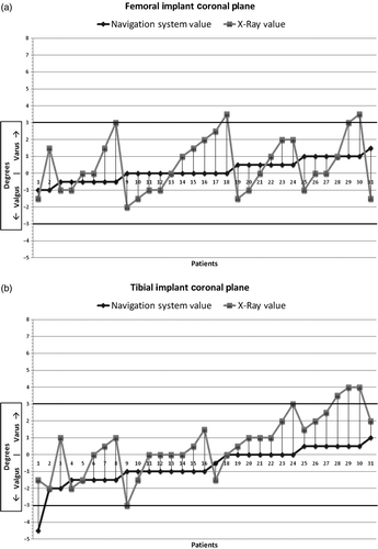
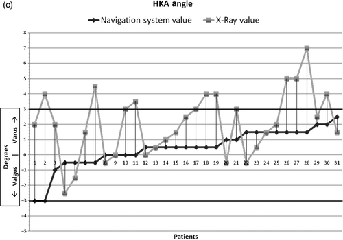
Significant deviations were observed when X-ray measurements were compared to intra-operative data collected from the navigation system. A significant difference was found in the coronal angle of the tibial implant (1.29° ± 1.35°; p < 0.0001) and in the HKA angle (1.59° ± 2.36; p = 0.0007). These values are varus relative to the navigation data, meaning the actual X-ray measurement was generally more varus than the value given by the navigation system. No significant difference was found for the coronal angle of the femoral implant (0.30° ± 1.74°; p = 0.33) (). A deviation of more than 3° was noted in the coronal plane of the tibial implant in 9.6% of the patients, in the coronal plane of the femoral implant in 6.4% of the patients, and in the HKA angle of 29% of the patients ().
Figure 2. Navigation values and X-ray measurements obtained for each patient for the femoral (a) and tibial (b) implant coronal plane angles and the HKA angle (c). Patients are sorted according to the value obtained intra-operatively from the navigation system, ranging from most valgus to most varus. Black diamonds represent values measured intra-operatively by the navigation system and gray squares represent X-ray measurements at follow-up. The graphs demonstrate the varus deviation in the actual measurements at follow-up compared to the navigation system values.

Table I. Paired t-test. Comparison of values measured by the navigation system and at follow-up clinic 6 weeks post-surgery.
Discussion
A long standing X-ray is regarded by some as the gold standard for assessing the mechanical axis of the lower limb Citation[15], Citation[16] and is a common method of post-operative axis assessment. This system has high reproducibility with high inter- and intra-observer reliability Citation[14], Citation[17].
Our study found significant differences between navigation data and post-operative weight-bearing X-rays. The percentage of outliers of more than 3° was 6.4%, 9.6% and 29% for the coronal femoral, coronal tibial and HKA angles, respectively. Several recent studies have discussed the improved results obtained with navigated TKA as compared to conventional TKA Citation[5–11], and it is a common assumption that CAOS decreases the rate of outliers from the 30% seen with conventional instrumentation Citation[6], Citation[7], Citation[18] to 10% Citation[6], Citation[5–11], Citation[19], Citation[20]. However, though these investigations did achieve better alignment when using CAOS techniques, several of them failed to attain the desired rate of only 10% outliers. Kim et al. demonstrated that 28% of cases were outliers in the mechanical axis, 16% in the coronal positioning of the tibial implant, and 13% in the coronal positioning of the femoral implant Citation[21]. For the mechanical axis, the rate of outliers was reported as 29% by Yau et al. Citation[22], 21% by Haaker et al. Citation[23], 16% by Jenny et al. Citation[24], and 52% by Maculé-Beneyto et al. Citation[25].
The majority of studies assessing the accuracy of navigation systems use radiographs to compare post-operative results of conventional and navigated TKA, the final limb alignment and coronal implant positions being measured in each case. However, these studies do not assess the accuracy of the navigation system, but rather how many outliers CAOS produces compared to the conventional method. We set out to determine the accuracy of the system itself by comparing the data provided intra-operatively by the navigation system with actual measurements obtained from long standing X-rays, and noted significant differences between the two sets of data.
Several factors may account for these differences. First, the data provided by the navigation system is for a patient under either regional or general anesthesia. Next, the intra-operative measurements are obtained with the CAOS system when the soft tissues surrounding the knee are open; the muscle tone in the post-operative setting might change the limb mechanical axis. Furthermore, since the measurements of the axes and implant positions are done on a long standing X-ray Citation[5–11], gravity and muscle tone in this position might change the limb mechanical axis as well. Additionally, intra-operative alignment as measured by a navigation system is measured in a femoral neutral rotation, while the apparent alignment obtained from a long standing X-ray could be influenced by the femoral rotation Citation[14]. The post-operative radiograph in our study was usually acquired during a 6-week follow-up visit. The flexion contracture that might persist at that time, as well as rotational malpositioning of the implant, might change the mechanical axis of the lower limb Citation[26–28]. Obtaining measurements at a longer-term follow-up may provide a more accurate assessment, since there would be less flexion contracture affecting the axis.
Some researchers have tried to explain the shortcomings of navigation systems. Catani et al. found deviations between the coronal cut and implant angles; they attributed these differences to cementation and recommended evaluating the implant positions before the cement hardens. Despite these deviations, their rate of outliers of more than 3° in the coronal femoral implant positioning was only 2% Citation[13]. Ensini et al. Citation[8] and Rauh et al. Citation[14] found a deviation of approximately 1° between intra-operative measurements with the navigation system and manual measurements performed on follow-up long standing X-rays. They assumed that the CAOS approach was more accurate than the manual measurements and attributed the differences to the human factor. Our own study raises the question of which is more accurate: the navigation data or the X-rays? Assuming flawless landmarking, it could well be the navigation data. However, the uncertainty of the landmarking accuracy makes the X-ray data more reproducible Citation[14], Citation[17]. Bäthis et al. demonstrated intra-operative cutting errors in the coronal and sagittal planes that were caused by technique and instrumentation. They observed deviations, in both femoral and tibial cuts, between the cutting-block position before sawing and the final resection plane afterwards. They claimed that a more stable fixation of the cutting blocks and more appropriate preparation instruments are needed Citation[29]. Two studies demonstrated that deviations in the mechanical axis following CAOS might be the result of inaccurate landmarking: Siston et al. found that a navigation system that relies on digitization of the epicondyles to establish femoral rotational alignment might cause axis inaccuracy Citation[30], while Yau et al. demonstrated that significant deviations in the limb axis were caused by intra-observer errors during the visual selection of anatomic landmarks Citation[31].
We believe that deviations from the intra-operative data could also be a result of software and navigation equipment quality. In a previous study conducted in our department on Sawbones, we demonstrated that intentional landmarking errors led to incorrect cuts (up to 2.6°) without being recognized by the navigation system Citation[32].
The studies that found deviations between actual measurements and those obtained with CAOS techniques claimed to have an average deviation of approximately 1° Citation[8], Citation[13], Citation[14], Citation[29], Citation[31], Citation[33]. Our mean deviation from the intra-operative data in the coronal tibial implant position and HKA angle is around 1.5°. Based on our data and that of others, we recommend that surgeons who rely on intra-operative data during CAOS for TKA accept no more than a 1.5° deviation from neutral. Following this guideline provides a much greater assurance that the final angle will remain within 3°. We also recommend that all surgeons measure the post-operative follow-up long standing X-rays to obtain a true evaluation of their patients’ alignment.
Acknowledgments
The authors would like to thank Maricar Alminiana and Laura Des Rosiers for their assistance during the project.
References
- Lotke P, Ecker M. Influence of positioning of prosthesis in total knee replacement. J Bone Joint Surg Am 1977; 59-A: 77–79
- Ritter MA, Faris PM, Keating EM, Meding JB. Postoperative alignment of total knee replacement. Its effect on survival. Clin Orthop Relat Res 1994; 299: 153–156
- Jeffery RS, Morris RW, Denham RA. Coronal alignment after total knee replacement. J Bone Joint Surg Br 1991; 73(5)709–714
- Merloz P. Computer-assisted knee replacement. European Instructional Course Lectures 2003; 8: 154–159
- Anderson KC, Buehler KC, Markel DC. Computer assisted navigation in total knee arthroplasty: Comparison with conventional methods. J Arthroplasty 2005; 20(7 Suppl 3)132–138
- Bäthis H, Perlick L, Tingart M, Lüring C, Zurakowski D, Grifka J. Alignment in total knee arthroplasty. A comparison of computer-assisted surgery with the conventional technique. J Bone Joint Surg Br 2004; 86(5)682–687
- Bolognesi M, Hofmann A. Computer navigation versus standard instrumentation for TKA: A single-surgeon experience. Clin Orthop Relat Res 2005; 440: 162–169
- Ensini A, Catani F, Leardini A, Romagnoli M, Giannini S. Alignments and clinical results in conventional and navigated total knee arthroplasty. Clin Orthop Relat Res 2007; 457: 156–162
- Sparmann M, Wolke B, Czupalla H, Banzer D, Zink A. Positioning of total knee arthroplasty with and without navigation support. A prospective, randomised study. J Bone Joint Surg Br 2003; 85(6)830–835
- Kim SJ, MacDonald M, Hernandez J, Wixson RL. Computer assisted navigation in total knee arthroplasty: Improved coronal alignment. J Arthroplasty 2005; 20(7 Suppl 3)123–131
- Stokl B, Nogler M, Rosiek R, Fischer M, Krismer M, Kessler O. Navigation improves accuracy of rotational alignment in total knee arthroplasty. Clin Orthop Relat Res 2004; 426: 180–186
- Biant LC, Yeoh K, Walker PM, Bruce WJ, Walsh WR. The accuracy of bone resections made during computer navigated total knee replacement. Do we resect what the computer plans we resect? Knee 2008; 15(3)238–241
- Catani F, Biasca N, Ensini A, Leardini A, Bianchi L, Digennaro V, Giannini S. Alignment deviation between bone resection and final implant positioning in computer-navigated total knee arthroplasty. J Bone Joint Surg Am 2008; 90(4)765–771
- Rauh MA, Boyle J, Mihalko WM, Phillips MJ, Beyers-Thering M, Krackow KA. Reliability of measuring long-standing lower extremity radiographs. Orthopedics 2007; 30(4)299–303
- Hinman RS, May RL, Crossley KM. Is there an alternative to the full-leg radiograph for determining knee joint alignment in osteoarthritis?. Arthritis Rheum 2006; 55: 306–313
- Sabharwal S, Zhao C. Assessment of lower limb alignment: Supine fluoroscopy compared with a standing full-length radiograph. J Bone Joint Surg Am 2008; 90(1)43–51
- Odenbring S, Berggren AM, Peil L. Roentgenographic assessment of the hip-knee-ankle axis in medial gonarthrosis. A study of reproducibility. Clin Orthop Relat Res 1993; 289: 195–196
- Brys DA, Lombardi AV Jr, Mallory TH, Vaughn BK. A comparison of intramedullary and extramedullary alignment systems for tibial component placement in total knee arthroplasty. Clin Orthop Relat Res 1991; 263: 175–179
- Matsumoto T, Tsumura N, Kurosaka M, Muratsu H, Kuroda R, Ishimoto K, Tsujimoto K, Shiba R. Prosthetic alignment and sizing in computer-assisted total knee arthroplasty. Int Orthop 2004; 28(5)282–285
- Matziolis G, Krocker D, Weiss U, Tohtz S, Perka C. A prospective, randomized study of computer-assisted and conventional total knee arthroplasty. Three-dimensional evaluation of implant alignment and rotation. J Bone Joint Surg Am 2007; 89(2)236–243
- Kim YH, Kim JS, Yoon SH. Alignment and orientation of the components in total knee replacement with and without navigation support: A prospective, randomized study. J Bone Joint Surg Br 2007; 89(4)471–476
- Yau WP, Chiu KY, Zuo JL, Tang WM, Ng TP. Computer navigation did not improve alignment in a lower-volume total knee practice. Clin Orthop Relat Res 2008; 466: 935–945
- Haaker RG, Stockheim M, Kamp M, Proff G, Breitenfelder J, Ottersbach A. Computer-assisted navigation increases precision of component placement in total knee arthroplasty. Clin Orthop Relat Res 2005; 433: 152–159
- Jenny JY, Boeri C. Computer-assisted implantation of total knee prostheses: A case-control comparative study with classical instrumentation. Comput Aided Surg 2001; 6(4)217–220
- Maculé-Beneyto F, Hernández-Vaquero D, Segur-Vilalta JM, Colomina-Rodríguez R, Hinarejos-Gomez E, García-Forcada I, Seral-Garcia B. Navigation in total knee arthroplasty. A multicenter study. Int Orthop 2006; 30(6)536–540
- Loner JH, Laird MT, Stuchin SA. Effect of rotation and knee flexion on radiographic alignment in total knee arthroplasties. Clin Orthop Relat Res 1996; 331: 102–106
- Stricker SJ, Faustgen JP. Radiographic measurement of bowleg deformity: Variability due to method and limb rotation. J Pediatr Orthop 1994; 14(2)147–151
- Lohman KN, Rayhel HE, Shneiderwind WP, Danoff JV. Static measurement of tibia vara. Reliability and effect of lower extremity position. Phys Ther 1987; 67(2)196–202
- Bäthis H, Perlick L, Tingart M, Perlick C, Lüring C, Grifka J. Intraoperative cutting errors in total knee arthroplasty. Arch Orthop Trauma Surg 2005; 125(1)16–20
- Siston RA, Patel JJ, Goodman SB, Delp SL, Giori NJ. The variability of femoral rotational alignment in total knee arthroplasty. J Bone Joint Surg Am 2005; 87(10)2276–2280
- Yau WP, Leung A, Chiu KY, Tang WM, Ng TP. Intraobserver errors in obtaining visually selected anatomic landmarks during registration process in nonimage-based navigation-assisted total knee arthroplasty: A cadaveric experiment. J Arthroplasty 2005; 20(5)591–601
- Brin YS, Livshetz I, Antoniou J, Greenberg-Dotan S, Zukor DJ. Precise landmarking in computer assisted total knee arthroplasty is critical to final alignment. J Orthop Res Mar 22, 2010, [Epub ahead of print]
- Yau WP, Leung A, Liu KG, Yan CH, Wong LL, Chiu KY. Interobserver and intra-observer errors in obtaining visually selected anatomical landmarks during registration process in non-image-based navigation-assisted total knee arthroplasty. J Arthroplasty 2007; 22(8)1150–1161
