Abstract
The learning curve for computer navigated total knee arthroplasty (TKA) is not well defined. We collected data prospectively on a consultant surgeon's first 50 navigated TKAs. Over the same period, matching data was taken from 50 consecutive cases performed by an expert who has performed over 1000 navigated TKAs. From the first case, the novice navigator was able to achieve the same standard as the expert in terms of post-implant mechanical alignment in the coronal and sagittal planes. Equally, at 6 weeks and one year post-surgery there was no significant difference in the mean Oxford score, mechanical axis and range of movement for the two groups of patients. Operative time was significantly longer for the novice surgeon in the first 20 cases (92 versus 73 min, p < 0.001), but by the final 20 cases there was no difference (72 versus 74 min, p = 0.944). This study shows that the learning curve for navigated TKA is approximately 20 cases and that a beginner can reproduce the results of an expert from the outset.
Introduction
There is a growing cohort of evidence supporting the use of computer navigation in total knee arthroplasty (TKA). Research has repeatedly shown that using navigation results in more accurate and more consistent implant positioning and restoration of the mechanical axis Citation[1–3], both key factors in determining the long-term performance of the prosthesis Citation[4–8]. These encouraging results have led to an increasing number of orthopaedic surgeons using the technology in their practice. Despite this, however, the use of computer navigation for TKA remains relatively uncommon in the UK. Data from the National Joint Registry of England & Wales shows that only 2% of TKAs were performed using navigation in 2008 Citation[9].
It is well recognized that surgeons wishing to adopt any new practice undergo a learning curve Citation[10]. The nature and length of this learning curve is multi-factorial, but research suggests that for some operations it can be several years in duration Citation[11], Citation[12]. This, quite appropriately, causes a degree of apprehension and caution among surgeons and can often be a major factor in their reluctance to embrace new techniques. Indeed, the General Medical Council enquiry into the Bristol Paediatric Surgical Unit stated that “patients should not be exposed to surgeons operating during the early phase of their learning curves” Citation[13].
Computer navigated TKA offers an inherent advantage to surgeons learning the technique in that it provides constant visual and numerical feedback throughout the procedure–so-called “concurrent extrinsic” feedback Citation[14]. On this basis, we hypothesized that the learning curve for navigation would be significantly shorter than that described for conventional operative techniques Citation[15]. This study assesses the results of a newly appointed consultant (with experience of over 250 conventional TKA procedures) performing his first 50 computer navigated TKAs and compares the outcomes to those of an expert navigator over the same period.
Patients and methods
Demographics
All the operations in this study were performed at the Golden Jubilee National Hospital (NHS Scotland, UK) over a 6-month period between April and September 2007. The department is an NHS national elective orthopaedic surgery unit specializing in lower limb arthroplasty. The caseload comes from throughout Scotland. We recorded standard demographic information including age, gender, BMI and operative side for all patients.
Study design
Data was collected prospectively on the first 50 consecutive computer navigated primary TKAs performed by a single consultant with no previous experience of navigation (J.B., the corresponding author). This consultant had previously performed over 250 non-navigated primary TKAs. Over the same period, matching data were collected on 50 consecutive computer navigated primary TKAs performed by the senior author (F.P.), who is an expert navigator with experience of over 1000 navigated primary TKAs Citation[16]. Neither surgeon used a non-navigated technique during the study. The operating theater sessions were arranged such that both surgeons used the same theater with the same theater staff teams.
Exclusion criteria
The only exclusion criterion was if a junior trainee and not the consultant performed the operation during the 6-month period. For the experienced group, 25 cases were excluded, which meant that a case series of 75 was required to obtain 50 cases for comparison. There were no cases performed by a junior trainee in the novice group.
Surgical technique
A similar operative technique was employed by both surgeons. A tourniquet was used in all cases. The surgeons used a standard midline incision (not minimally invasive) with medial and lateral para-patellar approaches as required. Both surgeons used identical instrument sets and implanted the Columbus® fixed-bearing cemented TKR prosthesis (B. Braun Aesculap, Tuttlingen, Germany). The OrthoPilot® CT-free computer navigation system (B. Braun Aesculap) with active tracking used throughout. The KneeSuite TKA software version 4.2 was installed and the “no soft-tissue management” option was used in all cases. Both surgeons used clips for skin closure.
Data collection
All patients attended a pre-assessment clinic prior to their operation and informed consent for computer navigated TKA was obtained. During their clinic visit all patients were asked to complete the Oxford Knee Score questionnaire. This is a validated marker of functional outcome Citation[17]. In addition, a weight-bearing digital Hip-Knee-Ankle (HKA) long-leg radiograph was acquired at this time using a standardized technique according to the recommendations of Siu et al. Citation[18]. From this, the coronal femoro-tibial mechanical (FTM) angle was measured using PACS software (Kodak Ltd., Hemel Hempstead, Hertfordshire, UK). This was defined as the angle between the center of the femoral head, the center of the knee and the center of the ankle joint. These points were identified according to the criteria described Moreland et al. Citation[19].
Outcome measures were recorded at the time of operation (OrthoPilot® data) and at 6-week and one-year follow-up clinic visits (clinical measurements).
At the time of operation, data on the patients’ pre-implant knee deformity (FTM angle and fixed flexion) were collected using the navigation system. The same measurements were repeated at the end of the procedure after the implants were sited and with the patella relocated. To ensure consistency, both surgeons recorded the data with the ankle elevated and no axial force applied through the knee. Operative time was recorded in minutes from tourniquet inflation to completion of wound dressing.
All patients were reviewed at a dual-discipline (physiotherapist and nursing) arthroplasty clinic 6 weeks post-surgery. At this time, a further HKA radiograph was acquired and the Oxford questionnaire was once again completed. The X-ray FTM angle was calculated as before. Patients were reviewed by a physiotherapist who recorded the active flexion and extension of the knee using a goniometer. Active maximum flexion was defined as the maximum flexion a patient could achieve without assistance and was on a positive scale. Active extension was recorded with the patient lying down and straightening the knee as much as possible without assistance. Failure to straighten the leg was recorded as a fixed flexion deformity. Therefore, positive values represent flexion and negative values hyperextension throughout the study. Similarly, valgus deformities were recorded as positive values, varus deformities as negative values, and 0° was aligned throughout the study.
Patients were again seen at the dual-discipline (physiotherapist and nursing) arthroplasty clinic after one year. At this time, further recordings of Oxford score and knee movement were made following the same protocol as used at the 6-week appointment.
Statistical methods
Data was analyzed using SPSS v.15 (SPSS Inc., Chicago, IL), GraphPad Prism v.5 (GraphPad Software, La Jolla, CA) and Microsoft Excel 2007 (Microsoft, Redmond, WA). Statistical analyses of the data were performed using Student's t-test (for parametric continuous variables), the Mann-Whitney test (for non-parametric continuous variables) and the chi-squared test. The Kolmogorov-Smirnov test was used to assess data for normal distribution. Statistical significance was assumed at p ≤ 0.05.
Results
Demographics
There was no significant difference between the novice- and expert-operated groups in terms of age, gender, operative side, BMI and pre-operative Oxford scores (). The pre-implant FTM angle recorded on OrthoPilot® showed that in the expert group there were patients with more severe varus and valgus deformities (p = 0.026), although the overall range was similar in both groups.
Table I. Patient demographics, pre-operative Oxford Knee Score and deformity.
Operation data
Operative times for the novice and expert surgeons are illustrated in . The novice surgeon took an average of 19 min longer than the expert during the first 20 cases (p < 0.001), but by the last 20 cases there was no significant difference (p = 0.944). When comparing the novice surgeon's first 20 and final 20 cases (), there was a significant reduction in the median operative time from 90 to 70 min (p < 0.001). Both surgeons showed no significant difference in post-implant extension and FTM angles from the outset (). All but two patients in the novice group and one patient in the expert group had post-implant FTM angles within ±2° of neutral ().
Figure 1. Comparison of operative time for novice and expert navigator for 50 sequential cases.
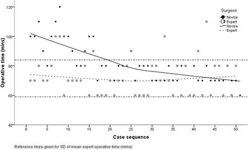
Figure 2. Comparison of operative time for novice navigator's first 20 and last 20 cases.
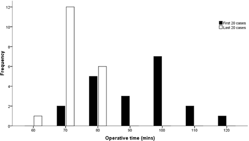
Figure 3. Post-implant coronal FTM angle recorded on OrthoPilot.
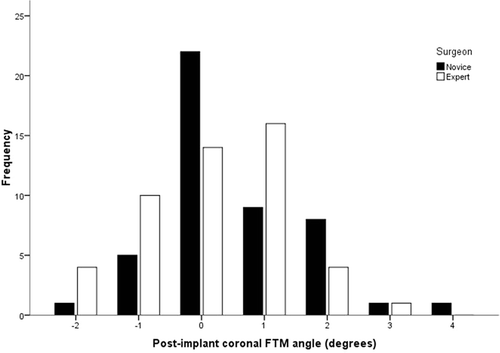
Table II. Comparison between novice and expert groups at operation. [Values are given as mean ± SD (range)]
Post-operative follow-up
There was no significant difference between the novice and expert surgeons’ 50 cases at 6 weeks or one year post-surgery in terms of Oxford score, X-ray FTM angle, knee maximum flexion, knee fixed flexion and overall total range of movement (ROM) (). shows the distribution of 6-week X-ray FTM angles.
Figure 4. 6-week post-operative HKA X-ray FTM angle.
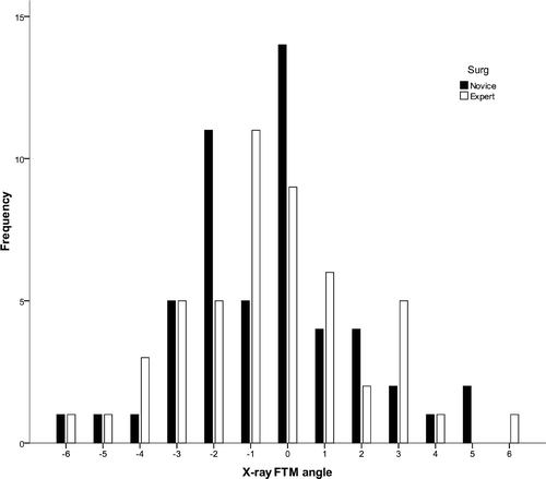
Table III. Outcomes at 6 weeks and one year follow-up. [Values are given as mean ± SD (range)]
The 50 case series was then subdivided into groups to help further evaluate the learning curve. We compared the novice navigator's first 20 cases with the expert's first 20 cases. Again, there was no statistical difference in any of the outcome measures. Finally, the novice's last 20 cases were compared to the expert's last 20 cases and no difference in the outcomes was demonstrated.
Further analysis was performed on just the novice surgeon's outcomes at one year, comparing his first 20 cases to his last 20 cases. This enabled us to see if the novice navigator's performance improved over the case series, independent of the expert's performance. Once again, no significant difference was demonstrated between the Oxford scores (mean: 23 ± 11 versus 22 ± 7, p = 0.673), knee fixed flexion (1° ± 3° versus 2° ± 4°, p = 0.556), knee maximum flexion (99° ± 10° versus 98° ± 13°, p = 0.825) and ROM (98° ± 10° versus 96° ± 15°, p = 0.722).
Discussion
The use of computer navigation for total knee arthroplasty has been shown to result in improved radiological and clinical outcomes Citation[20–24]. Despite this, only a small percentage of knee replacements performed in the UK currently use navigation. One factor underlying this is a common perception that computer navigation is a technically demanding and time-consuming technique with a long learning curve. In this study we have demonstrated that this is not the case: From the outset, a surgeon with no prior experience of computer navigated TKA was able to achieve outcomes equivalent to those of an expert user.
Operative time
One concern regarding the use of computer navigation for TKA is an increased operative time. Compared to conventional mechanical instrumentation, studies have shown that using navigation increases the operative time of TKA by approximately 10–20 min Citation[1], Citation[20], Citation[25–27], and surgeons learning to use navigation are likely to take longer still Citation[15], Citation[28]. Since prolonged operative time is associated with an increased complication rate Citation[29] and reduced cost-effectiveness, an awareness of the learning curve for navigation is of considerable value for surgeons considering adopting the technique.
We observed that the novice surgeon initially took approximately 20 min longer than the expert to complete the operation, with an average operative time of around 90 min. However, after 20 cases the novice surgeon was able to improve this significantly and consistently achieved the same operative times as the expert navigator (around 70 min). These operative times are comparable with those published for conventional TKA (60–114 min) Citation[1], Citation[3], Citation[30], Citation[31]. Our results correlate well with those of a recent multicenter study comparing experienced and beginner centers performing computer navigated TKA Citation[15]. In this study a learning curve for operative time of around 30 cases was reported, with the beginner centers initially taking approximately 10–20 min longer than the experienced centers. Our own study differs from the multicenter study in that it is the first to directly compare two surgeons working in a center experienced in computer navigation, and this may account for the shorter learning curve.
At the conclusion of the current study the novice navigator (J.B.) was invited to reflect on what had initially increased his operative times. He did not identify one particular issue that accounted for the difference, but felt that it was due to lack of familiarity with a new way of working, which would be experienced in the adoption of any new procedure. During the early use of the system the workflow was a distinct “step by step” process with each navigation screen being worked through in isolation, rather than a continuous flow in which each screen was understood in its relation to the overall procedure. There was a steady improvement as the workflow became embedded, placing each step in the context of the following steps and thus enabling the surgeon to have the correct instruments to hand, the limb in the correct position, etc., without having to check the next step. Also, increasing familiarization with the computer screen graphics over time meant that the information displayed could be assimilated more rapidly and the salient information extracted without having to check everything. This led to a decrease in hesitation, repetition or deviation throughout the operation, thus reducing time. Therefore, with regular use, not only was each individual step improved, but knowledge of the workflow made it a continuous process rather than a set of individual steps executed sequentially.
The only specific area where it was felt that acquisition of a new set of skills was required was the freehand orientation and placement of tracked instruments, linking the movement on the screen to actual movements in 3D space. A small increase in the operative time could be attributable to this.
Interestingly, all the above points could be rehearsed and learned in simulated conditions, such as a dry bone laboratory. Therefore, for a surgeon who already has TKA experience, an opportunity to familiarize himself with the software and workflow could reduce this initial period of increased operative time in either magnitude or duration.
Alignment
Accurate restoration of the mechanical axis is of key importance in ensuring implant longevity in TKA. It is thought that a mechanical axis within a range of ±3° varus/valgus is associated with a better long-term outcome Citation[32]. Studies have demonstrated that the post-operative coronal femoro-tibial mechanical angle achieved using navigation systems is significantly better than that obtained using conventional techniques. Even in the hands of experienced surgeons, the post-operative FTM angle when using conventional instrumentation exceeds ±3° of neutral alignment in around 25% of cases Citation[20], Citation[21], Citation[33].
The results from our series have shown that from the first case the surgeon new to navigation was able to achieve the same standard of limb alignment as the expert as measured both intra-operatively with the navigation system and post-operatively with HKA measurements. Moreover, 48 out of 50 (96%) cases performed by the novice surgeon had a post-implant navigation measured FTM angle within ±2° of neutral. This is comparable to the results reported by experienced navigators throughout the literature Citation[1], Citation[22].
It is noted that the pre-implant FTM angle measured on OrthoPilot® showed a small but statistically significant difference between the novice and expert groups. The most likely reason for this is that the 25 cases which the expert navigator gave to the trainee (and which were thus excluded from the study) had less deformity. Whilst this is a potential source of bias, the effect is likely to be negligible as the mean difference was less than 2° varus/valgus and the groups were equivalent in all other areas.
A possible limitation of this study is that the coronal alignment measurements were based on long-leg HKA X-rays. It is acknowledged that there are inherent inaccuracies in this method, including errors introduced by fixed flexion deformities. The current gold standard for recording limb alignment is CT imaging Citation[34]. However, within the context of this study, it was considered unethical to expose patients to the increased radiation dose required for CT. Moreover, plain X-rays are used in everyday practice in our institution and in the majority of similar studies, so we considered it the most relevant measurement technique for comparison purposes. To maximize accuracy and ensure reproducibility, a standardized protocol was used for all long-leg X-rays acquired in our study. This protocol followed the recommendations of Siu et al., who have previously described and validated the accuracy of the technique Citation[18].
Functional scores
An important outcome measure for TKA is functional scoring. This study demonstrated no difference between the 6-week and one-year Oxford Knee Scores obtained by the novice and expert surgeons throughout the study. The Oxford Knee Score is a validated marker of functional outcome and has been demonstrated to be sensitive to small clinical changes Citation[17]. However, we recognize that this study is limited by the relatively short follow-up period, as improvements in Oxford Knee Score can continue over a year post-operatively Citation[35]. Many of the benefits of improved alignment with computer navigation are only likely to become apparent in long-term follow-up, and perhaps the most valid comparison between the novice and expert surgeons would be the long-term Oxford Knee Scores achieved. Nevertheless, it is of interest that the initial prolonged operative period and learning curve had no effect on the medium-term functional outcome achieved.
The design of this study offers the significant advantage of being able to directly compare the performance of a beginner navigator to that of an expert within the same hospital and operating theater environment, and employing the same image-free navigation system. This meant that we were able to specifically assess the surgical learning curve and not that of the theater and nursing teams. The limitation of this design is that it compares two individual surgeons, and it is recognized that the learning curve may be different for other surgeons. However, given that our results are comparable with the learning curves described in similar research, we believe that the conclusions we have drawn can be applied generally.
This study supports the hypothesis that computer navigated total knee arthroplasty enjoys a short learning curve and that beginners can obtain good results from the outset. Furthermore, the study demonstrates that navigation can provide an accurate assessment tool for evaluating surgical performance. This advantage of computer navigation is highlighted by the lack of comparable research on the learning curve of TKA with standard instrumentation.
References
- Stulberg SD, Loan P, Sarin V. Computer-assisted navigation in total knee replacement: Results of an initial experience in thirty-five patients. J Bone Joint Surg Am 2002; 84-A(Suppl 2)90–98
- Mihalko WM, Krackow KA. Differences between extramedullary, intramedullary, and computer-aided surgery tibial alignment techniques for total knee arthroplasty. J Knee Surg 2006; 19(1)33–36
- Matziolis G, Krocker D, Weiss U, Tohtz S, Perka C. A prospective, randomized study of computer-assisted and conventional total knee arthroplasty. Three-dimensional evaluation of implant alignment and rotation. J Bone Joint Surg Am 2007; 89(2)236–243
- Delp SL, Stulberg SD, Davies B, Picard F, Leitner F. Computer assisted knee replacement. Clin Orthop Relat Res 1998; 354: 49–56
- Fehring TK, Odum S, Griffin WL, Mason JB, Nadaud M. Early failures in total knee arthroplasty. Clin Orthop Relat Res 2001; 392: 315–318
- Jeffery RS, Morris RW, Denham RA. Coronal alignment after total knee replacement. J Bone Joint Surg Br 1991; 73(5)709–714
- Lotke PA, Ecker ML. Influence of positioning of prosthesis in total knee replacement. J Bone Joint Surg Am 1977; 59(1)77–79
- Oswald MH, Jakob RP, Schneider E, Hoogewoud HM. Radiological analysis of normal axial alignment of femur and tibia in view of total knee arthroplasty. J Arthroplasty 1993; 8(4)419–426
- National Joint Registry of England and Wales. 6th Annual Report; 2009.
- Hopper AN, Jamison MH, Lewis WG. Learning curves in surgical practice. Postgrad Med J 2007; 83(986)777–779
- Bridgewater B, Grayson AD, Au J, Hassan R, Dihmis WC, Munsch C, Waterworth P. Improving mortality of coronary surgery over first four years of independent practice: Retrospective examination of prospectively collected data from 15 surgeons. BMJ 2004; 329(7463)421
- Sutton DN, Wayman J, Griffin SM. Learning curve for oesophageal cancer surgery. Br J Surg 1998; 85(10)1399–1402
- Jackson B. Learning from Bristol: The Report of the Public Inquiry into Children's Heart Surgery at the Bristol Royal Infirmary 1984–1995. Annex A. Chapter 14–Care in the operating theatre and the “learning curve”. 2001 http://www.bristol-inquiry.org.uk/final_report/annex_a/chapter_14_15.htm
- Gofton W, Dubrowski A, Tabloie F, Backstein D. The effect of computer navigation on trainee learning of surgical skills. J Bone Joint Surg Am 2007; 89(12)2819–2827
- Jenny JY, Miehlke RK, Giurea A. Learning curve in navigated total knee replacement. A multi-centre study comparing experienced and beginner centres. Knee 2008; 15(2)80–84
- Picard F. Computer Assisted Total Knee Arthroplasty. Validation of the Image Free Concept. Pro Business GmbH, Berlin 2007
- Dawson J, Fitzpatrick R, Murray D, Carr A. Questionnaire on the perceptions of patients about total knee replacement. J Bone Joint Surg Br 1998; 80(1)63–69
- Siu D, Cooke TD, Broekhoven LD, Lam M, Fisher B, Saunders G, Challis TW. A standardized technique for lower limb radiography. Practice, applications, and error analysis. Invest Radiol 1991; 26(1)71–77
- Moreland JR, Bassett LW, Hanker GJ. Radiographic analysis of the axial alignment of the lower extremity. J Bone Joint Surg Am 1987; 69(5)745–749
- Chauhan SK, Scott RG, Breidahl W, Beaver RJ. Computer-assisted knee arthroplasty versus a conventional jig-based technique. A randomised, prospective trial. J Bone Joint Surg Br 2004; 86(3)372–377
- Picard F, Deakin AH, Clarke JV, Dillon JM, Gregori A. Using navigation intraoperative measurements narrows range of outcomes in TKA. Clin Orthop Relat Res 2007; 463: 50–57
- Sparmann M, Wolke B, Czupalla H, Banzer D, Zink A. Positioning of total knee arthroplasty with and without navigation support. A prospective, randomised study. J Bone Joint Surg Br 2003; 85(6)830–835
- Swank ML. Computer-assisted surgery in total knee arthroplasty: recent advances. Surg Technol Int 2004; 12: 209–213
- Kinzl L, Gebhard F, Keppler P. [Total knee arthroplasty–navigation as the standard]. Chirurg 2004; 75(10)976–981
- Bolognesi M, Hofmann A. Computer navigation versus standard instrumentation for TKA: A single-surgeon experience. Clin Orthop Relat Res 2005; 440: 162–169
- Hart R, Janecek M, Chaker A, Bucek P. Total knee arthroplasty implanted with and without kinematic navigation. Int Orthop 2003; 27(6)366–369
- Jenny JY, Boeri C. Computer-assisted implantation of total knee prostheses: A case-control comparative study with classical instrumentation. Comput Aided Surg 2001; 6(4)217–220
- Walde TA, Burgdorf D, Walde HJ. Process optimization in navigated total knee arthroplasty. Orthopedics 2005; 28(10 Suppl)S1255–S1258
- Peersman G, Laskin R, Davis J, Peterson M, Richart T. Prolonged operative time correlates with increased infection rate after total knee arthroplasty. HSS Journal 2006; 2(1)70–72
- Schroer WC, Diesfeld PJ, Reedy ME, LeMarr AR. Evaluation of complications associated with six hundred mini-subvastus total knee arthroplasties. J Bone Joint Surg Am 2007; 89(Suppl 3)76–81
- Seon JK, Song EK. Navigation-assisted less invasive total knee arthroplasty compared with conventional total knee arthroplasty: A randomized prospective trial. J Arthroplasty 2006; 21(6)777–782
- Petersen TL, Engh GA. Radiographic assessment of knee alignment after total knee arthroplasty. J Arthroplasty 1988; 3(1)67–72
- Bäthis H, Perlick L, Tingart M, Lüring C, Zurakowski D, Grifka J. Alignment in total knee arthroplasty. A comparison of computer-assisted surgery with the conventional technique. J Bone Joint Surg Br 2004; 86(5)682–687
- Chauhan SK, Clark GW, Lloyd S, Scott RG, Breidahl W, Sikorski JM. Computer-assisted total knee replacement. A controlled cadaver study using a multi-parameter quantitative CT assessment of alignment (the Perth CT Protocol). J Bone Joint Surg Br 2004; 86(6)818–823
- Marx RG, Jones EC, Atwan NC, Closkey RF, Salvati EA, Sculco TP. Measuring improvement following total hip and knee arthroplasty using patient-based measures of outcome. J Bone Joint Surg Am 2005; 87(9)1999–2005