Abstract
This study retrospectively compares the postoperative alignment of imageless navigation assisted (INA) total knee arthroplasties (TKAs) using comprehensive gap balancing with that of conventional TKAs in 72 medial osteoarthritic varus knees with coronal plane anatomic variations of the distal femur and proximal tibia. The navigation group showed significantly lower postoperative differences in the mechanical axis (MA) of the lower limb (p = 0.003), with fewer outliers (p = 0.03), better femoral component positioning relative to the MA (p = 0.02), and less difference between the weight-bearing MA of the lower limb and the MA of the femur (p = 0.003) and tibia (p = 0.005). INA comprehensive gap balancing TKA provides a better correction of leg alignment and better orientation of components with respect to the MA in medial osteoarthritic varus knees with lateral bowing of the femoral shaft and external rotation of the femur and/or proximal tibia vara, thus indicating the superiority of this approach over the conventional technique in such situations.
Introduction
Since being introduced for knee surgery in 1997 Citation[1], navigation systems have found an important place in the armamentarium of knee surgeons. However, analysis of alignment and component orientation after navigated and conventional arthroplasties performed over the past decade reveals variable results. The findings include both significant and non-significant differences between the procedures Citation[2–11], with some groups advocating the navigation technique and others failing to find any improvement in outcome with this approach. Meta-analyses examining as many as 29 studies have been conducted, and the consensus of the majority of such reports suggests that use of navigation yields a significant improvement in terms of both accuracy and reproducibility of the prosthetic component orientation for all alignment outcomes at ±3° from neutral, with the exception of sagittal femoral alignment and tibial slope, for which statistical significance was not attained Citation[12], Citation[13]. In the majority of these studies, patients were randomly selected and compared as cases of total knee arthroplasty (TKA) in osteoarthritic (OA) varus knees. However, the present study reviewed only the comparative results for a small subset of OA varus knees which had anatomic variations in the coronal plane, such as lateral bowing of the femoral shaft with external rotation of the femur and/or proximal tibia vara, so as to provide evidence pertaining to one particular technique.
The importance of evaluating anatomic variations around the knee prior to TKA and their influence on the postoperative results and clinical outcomes has been emphasized by several authors Citation[14], Citation[15]. Theoretical assumptions have also been made regarding the potential pitfalls of using a navigation system in such situations, with two points being of apparent concern: First, the navigation system is programmed to cut the articular surfaces perpendicular to the mechanical axis (MA). However, in coronal plane anatomic variations of the distal femur and proximal tibia, which influence the rotation angles of the femoral and tibial rotational axes, a cut perpendicular to the MA may cause inappropriate bony resection and resultant post-TKA flexion instability Citation[16], Citation[17]. Secondly, the registration process simulates a non-weight-bearing situation, and the computer registers the MA of the femur and tibia separately, as guided by the kinematic and anatomic registration, the results of which also depend on the expertise of the surgeon in choosing the correct reference points. This may result in a discrepancy between the weight-bearing and actual MA of the lower limb.
The primary motivation for this study was to provide a clearer validation of the usefulness of navigation systems in cases featuring coronal plane anatomic variations around the knee by comparing the postoperative alignment results with those obtained using the conventional technique. It was hypothesized that the use of imageless navigation assisted TKA would result in improved component positioning and limb alignment and reduced MA differences as compared to results for the conventionally operated group. Additionally, we also tried to review the theoretical pitfalls of the navigation system when used in such situations.
Materials and methods
Patient selection and pre-operative radiographic assessment
All patients included in this retrospective review provided informed consent for the use of their data for research purposes.
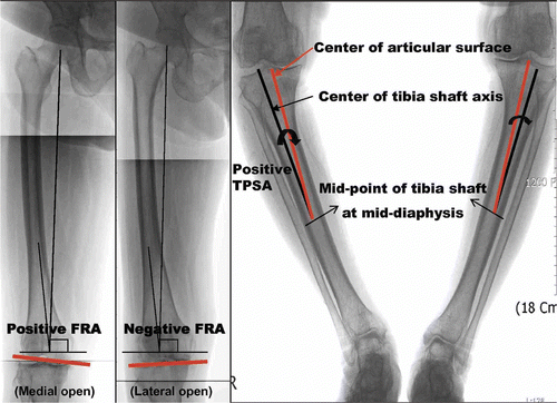
Preoperative full weight-bearing antero-posterior (AP) radiographs of 190 knees that underwent TKA for varus osteoarthritis between March 2005 and December 2008 were retrospectively reviewed to assess the distal femoral and proximal tibial coronal plane geometry. Patients were excluded if they had valgus gonarthrosis or post-traumatic deformity around the knee joint, or if the knee was found to have been in flexion or rotation when the radiograph was acquired. Two parameters were assessed: the femoral resection angle (FRA) and the tibial plateau shift angle (TPSA). The FRA is the angle between a line perpendicular to the mechanical axis of the femur and the tangent to the distal femoral condyles, with a medial open or positive FRA being seen in normal knees. The lateral bowing of the femoral shaft and the external rotation of the femur in the coronal plane may be seen as distal femoral varus configuration, with the radiographs depicting a negative FRA (). The TPSA is the angle between the central line of the proximal diaphysis of the tibia and the line from the center-point of the tibial articular surface to the center of the midpoint of the tibial shaft at mid-diaphysis. A positive TPSA represents lateral shifting of the tibial shaft axis relative to the center of the tibial articular surface (), with values greater than 2 signifying proximal tibia vara. Post-assessment, 72 knees (38.4%) were ultimately included in the study, having an FRA < 0o (46 knees, 24.2%) and/or a TPSA > 2o (41 knees, 21.6%).
Figure 1. Full-length weight-bearing AP radiographs showing the measurement of the femoral resection angle (FRA) and the tibial plateau shift angle (TPSA) to determine the coronal plane anatomic variations of the distal femur and proximal tibia, which include a negative FRA (lateral open) and/or positive TPSA (>2°, lateral shift of tibia shaft axis).
Operative procedure review and group allocation
All 72 TKAs were performed by the senior author (K.J.O.). Forty-three knees were operated using the conventional technique: 18 were implanted with the NexGen® LPS-Flex (Zimmer, Warsaw, IN) prosthesis, 10 with the Genesis™ II (Smith & Nephew, Memphis, TN), and 15 with the Sigma RPF-Flex (Depuy, Warsaw, IN). The remaining 29 knees were implanted with e.motion UC (Ultra-Congruency) implants (B. Braun Aesculap, Tuttlingen, Germany) using an imageless navigation system (OrthoPilot®, B. Braun Aesculap) for guidance.
Surgical technique
Over time, conventional instrument systems have blurred distinctions and adopted aspects of both measured resection and gap balancing philosophies Citation[18]. We used a blend of both ideologies, with a preference for the measured resection approach in the conventional group, and performed implantation using standard femoral intramedullary and tibial extramedullary guides. The distal femoral valgus angle was arbitrarily set depending on the preoperative differences in the anatomical and mechanical axes; sagittal placement was kept parallel to the anterior femoral cortex and rotational alignment parallel to the transepicondylar axis. The desired tibial alignment was 90° to the MA in the frontal plane and along the lateral border of the medial third of the tibial tubercle for rotational alignment.
In the navigation group, we comprehensively used the gap balancing approach. Input was acquired in the form of kinematic and anatomic registration of data, the initial step being the determination of the centers of the hip, knee and ankle joints using the kinematic registration process. This technique is a novel, surgeon-independent innovation for determining the center of rotation of these joints. Anatomic registration of the knee and ankle joints is then performed; this increases the accuracy of the calculation of the joint centers established initially with the kinematic registration Citation[19]. Together, these two processes result in the determination of more optimized centers of rotation followed by automatic calculation of the MA of the extremity in the frontal and sagittal planes Citation[20]. The tibial resection was performed first, after which a “four-point contact” alignment block was applied which rested on the distal and posterior femoral condyles. This feature is especially favorable for determining the bony geometry of the distal femur and the appropriate balancing of the collaterals during the subsequent step in knees with anatomic variations. Subsequently, the joint space in flexion and extension was measured, using a ligament tensor, to determine precisely the gap under constant ligament tension. Balancing of the collateral ligaments in flexion/extension as well as medial/lateral prior to bone resection was performed. The goal was to have the medial and lateral collateral ligaments measure within 2–3 mm of one another; accordingly, a soft tissue release was performed until the ligament measurements were acceptable. After balancing the two collaterals, an interactive 3D simulation “tuning phase” for virtual planning of the femoral component was conducted. The basic idea was to predict the flexion-extension gaps for different femoral resection plans, component rotations and implant sizes, which ultimately allowed us to adjust and predict the level, size, anterior/posterior translation, varus/valgus, and rotation of the femoral component, and the tibial insert size. This step is crucial and precedes the actual execution of the femoral cuts, allowing a simulation of the ideal outcome for knees with the specified anatomic variations and well-balanced implants.
Assessment of postoperative alignment
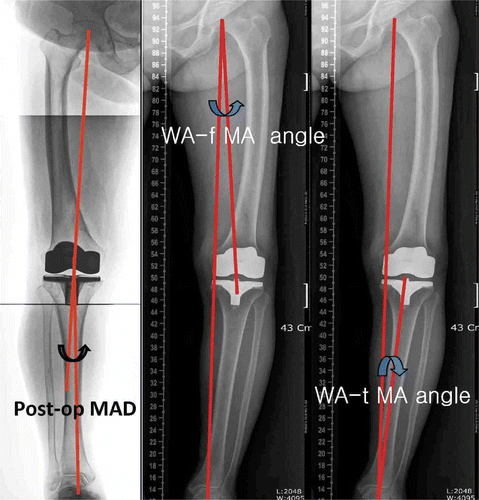
Postoperative radiographic evaluation of 7 parameters was performed on full-length weight-bearing AP radiographs (). The parameters evaluated were the angles suggested by the Knee Society Citation[21]: the femoral and tibial component angles on an AP view (α, β) and the femoral component position relative to the MA on an AP view (θ). Other parameters measured were the postoperative weight-bearing MA difference (MAD) angle of the lower limb (the difference between the postoperative MA of the femur and tibia), the weight-bearing mechanical axis (WA) of the lower limb (the line connecting the center of the femoral head and the center of the talar dome), and differences between the WA and MA of the femur (fWA) and tibia (tWA) separately (WA-f MA, WA-t MA, respectively).
Figure 2. Full-length weight-bearing AP radiographs showing the measurement of postoperative parameters: the postoperative mechanical axis difference (MAD) and differences between the weight-bearing mechanical axis of the lower limb (WA) and the mechanical axes of the femur (f WA) and tibia (tWA) separately (WA-f MA, WA-t MA, respectively).
Statistical analysis
Statistical analysis was performed using SAS version 9.13 (SAS Institute Inc., Cary, NC). The mean and standard deviations of all measurements were calculated. Independent t-tests were used to compare differences in mean angular values for the two groups. To compute the rate of optimally implanted prostheses for the two groups, we used the chi-square test. The differences were considered to be of statistical significance at a level of p < 0.05.
Results
The patients in the imageless navigation assisted (INA) and conventional study groups did not differ significantly in their demographic data and preoperative varus malalignment. The mean preoperative varus MA was higher in the navigation group, but this was not statistically significant ().
Table I. Preoperative details of study groups, with data expressed as mean ± standard deviation and ranges for continuous characteristics and absolute and relative frequencies for categorical endpoints.
Femoral and tibial component alignment
The femoral component showed a mean angle of 97.41 ± 1.81° in the coronal plane for the navigation group and 96.08 ± 1.82° for the conventional group, which was a statistically significant difference (p = 0.003). There were 4 navigated and 3 conventionally placed components outside the determined level of tolerance of ± 3° (p = 0.58). The tibial component showed a mean angle of 89.92 ± 1.76° in the coronal plane for the navigation group and 88.61 ± 1.70° for the conventional group, which was not statistically significant (p = 0.145). There were 5 navigated and 6 conventionally placed components outside the determined level of tolerance of ±3° (p = 0.53). The mean femoral component position with respect to the MA was 90.07 ± 1.19° in the navigation group and 89.14 ± 2.0° in the conventional group, which was statistically significant (p = 0.02). There were 1 navigated and 6 conventionally implanted prostheses outside the determined level of tolerance of ±3° (p = 0.51) (, ).
Figure 3. Graphs showing the total number of outliers in the two groups with respect to coronal femoral component angle (α), coronal tibial component angle (β), femoral component position relative to the mechanical axis (θ) and postoperative mechanical axis difference (MAD). p-values are shown for knees which were ±3° outside the tolerance limit. (* = statistically significant difference).
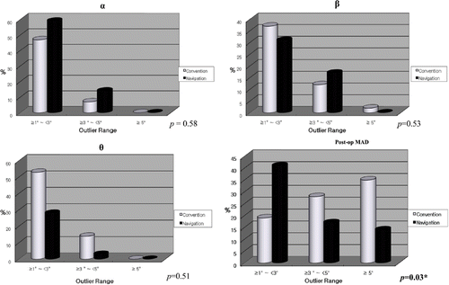
Table II. Postoperative details of study groups, with data expressed as mean ± standard deviation. p-values are also shown (* indicates a significant difference).
Mechanical axis
The navigation group showed an MAD of 2.13 ± 3.94° as compared to 4.07 ± 4.00° for the conventional group. There were 9 navigated and 27 conventionally implanted prostheses outside the determined level of tolerance of ±3° (p = 0.03). The conventional group had a greater tendency towards residual postoperative varus alignment as compared to the navigation group (p = 0.04) (, ).
Discrepancy between mechanical axes
The mean WA-f MA angle was 0.45 ± 1.63° for the navigation group and 1.67 ± 1.67° for the conventional group. The mean WA-t MA angle was 0.68 ± 2.19° for the navigation group and 2.23 ± 2.26° for the conventional group. Both these results were found to be statistically significant (p = 0.003 and 0.005, respectively) ().
Discussion
Previous reports have suggested that use of a navigation system enables severe varus deformities of up to 35° to be effectively corrected to within −2.0 to +3.0° of the mechanical axis Citation[22], Citation[23]; however, in these reports the varus OA knees were randomly selected and compared. In the present study, we specifically tried to review the usefulness of navigation systems in cases of varus OA knees with coronal plane anatomic variations, the preoperative assessment of which has consequences for the results of TKA procedures Citation[14].
To assess the anatomic variations, several parameters have been established. Kurosaka et al. Citation[24] described 8 parameters on AP radiographs and demonstrated the differences in anatomic configurations in normal and varus OA knees. Similarly, Nagamine et al. Citation[14] used 6 parameters in patients of Asian ethnicity to describe the effect of anatomic variations on the results of TKA, and further emphasized the importance of considering these factors preoperatively. Based on these considerations, theoretical assumptions have been made concerning the potential pitfalls of using navigation systems in procedures on knees with anatomic variations such as lateral bowing of the femoral shaft with external rotation of the femur Citation[17].
Knee flexion and rotation may cause apparent changes in the coronal plane, increased valgus in internal rotation, and increased varus in external rotation Citation[25]. Radiographs showing the knee in flexion or rotation were therefore excluded from the study during the patient selection process. To identify the anatomic variations around the knee, we measured the FRA and TPSA. The FRA is not only the resection angle of the distal femur, but also the external rotation angle of the femoral rotational axis relative to the posterior condylar line when soft tissue balance is obtained, with a negative FRA suggesting a distal femoral varus configuration Citation[14] (). The FRA was <0o in 24.2% of knees in our series. An FRA of less than 0° may lead to the distal femoral cut being made too far in the valgus position. Such valgus cuts, along with medial soft tissue release, causing an external rotation of the femur relative to the tibia, may lead to varus instability in flexion Citation[17]. The TPSA, on the other hand, demonstrated the location of the central point of the tibial articular surface relative to the central line of the proximal diaphysis of the tibia, with values greater than 2 being suggestive of proximal tibial vara Citation[14] (). The TPSA was greater than 2° in 21.6% of knees in our series. In cases with a positive TPSA, the mechanical axis does not coincide with the anatomic axis of the tibial shaft, but passes through the medial side of the knee joint; this can result in more bone resection on the lateral side and a consequent postoperative valgus malalignment Citation[16].
The coronal plane femoral component angle with respect to the anatomic axis was significantly better in the conventional group of our study (p = 0.003). No significant differences were found in the tibial component angles. More importantly, the femoral component position with respect to the MA was significantly better in the navigation group (p = 0.02). When using the conventional technique, we performed the femoral resection first, and the standard intramedullary femoral alignment guide was set arbitrarily, depending on the preoperative difference in the anatomical axis and MA. The planned bone cuts are taken in the frame of reference of the axis set by the intramedullary guide, and thus the femoral component angles were more satisfactory in relation to the anatomical axis. However, the effects of coronal plane anatomic variations on the resultant MA of the lower limb after final placement of the components were not taken into consideration. Thus, the resultant bone cuts may induce valgus malalignment and postoperative instability. In contrast, in the navigation assisted procedure, it is the MA that is set based on the center of the femoral head relative to the intercondylar notch, and the tibial and femoral bone cuts are planned with reference to the MA generated by the navigation system. Thus, in spite of the fact that anatomic registration may appear to be below par theoretically as a result of the anatomic variations, the system provides a more satisfactory final MA by virtue of a more optimized kinematic registration process. The assertions regarding the potential pitfalls of using a navigation system, in view of the anatomic variations around the knee that affect the rotation angles of the femoral and tibial rotational axes, may apparently hold true in relation to the anatomical axis; however, the final components were found to be significantly better placed in relation to the MA. Also, the implantation result, as well as the resulting soft tissue situation, is simulated intraoperatively by the navigation system with an interactive “tuning phase” prior to implantation, which ultimately results in adequately balanced gaps in both flexion and extension, a satisfactory final postoperative leg axis, and well-aligned components with respect to the MA in all cases.
The value of well-aligned components should not be underestimated, and improved component alignment relative to the MA has been shown to increase survival of implanted knee prostheses. The relationship between implant malalignment and longevity has been described previously, and deviations in coronal alignment greater than 3° of varus have been correlated with poorer implant survival Citation[26]. Rand and Coventry found a 90% ten-year survival for knee implants when the MA was less than 4o of varus/valgus malalignment; survival fell to 71–73% in knees with values greater than 4° Citation[27]. Shifts in the MA away from the neutral position are associated with patterns of femoro-tibial tracking that are in turn associated with abnormal stresses at the bearing surface which can lead to accelerated wear and decreased longevity Citation[28], Citation[29]. The discrepancy between the weight-bearing and actual MA of the lower limb is a potential pitfall for both conventional and INA techniques, as both are performed in a non-weight-bearing context. However, the MAD angles and the differences between the WA and MA of the femur and tibia were found to be significantly smaller in the navigated group. Thus, a more consistent and optimized MA is achieved at the end of navigation assisted TKA that is favorable for implant survival.
In our study, we tried to focus only on the coronal plane anatomic variations around the knee and their effect on the postoperative results. Nevertheless, we also used implants from various manufactures with different tibial slopes in the conventional group and consequently did not measure postoperative sagittal component alignment in the two comparison groups. Further studies are required to measure the effects of these anatomic variations on the component angles in the sagittal plane. The comparatively low number of patients in the two groups may be a weak point of our study. The small number arises from the low prevalence of such variations around the knee in the general population, as previously suggested by others Citation[16]. To overcome this, to a certain extent, we combined both groups (FRA and TPSA) together, and at the conclusion of the study the patient number proved sufficient to detect statistically significant differences that support our conclusions.
Among knee surgeons there is a clear consensus regarding the competency of navigation systems to effectively recreate an acceptable mechanical axis in the presence of significant extra-articular anatomical deformities and/or intramedullary hardware Citation[30], Citation[31]. We further support the notion that use of navigation systems may not always result in more accurate orientation and alignment of the components compared to that achieved with conventional TKA, other than by decreasing the total number of outliers, but knee surgeons should clarify and precisely select the indications for use of such systems. The present study provides more clear evidence regarding the superiority of a navigation assisted approach over the conventional procedure for knees with lateral bowing of the femoral shaft with external rotation of the femur and/or proximal tibia vara, which are often observed in medial OA varus knees, in achieving more satisfactory and accurate final MA alignment, reducing the number of outliers and overcoming the effects of potential pitfalls. Further longitudinal studies are required to determine the effect of these results on the long-term clinical outcome.
References
- Krackow KA, Bayers-Thering M, Phillips MJ, Mihalko M. A new technique for determining proper mechanical axis alignment during total knee arthroplasty: Progress toward computer assisted TKA. Orthopedics 1999; 22: 698–702
- Picard F. Early experience with the Aesculap OrthoPilot Knee Navigation System. Presentation at the Fourth Annual North American Program on Computer Assisted Orthopaedic Surgery (CAOS/USA), Pittsburgh, PA, June 2000.
- Sparmann M, Wolke B, Czupalla H, Banzer D, Zink A. Positioning of total knee arthroplasty with and without navigation support: A prospective, randomised study. J Bone Joint Surg Br 2003; 85-B: 830–835
- Hart R, Janeček M, Chaker A, Buček P. Total knee arthroplasty implanted with and without kinematic navigation. Int Orthop 2003; 27: 366–369
- Mielke RK, Clemens U, Jens JH, Kershally S. [Navigation in knee endoprosthesis implantation – preliminary experiences and prospective comparative study with conventional implantation technique] (in German). Z Orthop Ihre Grenzgeb 2001; 139(2)109–116
- Bäthis H, Perlick L, Tingart M, Lüring C, Zurakowski D, Grifka J. Alignment in total knee arthroplasty. A comparison of computer assisted surgery with the conventional technique. J Bone Joint Surg Br 2004; 86-B: 682–687
- Tingart M, Lüring C, Bäthis H, Beckmann J, Grifka J, Perlick L. Computer-assisted total knee arthroplasty versus the conventional technique: How precise is navigation in clinical routine?. Knee Surg Sports Traumatol Arthrosc 2008; 16: 44–50
- Kim YH, Kim JS, Choi Y, Kwon OR. Computer-assisted surgical navigation does not improve the alignment and orientation of the components in total knee arthroplasty. J Bone Joint Surg Am 2009; 91: 14–19
- Kim YH, Kim JS, Yoon SH. Alignment and orientation of the components in total knee replacement with and without navigation support: A prospective randomised study. J Bone Joint Surg Br 2007; 89-B: 471–476
- Lützner J, Krummenauer F, Wolf C, Günther KP, Kirschner S. Computer-assisted and conventional total knee replacement. A comparative, prospective, randomized study with radiological and CT evaluation. J Bone Joint Surg Br 2008; 90-B: 1039–1044
- Yau WP, Chiu KY, Tang WM. Computer navigation did not improve alignment in a lower-volume total knee practice. Clin Orthop Relat Res 2008; 466: 935–945
- Mason JB, Fehring TK, Estok R, Banel D, Fahrbach K. Meta-analysis of alignment outcomes in computer-assisted total knee arthroplasty surgery. J Arthroplasty 2007; 22(8)1097–1106
- Bäthis H, Shafizadeh S, Paffrath T, Simanski C, Grifka J, Lüring C. [Are computer assisted total knee replacements more accurately placed? A meta-analysis of comparative studies] (in German). Orthopade 2006; 35: 1056–1065
- Nagamine R, Miura H, Bravo CV, Urabe K, Matsuda S, Miyanishi K, Hirata G, Iwamoto Y. Anatomic variations should be considered in total knee arthroplasty. J Orthop Sci 2000; 5: 232–237
- McGrath MS, Suda AJ, Bonutti PM, Zywiel MG, Marker DR, Seyler TM, Mont MA. Techniques for managing anatomic variations in primary total knee arthroplasty. Expert Rev Med Devices 2009; 6(1)75–93
- Matsuda S, Mizu-uchi H, Miura H, Nagamine R, Urabe K, Iwamoto Y. Tibial shaft axis does not always serve as a correct coronal landmark in total knee arthroplasty for varus knees. J Arthroplasty 2003; 18: 56–62
- Nagamine R, Kondo K, Ikemura S, Shiranita A, Nakashima S, Hara T, Ihara H, Sugioka Y. Distal femoral cut perpendicular to the mechanical axis may induce varus instability in flexion in medial osteoarthritic knees with varus deformity in total knee arthroplasty: A pitfall of the navigation system. J Orthop Sci 2004; 9: 555–559
- Vail TP, Lang JE. Surgical techniques and instrumentation in total knee arthroplasty. Surgery of the Knee, 4th, WN Scott. Churchill Livingstone, Philadelphia 2006; 2: 1457–1470
- Stulberg SD, Loan P, Sarin V. Computer assisted navigation in total knee replacement: Results of an initial experience in thirty-five patients. J Bone Joint Surg Am 2002; 84: 90–98
- Saragaglia D, Picard F, Chaussard C, Montbarbon E, Leitner F, Cinquin P. [Computer-assisted knee arthroplasty: Comparison with a conventional procedure. Results of 50 cases in a prospective, randomized study] (in French). Rev Chir Orthop Reparatrice Appar Mot 2001; 87: 18–28
- Ewald FC. The Knee Society total knee arthroplasty roentgenographic evaluation and scoring system. Clin Orthop Relat Res 1989; 248: 9–12
- Picard F, Deakin AH, Clarke JV, Dillon JM, Gregori A. Using navigation intraoperative measurements narrows range of outcomes in TKA. Clin Orthop Relat Res 2007; 463: 50–57
- Sampath SAC, Voon SH, Sangster M, Davies H. The statistical relationship between varus deformity, surgeon's experience, BMI and tourniquet time for computer assisted total knee replacements. The Knee 2009; 16: 121–124
- Kurosaka M, Hashimoto M, Yoshiya S et al. Radiographic assessment of static lower limb alignment of varus osteoarthritic knees. In: Transactions of the 40th annual meeting of the Orthopaedic Research Society, New Orleans, February 1994. p 675.
- Swanson KE, Stocks GW, Warren PD, Hazel MR, Janssen HF. Does axial limb rotation affect the alignment measurements in deformed limbs?. Clin Orthop Relat Res 2000; 371: 246–252
- Jeffery RS, Morris RW, Denham RA. Coronal alignment after total knee replacement. J Bone Joint Surg Br 1991; 73: 709–714
- Rand JA, Coventry MB. Ten year evaluation of geometric total knee arthroplasty. Clin Orthop Relat Res 1988; 232: 168–173
- Taylor M, Barrett DS. Explicit finite element simulation of eccentric loading in total knee replacement. Clin Orthop Relat Res 2003; 414: 162–171
- Perillo-Marcone A, Barrett DS, Taylor M. The importance of tibial alignment: finite element analysis of tibial malalignment. J Arthroplasty 2000; 15: 1020–1027
- Klein GR, Austin MS, Smith EB, Hozack WJ. Total knee arthroplasty using computer-assisted navigation in patients with deformities of the femur and tibia – case report. J Arthroplasty 2006; 21(2)284–288
- Bottros J, Klika AK, Lee HH, Polousky J, Barsoum WK. The use of navigation in total knee arthroplasty for patients with extra-articular deformity. J Arthroplasty 2008; 23(1)74–78