Abstract
Introduction: A novel computerized algorithm for hip joint motion simulation and collision detection, called the Equidistant Method, has been developed. This was compared to three pre-existing methods having different properties regarding definition of the hip joint center and behavior after collision detection. It was proposed that the Equidistant Method would be most accurate in detecting the location and extent of femoroacetabular impingement.
Materials and Methods: Five plastic pelves and ten plastic femora with modified acetabula and head-neck junctions, allowing for 50 different morphologic combinations, were examined, along with six cadaver hips. First, motions along anatomically relevant paths were performed. These motions were tracked by a navigation system and impingement locations were digitized with a pointer. Subsequently, previously generated 3D models of all the specimens, together with the recorded anatomic motion paths, were applied to all four simulation algorithms implemented in a diagnostic computer application. Collisions were detected within the motion paths, and the linear and angular differences regarding the location as well as the size of the detected impingement areas were compared and analyzed.
Results: The Equidistant Method detected impingement with significantly higher linear and angular accuracy compared to the other methods (p < 0.05). The size of the detected impingement area was smaller than that detected with the other methods, but this difference was not statistically significant.
Conclusions: The increased accuracy of the Equidistant Method is achieved by implementing a dynamic hip joint center, more closely resembling the natural characteristics of the hip joint. Clinical application of this algorithm might serve as a diagnostic adjunct and support in the planning of joint-preserving surgery in patients with femoroacetabular impingement.
Introduction
Femoroacetabular impingement (FAI) is a known pre-arthrotic deformity of the hip joint. Specific and characteristic pathomorphologic femoral and acetabular malformations associated with FAI have been identified Citation[1–7]. Consequently, in accordance with the clinical presentation of the patient, diagnosis of FAI is established by assessing these malformations on plain radiographs (anterior-posterior view of the pelvis, false profile or axial views of the hip), computed tomography (CT) and magnetic resonance imaging (MRI) Citation[8]. However, assessment of the complex three-dimensional (3D) pathomorphology and the hip joint's function, as well as depiction of the exact zone of impingement, cannot be achieved accurately with these methods. The ability to do so might be a tremendous adjunct to diagnosis and planning of joint-preserving surgery, whether performed through open dislocation or arthroscopically Citation[9–12].
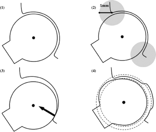
Three algorithms for hip joint simulation including collision detection already exist. All three compute virtual bony contacts based on hierarchical structures for object interference detection. Collisions between 3D polygon models defined by sets of vertices and triangles can be determined Citation[13]. However, based on a predefined center of rotation around which the femoral head moves during motion simulation, these hip joint simulations exhibit different properties in responding to detected collisions and additional translations (). The Simple Method collects all virtual acetabular impingement occurring during motion simulation; the Constrained Method only evaluates collision points within a maximum perimeter of 5 mm from the acetabular rim, while intra-articular impingement is neglected; and the Translated Method computes a displacement vector according to the detected impingement and subsequently performs an additional translation of the rotation center perpendicular to the detected intra-articular impingement area.
Figure 1. This schematic drawing of the femur and acetabulum explains the different hip joint simulation methods: (1) The Simple Method with a fixed, determined center of rotation, detecting any kind of impingement; (2) The Constrained Method with a 5-mm detection perimeter for impingement at the acetabular rim; (3) The Translated Method with an additional translation vector perpendicular to any detected intra-articular impingement area; and (4) The Equidistant Method with a computed acetabular and femoral sphere, maintaining a dynamic center of rotation and equidistant joint space.
For this study, a novel hip joint simulation method called the Equidistant Method was developed, which uses a dynamic hip joint center (HJC) during motion. It was hypothesized that the Equidistant Method would detect the location and the extent of osseous FAI in a more accurate and reliable manner than the other existing hip joint simulation algorithms.
Methods
The Equidistant Method
The Equidistant Method determines an equidistant joint space between the femur and acetabulum in every motion step (). Thus, during the initialization step, computation of a best-fitting sphere approximates the acetabulum. In addition, the acetabular opening plane is estimated by calculating a best-fitting plane derived from the acetabular rim points. Using this plane, the algorithm is capable of distinguishing femoral vertices located inside the acetabulum from those situated outside. Consequently, for each motion conducted with the femur, the femoral vertices lying inside the acetabulum can be determined and used to compute a best-fitting femoral sphere. The resulting additional femoral translation for maintenance of the equidistant joint space is calculated as the vector between the centers of the femoral and acetabular spheres. To improve the accuracy of the spherical approximations, vertices from the acetabular fossa and femoral fovea are excluded.
Specimens and 3D model generation
Five sawbone pelves (complete pelvis model 4060, Synbone AG, Malans, Switzerland) and 10 sawbone femurs (right femur model 2200, left femur model 2350, Synbone AG) were investigated. The head-neck junction of each femur and the acetabular rim of each pelvis were remodeled with epoxy to simulate different pathologic morphologies (cam and pincer impingement). To reproduce the intra-articular anatomy, 2-mm felt pads were glued into the acetabulum to simulate the cartilage. This method has proven to be feasible in previously conducted and validated studies Citation[14], Citation[15] and allowed for a good fit of the femoral head into the acetabulum. Conventional antero-posterior pelvic X-rays were obtained and standard radiographic pelvic and femoral parameters were measured using the software tool Hip2Norm Citation[16] in order to assess the relevance of the artificial pathologic morphologies. Combinations of the altered femora and pelves allowed for 50 different morphologic configurations. Subsequently, using a tracked hand-held laser scanner (Steinbichler Optotechnik GmbH, Neubeuern, Germany), virtual 3D models of these sawbones were generated.
In addition to the sawbones, three fresh frozen cadaveric pelves (six hip joints) were also included in this study. A CT scan of the specimens was obtained to reconstruct a virtual 3D model using the Amira visualization software (Visage Imaging, Inc., San Diego, CA).
Registration, tracking and conversion
Anatomical coordinate systems
The pelves were fixed in a table-mounted vice, then dynamic reference bases (DRBs) were attached to the pelvis and femur. A restricted surface matching Citation[17] was performed to register the sawbones to their 3D models, and a femoral and pelvic coordinate system was defined based on anatomical landmarks. Digitization of the left and right antero-superior iliac spine (ASIS) and the pubic tubercles enabled calculation of the anterior pelvic plane (APP) and the pelvic coordinate system Citation[18], Citation[19]. On the femoral side, the posterior condyles and the medial and lateral epicondyles were acquired for computation of the knee center. Additionally, the femoral head center was defined by repetitive pivoting of the hip joint. With the help of these landmarks the femoral coordinate system was established Citation[20].
Motion tracking and conversion
Following registration, anatomically relevant hip motions were performed and the pelvic and femoral DRBs were tracked by an Optotrak camera (Northern Digital, Inc., Waterloo, Ontario). These motions corresponded to the impingement tests conducted intraoperatively during open dislocation procedures Citation[21]. Anterior impingement was provoked with a combination of flexion, internal rotation and adduction, while posterior impingement was elicited with a combination of extension, external rotation and abduction. All motions were performed without load bearing. Motion paths were recorded by a MARVIN navigation application Citation[22]. Whenever osseous impingement between the head-neck junction and the acetabular rim occurred, the exact impingement location was digitized with a tracked pointer. To obtain a correlation between the DRB positions and the impingement points, the information was stored with time-stamps. For the plastic hip joints, five anterior and five posterior impingement situations were digitized, while for the cadaver specimens, ten anterior and ten posterior impingement situations were digitized.
Simulation analysis
Based on the established femoral and pelvic anatomical coordinate systems, the recorded DRB transformations were converted to anatomical motion paths (extension/flexion, abduction/adduction, internal/external rotation). These motion paths were then applied to the four hip joint simulation methods. During the simulation analysis the femur model was positioned according to each recorded motion step. Interference detection was then performed based on a collision detection algorithm for polygon meshes Citation[13]. The simulation results were compared to the manually acquired results. To assess linear accuracy, the distance between the virtual femoral head and the manually digitized impingement point was determined. To determine angular accuracy, the smallest additional virtual femoral rotation required to provoke a collision between the acetabulum and femur exactly at the digitized point was computed. Furthermore, the size of the computed acetabular impingement area whenever an impingement point was digitized was compared between methods (). Finally, the incidence of false-positive computer-detected impingement during impingement-free motion paths was evaluated in all hip joints.
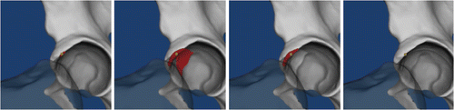
Figure 2. A 3D model of the pelvis for each simulation method is shown (from left to right: the Equidistant, Simple, Constrained and Translated methods). The femur has been rendered translucent for better visualization of the detected impingement zones at the acetabulum, which are colored red. The manually digitized impingement zones are shown by the yellow superimposed dots. The different properties of the methods regarding detection of location and size of impingement area are depicted, and the relation of the digitized point to the impingement area becomes apparent.
Statistical analysis
The Kolmogorov-Smirnov test was used to determine normal distribution in all measurement samples. Based on its result, a Kruskal-Wallis test for not normally distributed parameters was performed. Once a significant difference was detected, post hoc testing consisted of Mann-Whitney U-tests among the methods, with the significance level set to p < 0.05. A binary classification test was performed to measure the performance of all four hip joint simulation strategies with respect to the simulation of impingement-free motions and the detection of real impingement. For statistical analysis SPSS Statistics (SPSS 17.0; SPSS Inc., Chicago, IL) was used.
Results
The basic radiographic parameters of the sawbone pelves assessed with the Hip2Norm application show the great variety of artificially generated pathomorphologies ( and ).
Table I. After modification of the acetabular rim and the femoral head-neck junction using epoxy, classic radiographic parameters were measured by the Hip2Norm software. These reflect the wide variety of the artificially created pathomorphologies, as shown here.
Table II. The femoral alpha angles of the epoxy-modified sawbones. The range from normal to severely pathologic angles becomes apparent.
The Kruskal-Wallis test detected a significant variance between the methods in all assessed parameters. In more detail, the post hoc analysis showed that the Equidistant Method required significantly less additional virtual femoral rotation to provoke a collision between the acetabulum and femur compared to the other methods ( and ). Furthermore, the distance between the virtual femoral head and the manually digitized impingement point was significantly smaller for the Equidistant Method (Tables III and IV). The computed acetabular impingement area for the sawbones was smallest with the Equidistant Method, and featured the smallest standard deviation (). Statistical significance was detected in comparison to the Simple and Constrained methods; there was no significant difference compared to the Translated Method. In the cadaver set-up, the acetabular impingement area calculated by the Equidistant Method was significantly smaller compared to that calculated using the other methods ().
Table III. Statistical comparison of the sawbone results. A Kruskal-Wallis Test was performed for comparison of independent groups. For post hoc analysis, the single groups were compared using a Mann-Whitney U-Test.
Table IV. Statistical comparison of the cadaver results. A Kruskal-Wallis Test was performed for comparison of independent groups. For post hoc analysis, the single groups were compared using a Mann-Whitney U-Test.
Table V. Sensitivity and specificity of the methods were assessed with a binary classification test.
The binary classification test proved that the Equidistant Method showed the highest sensitivity for detection of impingement (). Regarding the specificity, the Equidistant Method was the most specific, except when compared to the Translated Method in the sawbone set-up ().
Discussion
A new hip joint simulation for assessment of femoroacetabular impingement, called the Equidistant Method, has been developed. In an in vitro study, this method was compared to three other pre-existing hip joint simulations.
The results confirmed the hypothesis that the Equidistant Method would be the most accurate and reliable hip joint simulation for detection of location and extent of FAI. With regard to the linear simulation error, the superiority of the Equidistant Method was demonstrated by the significantly smaller calculated distance between the digitized impingement point and the femoral head in comparison to the distances calculated using the other methods. As for the angular simulation error, the Equidistant Method required significantly less additional femoral rotation to provoke impingement exactly at the digitized point, as compared to all the other methods. Matching the average area size of the computed acetabular impingement area, it showed that the Equidistant Method calculated significantly smaller impingement areas compared to the Simple and Constrained methods. However, even though a smaller area was computed, there was no significant difference in comparison to the Constrained and Translated methods.
Interpretation of the results leads to several assumptions. The Simple Method, which uses a fixed center of rotation, computes significantly larger impingement areas than the other methods. This is due to the fact that this simulation method rotates around a fixed hip joint center. Thus, intra-articular impingement occurs during motion (). This does not reflect motion and impingement behavior in the in vivo situation. Whereas the Constrained Method merely ignores the intra-articular impingement by defining a 5-mm detection perimeter around the acetabular rim, it is evident that simulation methods employing a dynamic center of rotation such as the Equidistant and Translated methods actively avoid computation of intra-articular impingement and hence seem to be more accurate in the detection of FAI. Comparing the Equidistant and Translated methods with regard to the other parameters assessed, it becomes apparent that the Equidistant Method is superior. Linear and angular error measurements computed by the Translated Method are almost identical to those of the Constrained and Simple methods, and thus significantly inferior to those obtained using the Equidistant Method. These conclusions are also confirmed by the binary classification test, where the Equidistant Method is the most sensitive method and where the Equidistant and Translated methods reach approximately equal specificity. The good specificity of the Translated Method is a consequence of the properties employed to avoid impingement, by translating the femoral head in the opposite direction to the detected collision.
Reflecting upon the results obtained in this study, further important conclusions can be drawn. As mentioned above, definition of the HJC is crucial for accurate simulation of hip joint range of motion and also for precise detection of collisions. The main consideration to be kept in mind is that, although mathematically favored, the “ball and socket” configuration of the hip joint is really only a technical approximation. In an anatomical study, Menschik Citation[23] showed that the hip joint actually exhibits a conchoid morphology. In light of this finding, previous attempts at diagnosis and assessment of femoroacetabular impingement Citation[14], Citation[15], Citation[24] must be discussed. Kang et al. Citation[24] tried to incorporate this knowledge into their application, but two major concerns arise when reviewing their methodology. To find the HJC, they performed limited motions within a 3D model of the joint, defining a constant joint space between the femoral head and the pelvis. The point where no collision could be detected during motion was their HJC, and future motion analysis was performed around this center. Thus, although they attempted to take into consideration the fact that the hip joint does not consist of two truly spherical components, they ultimately defined a spherical femoral head and used a “fixed” center of rotation. Secondly, the results obtained were not validated, since the mere clinical perception of range of motion of a single surgeon was defined as the reference for comparison with their simulation results. In an earlier approach introduced by the current authors’ group Citation[14], Citation[15], another problem became evident: despite the thoroughly defined center of rotation, a rotational error of up to 5.0-5.6° occurred. The simulation method tested in this study also employed a fixed center of rotation without performing additional translation movements. Both studies indicate that the simplified mathematical assumption of the hip as a rigid “ball and socket” joint with congruent components introduces artifacts and delivers inaccurate results during range-of-motion simulation and collision detection. This has been confirmed by a recently published study by Gilles et al. Citation[25], who were able to show that the hip joint does not move around a fixed center of rotation, and that, as a result of the conchoid morphology of the femur, movement of the hip joint is associated with a certain amount of translation.
In light of these findings, some limitations of the present study have to be discussed. First, emphasis was placed on the investigation of osseous impingement, and our imaging method of choice was therefore CT, due to its enhanced ability to depict osseous structures. It must therefore be stated that soft tissue influence deriving from, for example, the acetabular labrum was not considered during detection of impingement. This was deemed an advantage in the plastic bones, since modification of the acetabula and femoral head junctions allowed the creation of a wide pattern of bony morphologies ranging from dysplasia-like hips to extreme mixed-type impingement (see Tables I and II). Thus, the absence of any periarticular soft tissue allowed for exact visualization and digitization of the provoked femoroacetabular impingement. In the cadaver tests, the neglect of soft tissue and labrum might be a source of error. However, since the labrum itself, in contrast to bony malformation, is not involved in the pathogenesis of FAI but is rather prone to secondary injury during the progression of disease Citation[2], Citation[3], Citation[11], Citation[26], we accepted this limitation.
Furthermore, we performed all the measurements and simulation without load bearing. This is in accordance with the clinically and intraoperatively performed impingement tests, in which the examiner tries to provoke, localize and quantify impingement with the patient in the supine position. Changes in joint biomechanics due to load-dependent deformation Citation[23] of the articular cartilage are not taken into account.
In addition, the 3D models of the sawbones generated from data obtained with the laser scanner had to be simplified to reduce the number of vertices and thus the computational load. Hence, a certain degree of error might be assumed. Finally, the restricted surface matching used in this study is a commonly known source of error in surgical navigation applications Citation[17]. Ultimately, however, the aforementioned limitations impair all the applied simulation methods to similar extents, without favoring one method over the others.
Conclusions
An algorithm has been developed that is capable of detecting the site and extent of FAI more accurately than the previously described methods. It has been validated in cadaver and sawbone experiments. Despite several limitations, the authors believe that clinical application of this algorithm might aid in the understanding of the dynamic 3D pathology which confronts a surgeon when assessing a patient with FAI. Hence, it may support decision-making during the planning of an osteochondroplasty procedure, providing a superior image of the underlying structural and functional deformities as compared to traditional examination and imaging methods.
Acknowledgments
The authors would like to thank Dr. Steffen Ross (Institute of Forensic Medicine, University of Bern, Switzerland) for his valuable clinical and technical contributions. This research was supported by external funding from BrainLAB AG, Feldkirchen, Germany, and the NCCR “Computer aided and image-guided medical interventions” program of the Swiss National Science Foundation.
References
- Ecker TM, Tannast M, Puls M, Siebenrock KA, Murphy SB. Pathomorphologic alterations predict presence or absence of hip osteoarthrosis. Clin Orthop Relat Res 2007; 465: 46–52
- Ganz R, Parvizi J, Beck M, Leunig M, Notzli H, Siebenrock KA. Femoroacetabular impingement: A cause for osteoarthritis of the hip. Clin Orthop Relat Res 2003; 417: 112–120
- Jaeger M, Wild A, Westhoff B, Krauspe R. Femoroacetabular impingement caused by a femoral osseous head–neck bump deformity: Clinical, radiographical and experimental results. J Orthop Sci 2004; 9: 256–263
- Pitt MJ, Graham AR, Shipman JH, Birkby W. Herniation pit of the femoral neck. AJR Am J Roentgenol 1982; 138(6)1115–1121
- Siebenrock KA, Schoeniger R, Ganz R. Anterior femoro-acetabular impingement due to acetabular retroversion. Treatment with periacetabular osteotomy. J Bone Joint Surg Am 2003; 85-A(2)278–286
- Tonnis D, Heinecke A. Acetabular and femoral anteversion: Relationship with osteoarthritis of the hip. J Bone Joint Surg Am 1999; 81(12)1747–1770
- Wagner S, Hofstetter W, Chiquet M, Mainil-Varlet P, Stauffer E, Jansson V, Siebenrock KA, [Femoral-acetabular impingement: A pre-arthrotic deformity?] [German]. Z Orthop Ihre Grenzgeb 2006;144(3):248–251
- Tannast M, Siebenrock KA, Anderson SE. [Femoroacetabular impingement: Radiographic diagnosis – what the radiologist should know] [German]. Radiologia 2008; 50(4)271–284
- Beck M, Leunig M, Parvizi J, Boutier V, Wyss D, Ganz R. Anterior femoroacetabular impingement: Part II. Midterm results of surgical treatment. Clin Orthop Relat Res 2004; 418: 67–73
- Larson CM, Giveans MR. Arthroscopic management of femoroacetabular impingement: Early outcomes measures. Arthroscopy 2008; 24(5)540–546
- Murphy S, Tannast M, Kim YJ, Buly R, Millis MB. Debridement of the adult hip for femoroacetabular impingement: Indications and preliminary clinical results. Clin Orthop Relat Res 2004; 429: 178–181
- Philippon MJ, Stubbs AJ, Schenker ML, Maxwell RB, Ganz R, Leunig M. Arthroscopic management of femoroacetabular impingement: Osteoplasty technique and literature review. Am J Sports Med 2007; 35(9)1571–1580
- Gotschalk S, Lin M, Manocha D, OBB Tree – a hierarchical structure for rapid interference detection. In: Proceedings of SIGGRAPH ’96, New Orleans, LA, August 1996. pp 171–180
- Kubiak-Langer M, Tannast M, Murphy SB, Siebenrock KA, Langlotz F. Range of motion in anterior femoroacetabular impingement. Clin Orthop Relat Res 2007; 458: 117–124
- Tannast M, Kubiak-Langer M, Langlotz F, Puls M, Murphy SB, Siebenrock KA. Noninvasive three-dimensional assessment of femoroacetabular impingement. J Orthop Res 2007; 25(1)122–131
- Tannast M, Mistry S, Steppacher SD, Reichenbach S, Langlotz F, Siebenrock KA, Zheng G. Radiographic analysis of femoroacetabular impingement with Hip2Norm – reliable and validated. J Orthop Res 2008; 26(9)1199–1205
- Bächler R, Bunke H, Nolte LP. Restricted surface matching – numerical optimization and technical evaluation. Comput Aided Surg, 2001; 6(3)143–152
- DiGioia AM, Jaramaz B, Blackwell M, Simon DA, Morgan F, Moody JE, Nikou C, Colgan BD, Aston CA, Labarca RS, et al. The Otto Aufranc Award. Image guided navigation system to measure intraoperatively acetabular implant alignment. Clin Orthop Relat Res 1998, 355: 8–22
- Tannast M, Langlotz U, Siebenrock KA, Wiese M, Bernsmann K, Langlotz F. Anatomic referencing of cup orientation in total hip arthroplasty. Clin Orthop Relat Res 2005; 436: 144–150
- Murphy SB, Simon SR, Kijewski PK, Wilkinson RH, Griscom NT. Femoral anteversion. J Bone Joint Surg Am 1987; 69(8)1169–1176
- Beck M, Fucentese SF, Staub L, Siebenrock K. [Surgical dislocation of the hip for the treatment of femoroacetabular impingement. Technique and results.] [German]. Orthopade 2009; 38(5)412–418
- Rudolph T, Puls M, MARVIN: A medical research application framework based on open source software. Computed Methods Programs Biomed 2008;91:165-174
- Menschik F. The hip joint as a conchoid shape. J Biomech 1997; 30(9)971–973
- Kang M, Sadri H, Magnenat-Thalman N, Computer-assisted pre-operative planning for hip joint preserving surgery. In: Proceedings of the 5th Annual Meeting of the International Society for Computer Assisted Orthopaedic Surgery (CAOS-International), Helsinki, Finland, June 2005
- Gilles B, Christophe FK, Magnenat-Thalmann N, Becker CD, Duc SR, Menetrey J, Hoffmeyer P. MRI-based assessment of hip joint translations. J Biomech 2009; 42(9)1201–1205
- Tannast M, Goricki D, Beck M, Murphy SB, Siebenrock KA. Hip damage occurs at the zone of femoroacetabular impingement. Clin Orthop Relat Res 2008; 466(2)273–280