Abstract
Objective: To investigate the value of CT 3D reconstruction in the diagnosis and treatment of incisional hernia and the related factor of abdominal cavity volume.
Methods: Abdominal wall defect and herniary volume were measured using 3D reconstruction based on plain CT scans in 17 patients with incisional hernias.
Results: The herniary diameter, area and volume could be measured in the 17 patients and the abdominal cavity volume was also measured in 10 patients using the 3D reconstruction technique. The correlation indices of the abdominal cavity volume with the patient's height, weight and body mass index (BMI) were all less than 0.01.
Conclusion: Herniary area and volume and abdominal cavity volume can be accurately calculated through CT 3D reconstruction. The patch area should be more than 5 times as large as the defect area; combined with the perioperative overlap margin measurement method, this results in more scientific surgical management. The ratio of the herniary volume to the abdominal cavity volume may be conducive to preoperative assessment of the risk of abdominal compartment syndrome (ACS); however, the ratio that may lead to postoperative ACS remains to be determined. There are correlations of abdominal cavity volume with patient height, weight and BMI, especially with weight. We therefore propose that the abdominal cavity volume should be evaluated with internationally accepted indices.
Introduction
CT 3D reconstruction for hernia repair makes use of information concerning the herniary structure, area and volume that is derived from image data using the CT software. At present, synthetic patches are widely used in tension-free repair of abdominal wall hernias. The use of a patch overlap is an effective means of preventing hernia recurrence; however, the extent of the overlap that is sufficient (3 or 5 cm) has been a matter of some concern. Amid Citation[1] reported that patches are subject to a certain degree of shrinkage in the body, and a relative small patch is thus an important factor in the postoperative recurrence of a hernia. Therefore, this study investigated how to select the patches from a size perspective so as to reduce the risk of postoperative recurrence of a hernia. Use of this approach in combination with the conventional perioperative overlap margin measurement method results in a more scientific surgical management.
Table I. Herniary diameter, area and volume, and abdominal cavity volume measured using CT 3D reconstruction.
In patients with very large incisional hernias, the abdominal cavity volume is reduced because the abdominal cavity contents enter the hernia sac. When the hernia sac contents return to the abdominal cavity, difficulty in breathing and even respiratory failure or abdominal compartment syndrome (ACS) may occur. In this study, we briefly explored the measurement of abdominal cavity volume using internationally accepted indexes such as body mass index (BMI) and observed the ratio of herniary volume to abdominal volume in order to prognosticate ACS. To date, little research has been undertaken concerning the measurement of herniary area and volume or abdominal cavity volume, or the assessment of the risk of ACS using a quantitative indicator, namely the ratio of herniary volume to abdominal volume.
Materials and methods
Patients
The study group comprised 17 patients (14 males and 3 females, aged 47–77 years) who were treated for incisional hernia between November 2009 and March 2011. Based on the latest European Hernia Society classification of incisional hernia Citation[2], 2 patients were rated in M1, 3 patients in M2, one patient in M3-4, 2 patients in M4, 3 patients in L2, 5 patients in L3 and one patient in L2-3. All study methods were approved by the ethics committee of the General Hospital of PLA, and all patients enrolled in the study gave written formal consent prior to participation.
Table II. Volumes of hernia and abdominal cavity, along with height, weight and BMI for 10 patients.
Measurements
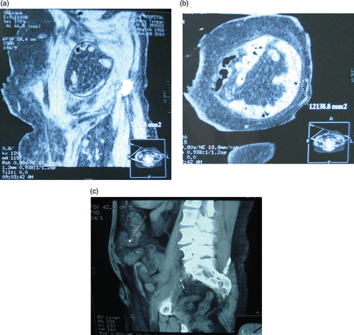
Multi-slice helical CT volume scans (whole abdominal plain CT scans with slice thickness of 0.75 cm) were performed with a GE LightSpeed scanner, and then 3D reconstruction was carried out using the associated ADW4.2 workstation. First, multi-angle reconstruction indicated the section imaging of the hernia ring and sac. The required measurement range was then marked, after which the software would automatically measure and display the herniary areas ().
Figure 1. (a) Marking the section of the hernia ring. (b) Measuring the area of the hernia ring. (c) Hernia on the norma sagittalis.

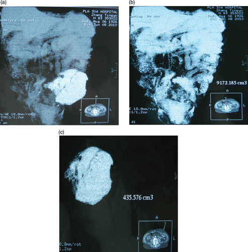
The required measurement-voluminal range was marked with Add Structure software. The abdominal cavity volume includes the liver, spleen, greater omentum, gastrointestinal tract and uterus. The volumes were then measured with measurement-voluminal software ().
Treatment techniques
The intraperitoneal onlay mesh (IPOM) technique was performed in 9 patients, the sublay technique in 7 patients, and the onlay technique in one patient.
Results
Based on the latest European Hernia Society classification of incisional hernia, the results were classified as shown in Tables .
Table III. Herniary area, patch sizes and surgical technique for 17 patients.
CT 3D reconstruction could accurately measure herniary diameter, area and volume in all 17 patients, and the abdominal cavity volume in 10 patients. The abdominal cavity volume could not be obtained for the other 7 patients because a whole abdominal plain CT scan was not performed in the early part of the study (). The hernia and abdominal cavity volumes, together with the height, weight and BMI of the 10 patients are shown in .
Correlation analysis of the abdominal cavity volume with the body height, weight and BMI of the 10 patients was performed with SPSS software (SPSS Inc., Chicago, IL). The correlation coefficients were 0.744, 0.935 and 0.789, respectively, all with t < 0.01, indicating good correlation, especially with body weight ().
Table IV. Supplementary statistical tables
The herniary areas, patch sizes and surgical techniques used are shown in . In this study, the patch sizes were 4.2–56.6 times as large as the herniary defect areas. The highest ratio of herniary volume to abdominal volume was 7.82%. No postoperative ACS occurred, and no hernia recurred in any of the 17 patients during the follow-up period of 1–14 months.
Discussion
Since 2005, at least five papers have been published concerning the application of CT 3D reconstruction (3D-CT) in the repair of abdominal wall hernias, including obturator hernias, slipped hernias, intermuscular hernias and hiatal hernias Citation[3–7]. These papers reported that CT 3D reconstruction could identify abdominal wall defects and hernia contents more clearly compared with plain CT scans, and that diagnosis of hernia was easy based on 3D-CT. In the patients with incisional hernias included in our study, 3D-CT could clearly display abdominal wall defects () and directly obtain an accurate herniary diameter (fidelity at 0.01 cm) and area (), which cannot be calculated using ordinary CT. Shrinkage varies according to the type of patch used, but is usually on the order of 15–40% Citation[8]. If the patch area after shrinkage is less than that of the defect area, the hernia will recur. At present, the patch size generally extends 3 or 5 cm beyond the herniary edge depending on the surgeon's experience during the operation. In this study, the patch size was determined from a size perspective, and the patch size was more than 4.2 times as large as the herniary defect area. Thus, even if the patch shrinkage is 40%, the patch area will still be larger than the defect area. We believe that a patch size more than 5 times larger than the herniary defect area is more reasonable. CT 3D reconstruction thus provides a basis for preoperative choice of patch size, which combined with the perioperative overlap margin measurement method results in more scientific surgical management.
Calculating the area of an abdominal wall defect is easy. Multi-slice helical CT volume scans were performed as previously described, followed by 3D reconstruction on the ADW4.2 post-processing workstation. First, multi-angle reconstruction indicated the section imaging of the hernia ring and sac. The required measurement range was then marked, after which the software would automatically measure and display the herniary areas (). Calculating herniary volumes is also easy. The required measurement-voluminal range was marked with Add Structure software, then the volumes were automatically measured and displayed using measurement-voluminal software. It can be seen from that 3D-CT can accurately measure volumes with a fidelity of 0.001 cm3.
Hernia repair does not only present a simple defect problem; hernial contents of different sizes are directly associated with perioperative treatment. For example, if the hernial volume is too large, postoperative ACS is likely to occur. At present, for very large hernias with a diameter of more than 10 cm, a great deal of attention is required to avoid postoperative ACS. Prognosticating ACS is currently based on clinical experience, but the question of whether the condition is related to a quantitative indicator, namely the ratio of herniary volume to abdominal volume, remains to be addressed. No ACS occurred in any of the patients in this study, which may reflect the smaller herniary volumes: the highest ratio of herniary volume to abdominal volume in the study group was 7.82%. The ratio of herniary volume to abdominal cavity volume that may lead to postoperative ACS has not yet been reported and will need to be investigated in studies with larger sample sizes.
Calculating the abdominal volume is more difficult, since it is necessary to define the abdominal contents. To accurately calculate the abdominal volume, the involved organs must first be marked. In this study, the abdominal cavity volume was taken to include the liver, spleen, greater omentum, gastrointestinal tract and uterus, because these are all either abdominal organs or abdominal interperitoneal viscera. The abdominal cavity volume varies with changes in the volume of abdominal gas and liquid. In addition, the abdominal cavity volume is also associated with extraperitoneal organs and tissues such as the kidney, large vessels, bladder and greater psoas muscle, to some degree, and the question arises as to whether these extraperitoneal organs and tissues should be included in the abdominal cavity volume. Therefore, the method of calculating the abdominal cavity volume requires further investigation. In this study, the average abdominal cavity volume was 9767.51 ± 594.810 cm3. It may also be asked whether the abdominal cavity volume greater in fat people. In this study, the correlation analysis of abdominal cavity volume with height, weight and BMI yielded correlation coefficients of 0.744, 0.935 and 0.789, respectively, all with t < 0.01, indicating good correlations, especially with body weight.
In summary, a patch size more than 5 times larger than the defect area is more reasonable; combined with the perioperative overlap margin measurement method, this results in more scientific surgical management. Correlations of the abdominal cavity volume with the patient's height, weight and BMI – especially with body weight – were found. No unified criteria have yet been established for calculating the abdominal cavity volume; however, we attempted to evaluate the abdominal cavity volume using internationally accepted indices. In this study, the highest ratio of herniary volume to abdominal volume was 7.82% and no postoperative ACS occurred in any patient. The ratio of herniary volume to abdominal cavity volume that may lead to postoperative ACS remains to be investigated.
Declaration of interest: The authors report no conflicts of interests.
References
- Amid PK. Classification of biomaterials and their related complications in abdominal wall hernia. Surgery 1997; 1: 15–21
- Muysoms FE, Miserez M, Berrevoet F, Campanelli G, Champault GG, Chelala E, Dietz UA, Eker HH, El Nakadi I, Hauters P, et al. Classification of primary and incisional abdominal wall hernias. Hernia 2009; 13: 407–414
- Zhang H, Cong JC, Chen CS. Ileum perforation due to delayed operation in obturator hernia: a case report and review of literatures. World J Gastroenterol 2010; 16: 126–130
- Shizukuishi T, Abe K, Takahashi M, Sakaguchi M, Aizawa T, Narata M, Maebayashi T, Fujii M, Tanaka I, Furuhashi S. Inguinal bladder hernia: Multi-planar reformation and 3-D reconstruction computed tomography images useful for diagnosis. Nephrology (Carlton) 2009; 14: 263
- Rogliani M, Silvi E, Arpino A, Gentile P. The Maltese cross technique: Umbilical reconstruction after dermolipectomy. J Plast Reconstr Aesthet Surg 2007; 60: 1036–1038
- Park HS, Kim JI, Kim MS, Kim SS, Cho SH, Park SH, Han JY, Kim JK. A case of transmesocolic hernia in elderly person without a history of operation. Korean J Gastroenterol 2006; 48: 286–289
- Polhill JL, Heniford BT. Re. Classification of hiatal hernias using dynamic three-dimensional reconstruction. Surg Innov 2006; 13: 209
- Li J-Y. Problems in incisional hernia repair with patch. Chinese J Surg 2007; 45: 1446–1448
