Abstract
Incorrect patellar resection during total knee arthroplasty can lead to anterior knee pain, patellar maltracking, patellofemoral impingement, patellar fracture, component loosening and reduced range of motion. A computer-assisted surgery (CAS) system was developed to improve the accuracy of the patellar cut. Twelve cadaveric knee specimens (6 pairs) were surgically prepared and the patella resected by two senior orthopaedic residents using either a conventional sawguide technique (right knee) or a computer-assisted sawguide technique (left knee). Multiple cuts and measurements were permitted for the conventional technique, to reflect the clinical situation, whereas only a single cut was permitted for the CAS technique. Prior training had been provided on artificial bones for both techniques. Custom marker arrays were mounted on the sawguide and patella. The user positioned the sawguide based on a real-time display that compared the current sawguide plane to the ideal resection. The resulting mediolateral and superoinferior resection angles and central thickness were measured from CT scans of the specimens, relative to the anterior surface of the patella. Both techniques resulted in symmetric cuts (<7°). Repeatability in the mediolateral direction was better for the CAS technique than for the conventional technique (p < 0.01). This study demonstrated that computer-assisted patellar resection is a feasible approach that can produce results equal to or better than those obtained with conventional techniques, even when the experimental conditions favor the conventional technique. Improvements in the CAS hardware could further improve the accuracy and usability of the system, resulting in reductions in postoperative complications. Patellar CAS could also serve as a valuable tool for feedback and training.
Introduction
The clinical consequences of improper patellar resection (i.e., the cutting of the kneecap during total knee arthroplasty to prepare for fitting of an artificial component) are not as well recognized as those associated with the tibia and femur, yet they can have a major impact on the patient's quality of life and the ultimate success of the surgery. Incorrect patellar resection, whether asymmetric or too thick or thin, has been linked to anterior knee pain (AKP) Citation[1-3], patellar maltracking Citation[4], patellofemoral impingement Citation[1], Citation[2], patellar fracture Citation[3], Citation[5], component loosening Citation[3] and reduced range of motion Citation[6], Citation[7].
Some surgeons resurface the patella while others do not, due in part to past problems with patellar loosening and fracture, and in part to the advent of more patella-friendly designs. Nevertheless, resurfacing the patella substantially decreases the likelihood of AKP, from an average incidence of 26% in patients without resurfacing to an average of 12% in patients with resurfacing, based on a meta-analysis of randomized controlled trials Citation[8]; revision surgery is likewise less common when the patella is resurfaced (3% versus 7%) Citation[8]. Secondary resurfacing, performed to address postoperative AKP in the unresurfaced patella, not only requires a second operation for the patient and the health care system, but is unsuccessful at resolving AKP in 60% of patients Citation[8]. These data support resurfacing the patella to reduce postoperative pain and complications and to reduce overall costs to the health care system. Once the decision is made to resurface the patella, it is important to resect it correctly.
The rate of asymmetry (i.e., a tilted cut, with different mediolateral or superoinferior thicknesses), although rarely reported, varies widely, averaging around 10% even amongst expert surgeons Citation[2], Citation[3], Citation[5], Citation[9]; the rate amongst less experienced surgeons is likely much higher. Maximum resection angles as high as 27° Citation[5] and 24° Citation[9] have been reported. Angles greater than 7° are considered asymmetric, based on a difference of more than 2 mm between the medial and lateral or superior and inferior thicknesses, as measured 15 mm in from each edge Citation[1-3], combined with the average patellar width of 46 mm reported in the literature Citation[10].
Although a variety of devices have been developed for patellar resection, many surgeons are dissatisfied with the accuracy of these devices and instead prefer to make the cut freehand. Our own studies on artificial bone models demonstrated disadvantages of each conventional technique (freehand, sawguide and reamer), signifying the need for a new technique Citation[11], Citation[12].
Computer-assisted surgery (CAS) systems have been shown to increase the accuracy of tibial and femoral cuts Citation[13], Citation[14]. It is reasonable to expect, therefore, that a CAS system for patellar resection would improve the accuracy of the patellar cut. Nevertheless, we are unaware of any commercial CAS system for patellar resection.
To address this need, our group developed a CAS system for patellar resection, as described below. Tests on artificial bone models in a simulated surgical setup showed that the CAS results were equivalent to or better than their conventional counterparts (p < 0.04) Citation[11], Citation[12]. Following further improvements to the hardware and software, the purpose of the present study was to test the system on cadaveric specimens. Although described as a stand-alone system, the expectation is that this system would be incorporated into a tibiofemoral CAS system for clinical use.
A patellar CAS system has several potential advantages: (1) By referencing off the anterior surface, the articulating geometry of the patella plays no role, avoiding the impact of patient sex and patellar deformity, which have been shown to affect resection accuracy Citation[9]; (2) superoinferior (SI) symmetry can be achieved as easily as mediolateral (ML) symmetry, which is important because SI asymmetry has a stronger correlation with AKP than ML symmetry Citation[2]; (3) patellar thickness, which affects range of motion and the likelihood of patellar fracture, can be controlled equally in all four quadrants; and (4) the CAS system can be used as a training tool, in or out of the operating room, to provide feedback on cut symmetry.
The purpose of this study was to compare computer-assisted and conventional patellar resection on cadaveric specimens. Our hypothesis was that the CAS system would be more accurate than the conventional techniques with respect to cut symmetry in the mediolateral and superoinferior directions, as well as with respect to the remaining bone thickness.
Materials and methods
A total of 12 cadaveric fresh frozen knee specimens (6 pairs: 5 male, 1 female; ages 60 to 87) were tested by two experimenters. The experimenters, both of whom were fourth-year orthopaedic residents, each tested three knee pairs, performing a computer-assisted sawguide cut on the left knee and a conventional sawguide cut on the right knee, the order of testing being randomized. Incorporating a standard surgical sawguide (Zimmer, Warsaw, IN) into our CAS system allowed us to make a direct comparison between the CAS and conventional techniques using the paired specimens. We also developed a computer-assisted freehand technique, but tests on artificial bones revealed that the sawguide CAS system was faster and more accurate, and was preferred by the users Citation[11], Citation[12]. A reamer CAS system was not possible without substantial hardware redesign because the reamer clamps onto the anterior surface where the CAS marker array is attached.
The original patellar thickness was determined in the CAS case by digitizing the apex of the patellar bone relative to the anterior surface; in the conventional case, the experimenter measured the thickness using calipers. In both cases, the desired bone remnant thickness was calculated as the original thickness minus the implant height, or a minimum thickness of 12 mm.
Following resection, in the conventional case the experimenter was permitted to measure the symmetry and thickness of the resulting patellar bone remnant and recut if desired (usually freehand) to reflect the clinical situation. In the CAS case, the experimenter was permitted only a single cut, based on the CAS navigation. This gave an inherent advantage to the conventional techniques, but was a better test of the CAS system.
The specimens were prepared using a medial parapatellar approach. The infrapatellar fat pad was removed in all cases. To identify the mediolateral axis in later CT scanning, the experimenters inserted 1-mm tantalum beads into the medial and lateral edges of the bone; these were subsequently hidden by soft tissue during testing. The patella was everted to perform the resection. Early osteoarthritis was detected on most of the specimens in both the patellofemoral and tibiofemoral joints; nonetheless, the specimens had primarily normal anatomy, e.g., without osteophytes or deformed geometry. The testing was approved by our institutional review board.
The CAS system hardware and software have been described in detail elsewhere Citation[11]. Briefly, the hardware consisted of a standard PC; a Spectra optoelectronic localizer (Northern Digital, Inc., Waterloo, Ontario, Canada; 0.25 mm RMS accuracy Citation[15]) similar to that found on most commercial navigation systems; a patellar marker array (); an instrument marker array (), digitizing pointer and cut-plane digitizing block (all purchased from Praxim SA, Grenoble, France); custom marker array attachments to suit our CAS system; and a conventional surgical sawguide for patellar resection (Zimmer, Warsaw, IN) (). We intentionally used a standard surgical instrument for the CAS technique, rather than developing a CAS-specific device, to retain the familiarity of the instruments to the surgeon and to allow for a direct comparison between the CAS and conventional techniques.
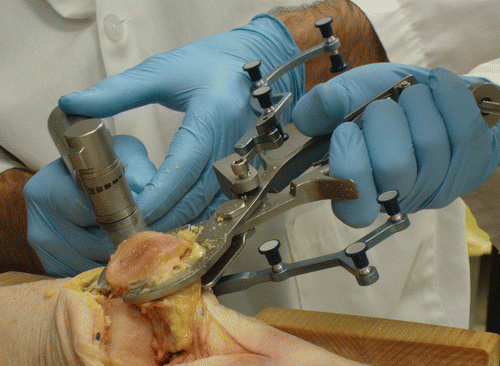
Figure 1. Resecting the patella with an oscillating saw after setting the orientation and placement of the computer-assisted sawguide. Note the “G” marker array mounted on the sawguide and the “Y” marker array mounted on the patella.
The flat, arrow-shaped baseplate portion of the patellar marker array, with three shallow spikes on the bottom Citation[11], Citation[16], was screwed securely into the bone using a screw 3 mm in diameter and 9 mm long positioned at the center of the patellar anterior surface. A marker array was then attached to the baseplate such that the three markers faced the localizer when the patella was everted. The baseplate and marker array system had been used clinically with the Praxim system to study patellar kinematics Citation[16]. At the end of each cadaveric experiment, the metal marker array base was replaced with an acrylic base having the same dimensions, such that the base appeared in the postoperative CT scans without creating metal artefacts.
The software was written in the graphical programming language LabView (Version 8.5; National Instruments, Austin, TX). The steps included (1) entering patient and surgeon information; (2) checking marker visibility; (3) digitizing a divot on the baseplate plus 10 points on the posterior surface of the patella (5 in the SI direction and 5 in the ML direction) to create an individualized schematic of the patella (this could be reduced to 3 points if the schematics were more generic); (4) planning the thickness of the desired cut and component size; (5) registering the saw plane of the sawguide (3 points); (6) navigating the sawguide relative to the planned resection in real time, with visual and numerical information showing the ideal and current saw plane in the SI and ML cross-sections (); (7) executing the cut (); and (8) recording the symmetry and thickness of the final resection plane. Since the ideal line was drawn parallel to the baseplate (at the desired thickness), it was important for the user to check early on that the baseplate was parallel to the anterior surface as intended. We made a specific effort to make the software user-friendly, both to ease the learning curve for users and to prepare for clinical implementation. A calibration step was performed with the digitizing pointer prior to each experimenter's set of tests. The software proceeded linearly step by step, but the user could jump to the marker visibility screen at any time, and a snapshot of the screen could be recorded at any time. Different images were presented to the user depending on whether the specimen was a right or left leg, whether the surgeon was right- or left-handed, and whether the incision was medial or lateral.
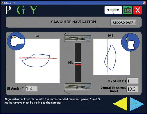
Figure 2. Real-time navigation screen for the computer-assisted technique. The current sawguide position and orientation (in red) are shown relative the resection goal (in black). Information is provided both visually and numerically. The SI view is relatively easy for the user to understand because the left/right rotations of the physical sawguide correspond to left/right rotations of the visual image. Since the ML view is along the leg, the mental mapping between the sawguide movements and the virtual representation is less obvious, and they were therefore represented in two different visual manners: in the center of the display, as the user moves the back of the sawguide up and down (thus changing the orientation in the ML plane), the red bar moves up and down accordingly; on the right is the schematic view. The blue circles show the orientation of the everted patella relative to the femur for the SI (left) and ML (right) views. The “G” marker array was mounted on the sawguide and the “Y” marker array was mounted on the patellar bone; both needed to be visible for the navigation to occur. “P” refers to the digitizing pointer used in earlier steps. The text at the bottom of the screen provides guidance to the user, although the users never needed this beyond the initial training.
CT scans were taken of each specimen to obtain detailed 3D information about the patellar cut (Siemens Somatom 64 CT scanner; slice thickness 0.6 mm, slice spacing 0.4 mm, in-plane resolution of 0.35 mm). The 3D reconstructed knees were viewed in all three planes using image analysis software (Amira, Version 5.2; Visage Imaging GmbH, Berlin, Germany). The mediolateral cross-sectional plane was defined by the previously inserted tantalum beads, perpendicular to the coronal plane containing the beads and the inferior pole of the patella; the superoinferior cross-sectional plane was perpendicular to the other two planes, passing through the inferior pole. The ML and SI cross-sections were saved as JPEG files and imported into ImageJ (Version 1.42; National Institutes of Health, Bethesda, MA) to measure the angle of the cut surface relative to the anterior bone surface as well as the central patellar thickness. The angle of the baseplate relative to the anterior surface was also measured. By measuring the resection surface relative to the bone for both the CAS and conventional cases, the reported angles are relative to the same reference point, the clinical goal. By measuring the angle of the resection surface to the baseplate, we could judge the experimenters’ ability to match the navigation goal, and also evaluate how well the baseplate represented the anterior surface.
The anterior surface was defined using a custom-written Matlab program (Version 7.5; The MathWorks, Inc., Natick, MA), based on our previous analysis of cut symmetry on clinical radiographs Citation[9]. Briefly, the x-y coordinates of the patellar contours were recorded in ImageJ, after which a least-squares line fit was performed on the central third of the top contour. The coordinates of the resection line endpoints in the axial and sagittal images were also recorded, allowing the angle and distance between the resection lines and anterior surface lines to be calculated.
The data were analyzed using two-way ANOVAs, with the factors of computer assistance or not and experimenter, with p < 0.05 required for significance. Our primary outcome measures were the differences in cut angle and thickness between the conventional and computer-assisted patellar resections. F-tests were used to compare standard deviations between the techniques. Shapiro-Wilk normality tests confirmed that all data conformed to a normal distribution. Statistical analyses were done using SPSS (Version 17.0; SPSS Inc., Chicago, IL).
Results
Both the computer-assisted and conventional sawguide cuts resulted in symmetric cuts (i.e., less than 7°) in both the mediolateral and superoinferior directions ( and ), except for one conventional cut that was on the borderline of asymmetry. Given these generally excellent symmetry results, there were no statistically significant differences between the means of the two techniques. However, the ML results were more repeatable, i.e., they had a smaller standard deviation, using the CAS approach (p < 0.01).
Figure 3. Mediolateral (ML) resection angles achieved using the conventional and computer-assisted sawguide relative to the anterior surface of the patella, as determined from CT scans. The CAS technique was more repeatable than the conventional technique (p < 0.01). The CAS results are also given relative to the baseplate attached to the anterior surface, which was the basis for the navigation. Multiple cuts and caliper measurements were allowed for the conventional case; for the CAS case only a single cut was allowed. Positive angles indicate lateral under-resection or medial over-resection. Angles less than 7° are considered symmetric.
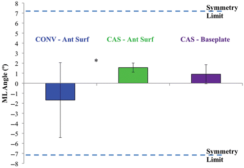
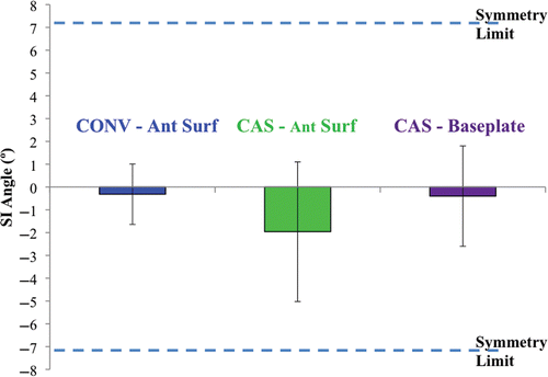
Figure 4. Superoinferior (SI) resection angles achieved using the conventional and computer-assisted sawguide relative to the anterior surface of the patella, as determined from CT scans. Positive angles indicate inferior under-resection or superior over-resection. Angles less than 7° are considered symmetric. While both experimenters were highly accurate in their navigation, the baseplate tended to be tilted superoinferiorly for one experimenter, resulting in lower accuracy relative to the bone. There were no statistically significant differences between the CAS and conventional results.
Mediolateral accuracy relative to the anterior surface of the patellar bone averaged 1.6° for CAS resection (SD 0.4°) compared to −1.7° for conventional resection (SD 3.7°). The results relative to the baseplate were similar for the CAS systems, indicating that the baseplate was a reasonable representation of the anterior surface in the ML direction ().
Superoinferior accuracy relative to the bone averaged −1.9° for CAS resection (SD 3.1°) versus −0.3° for conventional resection (SD 1.3°), with a clear distinction between experimenters (−4.2 ± 1.6° versus −0.4 ± 2.8°). Differences were related to the placement of the baseplate relative to the bone. Accuracy and repeatability of the CAS system relative to the CAS baseplate was excellent for both experimenters (−0.9 ± 1.2° and 0.3 ± 1.4°) and better than the values relative to the bone; this indicates that the baseplate was typically not parallel to the anterior surface in the SI direction for one of the experimenters.
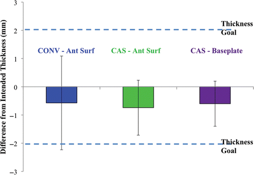
Thicknesses were comparable for both techniques (-0.7 ± 1.0° for CAS versus −0.6 ± 1.7° for the conventional technique), typically within 2 mm of the intended goal (). In both cases, the tendency was to over-resect the patellar bone slightly.
Figure 5. Difference from the intended central thickness for the conventional and CAS techniques, relative to the anterior surface and the baseplate. There were no statistically significant differences in the results.
Both experimenters experienced problems with line of sight between the marker arrays and the optoelectronic localizer, requiring some adjustments to the manner in which the experimenter held the patella, as well as periodic changing of the markers when they became contaminated with blood.
In two of the six CAS cases, one for each experimenter, the tendons prevented the sawguide from achieving the desired depth of cut. In the conventional case, this was not a problem since the experimenter was performing a second cut to fine-tune the symmetry in any case. In the CAS case, since only one cut was permitted, we adjusted the desired cut depth in the CAS system such that the displayed line on the navigation screen could be achieved physically with the sawguide. In some cases, the sawguide lost its grip or changed its orientation at the final stage of clamping, requiring the experimenter to repeat the navigation step; this difficulty was related to the sawguide itself rather than the CAS system.
Discussion
This study demonstrated the feasibility of a CAS system for patellar resection, achieving results equivalent to or better than those obtained with conventional sawguide resection. The greater repeatability of the mediolateral results indicates fewer outliers and a lower likelihood of asymmetric cuts. These results were achieved under experimental conditions that tended to favor the conventional system, including allowing multiple cuts (2–3) for the conventional system compared to just one for the CAS approach; training the experimenters through previous testing on artificial bone models with feedback from the CAS system; and using mostly male specimens with relatively normal bony geometry (a previous study demonstrated worse clinical symmetry for female knees and more deformed patellae Citation[9]). The conventional results reported here were better than those that have been reported clinically Citation[2], Citation[3], Citation[5], Citation[9]. Both the CAS and conventional techniques benefitted from the lack of time pressure compared to the operating room, allowing the experimenters to achieve the best possible symmetry, whether by caliper measurements and feel of the bone remnant, or by navigating as close as possible to the ideal line on the CAS monitor. Nevertheless, due to the line of sight and clamping issues, the experimenter did not necessarily strive for perfect symmetry or thickness when using the CAS system; likewise, with the conventional system, there is a limit to how well the bone can be shaved off to achieve perfect symmetry. A further benefit of a patellar CAS system is that resection could potentially be performed with minimal eversion of the patella.
Several issues arose that could lead to improvements in the CAS system. The first of these relates to the baseplate. The CAS system depends on this plate to accurately represent the anterior surface. While the experimenters were encouraged to check that it looked parallel, a different design of baseplate may lead to a more reliable representation of the anterior surface, resulting in better accuracy and fewer differences between experimenters, although the results obtained were still within the symmetry limit. Secondly, line-of-sight issues should be addressed, potentially through better angulation of the marker array, use of less contamination-prone marker material, and/or better training of the user to avoid touching the markers; we can also increase the angle at which the localizer accepts a marker position. Given that the sawguide was restricted in the depth direction due to the tendons, and sometimes lost its grip, leading to additional navigation time, a CAS-specific resection tool would be more appropriate. Ideally, the device would clamp securely onto the patella and then allow fine adjustments in orientation and thickness based on the navigation screen. In general, the tolerance for symmetry (±7°) is more lenient than the goals for thickness (±2 mm), while the performance of the system was reversed; therefore, additional efforts should be made to improve the accuracy of the resulting bone thickness. The experimenters found the software easy to understand and had no further suggestions for improvements.
In the experiments on artificial bones, the SI symmetry was always worse than the ML symmetry Citation[11], Citation[12]. In this experiment, the conventional sawguide SI symmetry was remarkably good. Although this may be due to differences between cadaveric and artificial bones, including guidance from the tendon locations, it could also be related to the training effect of practicing on the artificial bones. CAS has previously been shown to have a number of educational benefits, and can even improve the accuracy of a surgeon's conventional surgeries Citation[17] by focusing attention on the key steps and by providing real-time feedback.
Because of the extra steps performed specifically for the experiment, the total times required for the CAS and conventional techniques were not recorded in these cadaveric experiments. Tests on artificial bones showed that the CAS system took longer when only a single conventional cut was made, but when the conventional cut was measured and recuts were made, the times were comparable Citation[11], Citation[12]. Given further streamlining of the CAS procedure, CAS may be appropriate, even from a time perspective, if the surgeon routinely performs multiple cuts, especially if the patellar CAS system is well integrated with a tibiofemoral CAS system.
This study was limited by the small number of specimens and by having only two experimenters; in particular, this limited our power to detect statistically significant differences between the two methods. Nevertheless, these numbers were sufficient to reveal the key similarities and differences between the CAS and conventional techniques, and to generate ideas for further improvements. A larger study is warranted once the final hardware and software are complete. At that time, both the CAS and conventional options should be tested under more realistic time pressures and, if possible, using arthritically deformed specimens, particularly from females. A drawback of the current system is that it uses a bone screw to attach the baseplate. Patellar bone screws have been used clinically in kinematic studies Citation[16]; however, a non-invasive attachment mechanism, which is currently under development, would increase acceptance of the system.
Overall, these experiments demonstrated that CAS patellar resection could be a viable tool in the operating room for achieving symmetric cuts of a suitable thickness, thereby reducing postoperative complications in total knee arthroplasty and increasing patient satisfaction.
Acknowledgments
We wish to thank John Kornelson and Calvin Cockerline of the Department of Cell Biology and Anatomy for their assistance in the Anatomy Lab, and those who donated to the Body Donation Program. We would also like to thank Dr. Tak Fung for his statistical advice, and Karen Ho for her guidance on the image analysis.
Declaration of interest: Financial support for this research was provided by the Natural Sciences and Engineering Research Council of Canada (NSERC) and the University of Calgary.
References
- Baldini A, Anderson JA, Zampetti P, Pavlov H, Sculco TP. A new patellofemoral scoring system for total knee arthroplasty. Clin Orthop Relat Res 2006; 452: 150–154
- Baldini A, Anderson JA, Cerulli-Mariani P, Kalyvas J, Pavlov H, Sculco TP. Patellofemoral evaluation after total knee arthroplasty. Validation of a new weight-bearing axial radiographic view. J Bone Joint Surg Am 2007; 89: 1810–1817
- Pagnano MW, Trousdale RT. Asymmetric patella resurfacing in total knee arthroplasty. Am J Knee Surg 2000; 13: 228–233
- Anglin C, Brimacombe JM, Hodgson AJ, Masri BA, Greidanus NV, Tonetti J, Wilson DR. Determinants of patellar tracking in total knee arthroplasty. Clin Biomech 2008; 23: 900–910
- Chow JC, Goldstein WM, Gordon A, Levi G, Connor J, Baker J, Schwartz A, Chow B, Branson J, Patellar cut obliquity in the sagittal plane is predictive of peri-prosthetic patella fracture in cemented total knee arthroplasty. Presentation at the Annual Meeting of the American Academy of Orthopaedic Surgeons (AAOS), San Diego, CA, February 2007
- Bengs BC, Scott RD. The effect of patellar thickness on intraoperative knee flexion and patellar tracking in total knee arthroplasty. J Arthroplasty 2006; 21: 650–655
- Mihalko W, Fishkin Z, Krackow K. Patellofemoral overstuff and its relationship to flexion after total knee arthroplasty. Clin Orthop Relat Res 2006; 449: 283–287
- Helmy N, Anglin C, Greidanus NV, Masri BA. To resurface or not to resurface the patella in total knee arthroplasty. Clin Orthop Relat Res 2008; 466: 2775–2783
- Anglin C, Fu C, Hodgson AJ, Helmy N, Greidanus NV, Masri BA. Finding and defining the ideal patellar resection plane in total knee arthroplasty. J Biomech 2009; 42: 2307–2312
- Baldwin JL, House CK. Anatomic dimensions of the patella measured during total knee arthroplasty. J Arthroplasty 2005; 20: 250–257
- Fu C, Computer-assisted patellar resurfacing system for total knee arthroplasty. M.Sc. thesis, University of Calgary, Calgary, 2010
- Fu C, Wai J, Lee E, Myden C, Batuyong E, Hutchison C, Anglin C. Computer-assisted patellar resection system: Development and insights. J Orthop Res 2011, (In press)
- Novicoff WM, Saleh KJ, Mihalko WM, Wang XQ, Knaebel HP. Primary total knee arthroplasty: A comparison of computer-assisted and manual techniques. Instr Course Lect 2010; 59: 109–117
- Mason JB, Fehring TK, Estok R, Banel D, Fahrbach K. Meta-analysis of alignment outcomes in computer-assisted total knee arthroplasty surgery. J Arthroplasty 2007; 22: 1097–1106
- Northern Digital, Inc. Passive Polaris Spectra User Guide (4th edition). Waterloo, Ontario, Canada, 2008.
- Anglin C, Ho KC, Briard JL, de Lambilly C, Plaskos C, Nodwell E, Stindel E. In vivo patellar kinematics during total knee arthroplasty. Comput Aided Surg 2008; 13: 377–391
- Stulberg SD. Computer navigation as a teaching instrument in knee reconstruction surgery. J Knee Surg 2007; 20: 165–172