Abstract
Introduction: During the application of a hinged external elbow fixator, exact placement of the central pin remains difficult. Proper placement often necessitates multiple drilling attempts and fluoroscopic localization, which can be time consuming. We hypothesized that use of computerized navigation would enable a more precise placement of the central axis pin and would reduce the total number of drilling attempts.
Materials and Methods: Twelve elbow models incorporating soft tissue coverage were used in this study. First, the optimal placement trajectory (OPJ) of the axis pin was defined in the anterior-posterior (AP) and lateral planes of the elbow. Six elbows were used with the navigation system and the axis pin was inserted in combination with a conventional fluoroscopy system under constant two-dimensional guidance from the virtual images. The pins for the remaining six elbow specimens were implanted conventionally under fluoroscopic guidance. The distances and angular deviations from the OPJ position were measured, and the results for the conventional placement and computer navigation groups were compared. To determine the definitive axis pin placement, a CT scan of each elbow with 1-mm slice thickness was used and the results were measured based on the defined optimal pin placement. AP plane angulations and lateral plane distances were calculated in relation to the optimal insertion trajectory for each specimen. Finally, we counted the overall number of drilling attempts needed to find the optimal position for the axis pin.
Results: For the AP angulations, of the six elbows implanted using the conventional technique, half (n = 3) had deviations of ≥20° from the optimal axis. In contrast, in the navigated group, all cases (n = 6) were within 20° of the optimal axis in the AP plane. The mean AP angulation deviation in the conventional group was 20.5°, compared to 15° in the navigation group (p = 0.077). For the lateral distances, the mean distance from the drilling point to the point of optimal placement was 3.83 mm in the conventional group, versus 1.83 mm in the navigation group (p = 0.042). For all navigated cases, only one drilling attempt was necessary to achieve the desired position of the axial pin.
Conclusion: Compared with the conventional method of axis pin placement for an elbow fixator, two-dimensional navigation allows a reduction in the number of drilling attempts required. Furthermore, the accuracy in terms of AP angulation and lateral distance from a defined optimal placement is better when compared to that obtained with the conventional technique.
Introduction
The treatment of unstable elbow injuries remains complicated. While long-term immobilization can give rise to stiffness and contractures, early motion in the absence of adequate ligamentous guidance can lead to motion outside of the anatomic center of rotation and resultant failure of fracture reduction Citation[1]. Therefore, the clinical use of hinged or articulated external fixators for unstable elbow injuries has become popular since their invention in the 1990s.
The major benefit of such fixators, as compared to alternative reconstruction techniques, derives from the ability to begin active and passive range of motion postoperatively under constant ligamentous and articular stability. Recent design improvements have made the technical application of the Steinmann pin through the center of rotation of the elbow easier Citation[2]. However, some systems, such as the Compass Universal Hinge (Smith & Nephew), still necessitate central placement of this pin to ensure stable fixation and elbow motion in the anatomic center of rotation. Exact placement of the central pin intraoperatively remains technically challenging, and adequate application of hinged fixators with a complete reconstructed range of motion around the physiologic center of rotation is associated with a high surgical learning curve Citation[3]. Precise central pin placement may necessitate multiple drilling attempts; and furthermore, two-dimensional (2D) visualization under fluoroscopy can be both complex and time consuming.
Image-assisted navigation based on fluoroscopy has evolved rapidly and has been shown in several studies to increase accuracy and decrease radiation time in general drilling procedures Citation[4–7]. This technique allows for real-time visualization of the drill bit vector in multiple planes simultaneously. Increased accuracy of the drilling axis target has many advantages, particularly when precision is critical, as in the distal humerus with its associated anatomic relation to neural structures. A potential decrease in operative time also has important implications, including reduced anesthesia time, with associated lower patient morbidity, a potentially lower risk of infection, and lower operating room costs. Thus, alterations in procedures are constantly being sought to decrease the duration of the operation while maintaining or – ideally – improving the technical accuracy.
To test the accuracy of navigation-assisted central pin placement, we performed a comparative study evaluating pin placement under navigation versus conventional or freehand placement. We hypothesized that navigation would enable a more accurate placement of the axis pin with a lower total number of drilling attempts when compared to the conventional technique.
Material and methods
Experimental set-up
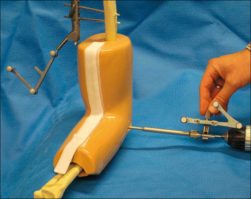
Twelve elbow models incorporating soft tissue coverage (Sawbones, Pacific Research Laboratories, Inc., Vashon, WA) were used in this study. The soft tissue coverage was around the elbow joint, whereas the proximal humerus and distal part of the lower arm were not covered. A radiolucent table was used, and the necessary reference marker for the navigation was securely fixed to a Steinmann pin inserted in the proximal humerus ().
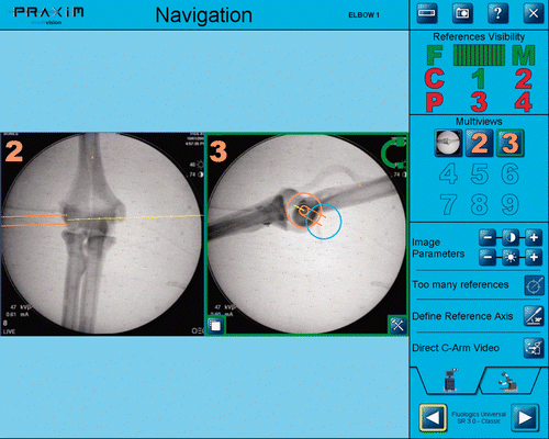
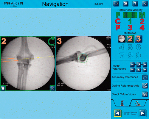
Initially, the optimal placement trajectory of the axis pin was defined in the anterior-posterior (AP) and lateral planes of the elbow, based on a conventional X-ray in both planes which was used as the reference for all sawbone testing conditions (both navigated and conventional) ( and ). The distances and angular deviations from this optimal position were measured as described below.
Testing protocol
A single operator (D.K.) performed all guide pin placements for both testing techniques.
Navigated technique
A commercially available navigation system (Praxim, Grenoble, France) was used in combination with a conventional fluoroscopy system (General Electric, USA). Initially, radiographs were taken in the AP and lateral views of the elbow joint to determine the anatomic center of rotation and transferred to the navigation system.
Planning of the center axis in both planes was achieved with the aid of the navigation system, which visualized the axis trajectory based on both the AP and lateral images. Once the central axis had been determined virtually, insertion of the central pin was carried out under 2D guidance from the virtual images. A 1.5-cm incision over the lateral soft tissue coverage above the epicondyle was made before the definitive drill application. Navigated drilling proceeded with the axis pin loaded into a navigated drill, which had already been registered to the pin size and length. Pin placement through the distal humerus was done under static 2D control without further fluoroscopic imaging.
Next, it was determined whether application of the external fixator system was possible with the related radiolucent parts (Compass Universal Hinge, Smith & Nephew, Memphis, TN). In all cases the axis pin was left in place and the complete fixator system applied to bring the elbow through a complete range of motion.
Conventional technique
In the conventional technique, the axis pin was drilled under fluoroscopic control using sequential AP and lateral views without navigated control. This approach followed that recommended in the technique guide, i.e., through the radiolucent parts of the external frame. Multiple drilling attempts were permitted until the pre-defined optimal placement trajectory was achieved, based on the operator's subjective reference in both fluoroscopic planes. The fixator application, including the range-of-motion test, was performed as in the navigated cases.
Measurements
Determination of the final axis pin placement in all the models was done after final pin removal (to avoid artifacts) using a computed tomography (CT) scan of each elbow joint (Optima CT 520, General Electric, Fairfield, CT). Using a 1-mm slice technique, reconstructions in the frontal and sagittal planes were performed in order to evaluate the position of the axis pin canal.
Based on the pre-defined optimal placement trajectory, variations in both the AP and lateral planes were measured as follows:
AP: Angular deviation <15°, <20°, <25°, <30° or >30° in relation to the optimal placement;
Lateral: Deviation in millimeters from the optimal insertion point.
The drill holes were identified on a CT scan, from which the number of drill holes needed and the exact placement of the pin were determined. Furthermore, in all cases the overall number of drilling attempts was counted and compared for the navigated and conventional groups.
Results
AP angulation
The AP deviations relative to the previously defined optimal placement trajectory for the six conventionally operated elbows and the six in the navigated group are shown in and . While three of the six conventional elbows deviated by more than 20°, there were no deviations greater than 19° in the navigated group. The mean AP angulation deviaton in the conventional group was 20.5°, compared to 15° in the navigated group (p = 0.077).
Table I. Deviation in AP angulation and lateral distance for conventional technique.
Table II. Deviation in AP angulation and lateral distance for navigated technique.
Lateral distance
The mean distance of the drilling point from the point of optimal placement was 3.83 mm in the conventional group, versus 1.83 mm in the navigated group (p = 0.042). Measurements in the lateral view revealed the distances from the optimal entry point for the individual elbows, as shown in and .
Drilling attempts
For all navigated cases, only one drill attempt was necessary to achieve the desired position of the axial pin.
Discussion
Since their first description in the literature in 1843, hinged external fixators of the elbow have undergone many design changes and have been applied for various indications. The modern elbow external fixator is based on anatomic ulnohumeral kinematics, approximating a single hinge joint while protecting bony and ligamentous repairs Citation[8]. It is indicated for patients with elbow instability resulting from bony or ligamentous etiologies Citation[9].
The fixators can be applied to patients with or without contractures, and allow early mobilization with correct alignment of the joint.
Surgical application of the hinged fixator is technically demanding and requires specialist knowledge and experience. The placement of the central axis pin is a crucial step in the use of such devices: slight mal-alignment of the pin can increase the stress in the surrounding articulating surfaces and ligaments, which is detrimental to the healing process Citation[10], Citation[11].
One potential complication of lateral percutaneous pin placement in the distal humerus is inadvertent damage to the radial nerve as a result of errant pin placement. The radial nerve passes close to the lateral head of the triceps and then lies between the brachialis and brachioradialis in the flexor compartment of the arm. Baumann et al. Citation[12] reported on three cases of radial nerve injury following application of a hinged elbow external fixator (Dynamic Joint Distractor II, Stryker Corporation) for instability of the elbow, with all three patients suffering a complete loss of radial nerve function Citation[12]. In all three cases the distance between the pin and the lateral epicondyle did not exceed 47 mm; it was previously reported that implantation of screws in the distal part of the humerus as near as 6 cm proximal to the lateral epicondyle is possible and safe Citation[13]. All of the pins in this series were implanted percutaneously using fluoroscopy; however, the authors recommended placing the pins through an open approach, inserting them under direct visualization.
Cheung et al. Citation[3] reported on complications related to the placement of an external fixator device and concluded that in order to minimize complications the application must be performed by an experienced surgeon, and the central pins should be placed under direct visualization.
The major outcome of this present study has been to demonstrate the accuracy of a navigation system used for placing the central pin of a hinged external elbow fixator. Our results suggest that finding the optimal placement trajectory with navigation is easier and requires fewer static radiographs, resulting in less radiation exposure for the patient and shorter operation time. The better accuracy achieved with navigation may also limit the risk of anatomic injuries; however, our study design was unable to address these issues.
There are several limitations to this study: We did not use human specimens for our scientific set-up, thereby limiting us with regard to describing our results in the context of neural structures and soft tissue damage. Additionally, we cannot make any profound statement regarding the comparison of time taken to create the optimal placement trajectory in the conventional or navigated groups. However, our results suggest that fewer pin placements are necessary with navigation, and that these attempts are more accurate, consequently leading to a reduction in possible complications.
Conclusion
In summary, this study focused on comparing accuracy between the navigated and conventional technique in the placement of the central pin of a hinged external elbow fixator. We believe that it serves as a good pilot for the design of a more rigorous study to compare surgical time, complications and cost.
Declaration of interest: The authors report no conflict of interest.
References
- Schep NW, De Haan J, Iordens GI, Tuinebreijer WE, Bronkhorst MW, De Vries MR, Goslings JC, Ham SJ, Rhemrev S, Roukema GR, et al. A hinged external fixator for complex elbow dislocations: A multicenter prospective cohort study. BMC Musculoskelet Disord 2011; 12: 130
- von Knoch F, Marsh JL, Steyers C, McKinley T, O'Rourke M, Bottlang M. A new articulated elbow external fixation technique for difficult elbow trauma. Iowa Orthop J 2001; 21: 13–19
- Cheung EV, O’Driscoll SW, Morrey BF. Complications of hinged external fixators of the elbow. JSES 2008; 17(3)447–453
- Belvedere C, Ensini A, Leardini A, Bianchi L, Catani F, Giannini S. Alignment of resection planes in total knee replacement obtained with the conventional technique, as assessed by a modern computer-based navigation system. Int J Med Robot 2007; 3(2)117–124
- Beckmann J, Stengel D, Tingart M, Götz J, Grifka J, Lüring C. Navigated cup implantation in hip arthroplasty. Acta Orthop 2009; 80(5)538–544
- Kim CW, Lee YP, Taylor W, Oygar A, Kim WK. Use of navigation-assisted fluoroscopy to decrease radiation exposure during minimally invasive spine surgery. Spine J 2008; 8(4)584–590
- Hankemeier S, Hüfner T, Wang G, Kendoff D, Zeichen J, Zheng G, Krettek C. Navigated open-wedge high tibial osteotomy: advantages and disadvantages compared to the conventional technique in a cadaver study. Knee Surg Sports Traumatol Arthrosc 2006; 14(10)917–921
- Tomaino MM, Sotereanos DG, Plakseychuk A. Technique for ensuring ulnohumeral reduction during application of the Richard compass elbow hinge. Am J Orthop 1997; 26: 646–647
- McKee MD, Bowden SH, King GJ, Patterson SD, Jupiter JB, Bamberger HB, Pakisma N. Management of recurrent, complex instability of the elbow with a hinged external fixator. J Bone Joint Surg Br 1998; 80: 1031–1036
- Stavlas P, Jensen SL, Søjbjerg JO. Kinematics of the ligamentous unstable elbow joint after application of a hinged external fixation device: A cadaveric study. JSES 2007; 16: 491–496
- Madey SM, Bottlang M, Steyers CM, Marsh JL, Brown TD. Hinged external fixation of the elbow: Optimal axis alignment to minimize motion resistance. J Orthop Trauma 2000; 14: 41–47
- Baumann G, Nagy L, Jost B. Radial nerve disruption following application of a hinged elbow external fixator: a report of three cases. J Bone Joint Surg Am 2011; 93(10)e51
- Gausepohl T, Koebke J, Penning D, Hobrecker S, Mader K. The anatomical base of unilateral external fixation in the upper limb. Injury 2000; 31(Suppl 1)11–20


