Abstract
Chondrosarcoma of the pelvis is difficult to treat due to the anatomical location and the high incidence of recurrence. Treatment is primarily surgical, and the surgical margins, based on MSTS criteria, have been shown to be predictive of disease recurrence and mortality. However, too-wide margins can decrease post-operative function. In the presented case, computer assisted surgery (CAS) was used to safely enable a joint-salvaging approach in a modified type 2/3 resection of a grade 2 chondrosarcoma of the os ischium and os pubis. The CAS navigation was vital to achieving the desired safe margins. The current follow-up period is 3.5 years, and the patient is disease-free, with no local recurrences or metastases having been detected. Post-operative function is excellent, with good MSTS and SF36 scores. This outcome is a good example of the value of CAS in certain cases.
Introduction
Bone tumor resections in the pelvic bone are known to be difficult due to the anatomical shape and surrounding structures Citation[1]. Chondrosarcomas located in the pelvis have a higher recurrence rate than those located in the long bones Citation[2], but treatment of chondrosarcomas in both the pelvis and the long bones is primarily surgical. Intermediate-grade and high-grade (2 and 3) chondrosarcomas of the pelvis are treated with wide resection, whereas therapy for grade 1 chondrosarcomas may differ, but radiotherapy and chemotherapy only have a role as adjuvants in cases with compromised margins or high-grade tumors Citation[3], Citation[4].
The type of resection is chosen according to the location and grade of the chondrosarcoma. These resections are described by the model proposed by Enneking and Dunham Citation[5]. Because of life-over-limb (or function) considerations, adequate margins are always the primary goal. Insufficiently wide margins, based on Musculo-Skeletal Tumor Society (MSTS) criteria, have been shown to be predictive of disease recurrence and mortality Citation[1], Citation[6], Citation[7]; however, margins that are too wide can decrease post-operative function. A joint-sparing approach is usually preferable to prosthesis placement or hip arthrodesis, especially in younger patients.
Ten-year survival of chondrosarcoma grade 2 in the pelvis has been reported to be around 75% Citation[1], Citation[6]. Recurrence has been reported in 44% of cases, with a median time to recurrence of 23 months Citation[6].
Computer assisted surgery (CAS) has proved to be a safe means of navigation in tumor resections, and its use has already been reported in resections of pelvic tumors Citation[8], Citation[9]. In the presented case, CAS made a joint-sparing approach possible, serving as a good example of the additional value which CAS can provide.
Case report
A 46-year-old woman presented with pain symptoms in the right hip region. Four years earlier, an X-ray of the pelvis, made at another hospital, had shown a small lesion near the right symphysis, in the upper ramus of the os pubis, which was interpreted as a cyst. There was no follow-up to these findings. Because of progressive stiffness of the right hip joint, an X-ray and MRI scan were performed 3.5 years later and a suspect lesion was found, whereupon the patient was referred to an orthopedic oncologist. A new MRI scan localized the lesion to both rami of the right os pubis with an expansion to the os ischium, close to the acetabulum. A needle biopsy, analyzed by a musculoskeletal pathologist, showed a grade 2 chondrosarcoma, which could be classified as a stage IIB lesion Citation[5], Citation[10]. The tumor length was 9.3 cm and its volume was calculated, using the method of Göbel et al. Citation[11], as 92.69 ml, with one episodical and one cylindrical arm. CT scans of the thorax and bone showed no metastases.
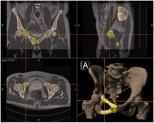
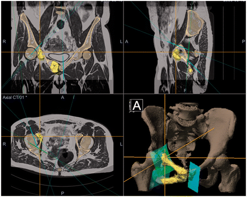
Because of the localization of the tumor near the hip joint and the lack of soft tissue involvement, the two options for resection were a joint-sparing local resection, or a segmental resection followed by reconstruction with a saddle prosthesis. A local resection (modified type 2/3 in the classification of Enneking et al. Citation[10]) was decided upon, partly resecting the frontal part of the right acetabulum to attain a safe margin. A pre-operative CT scan (Somatom Definition, Siemens, Erlangen, Germany) (120 kV, 447 mA, slice thickness 1 mm, FOV 326 mm) and an MRI scan (Avanto 1.5 T, Siemens, Erlangen, Germany) (T1 with/without contrast, T2 and FLAIR) were obtained for computer assisted surgery. The navigation was then prepared in advance of the surgery on the CAS system (Stryker Navigation System II with OrthoMap software, Stryker Mahwah, NJ) ( and ).
Figure 1. CT/MRI fusion with MRI-based tumor coloration using the OrthoMap Oncology Module (Stryker, Mahwah, NJ), showing the peri-acetabular part of the grade 2 chondrosarcoma.

Figure 2. Surgical planning of the resection planes in the OrthoMap Oncology Module with tumor coloration on the fused MRI/CT image. Patient information is digitally edited out in the bottom left panel. The bottom right panel shows a 3D rendering of the pelvic bone and the resection planes.

After preparing the patient for surgery, an illo-inguinal incision was made, with subsequent preparation from the symphysis in the lateral direction to the os ilium. The femoral neurovascular bundle was identified and marked. Two OrthoLock pins (Stryker) were placed in the right crista iliaca for the patient tracker. The CAS set-up was performed and an accuracy of 0.5 mm reported. The nervus obturatorius was identified and released, and with careful preparation the os ischium was uncovered. With the help of the navigation system, the deep resection plane was then identified and checked for a safe margin, and a Kirschner wire was placed to mark the plane. The symphysis joint was considered to be possibly contaminated, so a resection plane on the left os pubis was marked with a Kirschner wire with the help of CAS. Finally, the resection plane around the hip joint was identified with the CAS system and marked with diathermy.
The hip joint was cut along the marked planes with an osteotome, and the same method was used for the symphysis resection and os ischium resection plane. The tumor was then carefully removed, after cutting the hamstrings, while carefully preserving the n. obturatorius. A Marlex (polypropylene) mesh was used to reconstruct the inguinal channel, and the subcutis and cutis were closed in layers, with one deep drain being left in situ. After an uneventful wound healing period, the patient was mobilized using two elbow crutches.
The pathologist reported a macroscopic and microscopic R0 resection, with a minimum margin of 1 cm in the acetabulum resection plane. The tumor showed an infiltrative growing process with infiltration of the trabecular bone, and was reported as a grade 2 chondrosarcoma, as in the biopsy specimen.
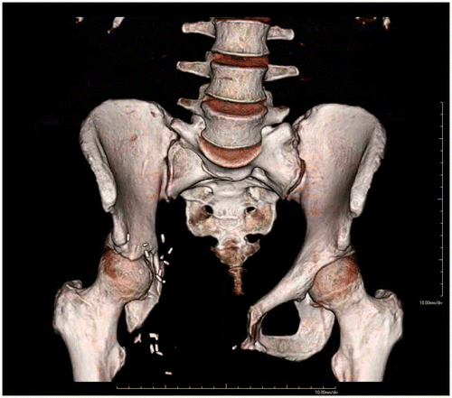
The current follow-up period is 3.5 years. The patient is disease-free, and no local recurrences or metastases have been detected. The follow-up was performed with MRI scans, multidetector CT scans (Siemens Somatom Definition) (120 kV, 662 mA, slice thickness 1 mm, FOV 440 mm) (), bone scans, and X-rays of the pelvis and thorax at specific intervals.
Function is currently good. The Musculo-Sketelal Tumor Society score (MSTS 1993) and the Short Form health survey (SF36 v2) score at 2 years post-surgery were excellent. The MSTS was 29/30, or 98%, while the SF36 was 42.1 PCS-36 and 36.2 MCS-36. After two years, the function of the right hip was 110/0/5° for flexion/extension, 20/0/10° for abduction/adduction and 30/0/20° for exorotation/endorotation. The patient is limited in end-range exorotation, and kneeling can be somewhat painful; however, she can walk unaided, has no pain, and has fully resumed her work.
Discussion
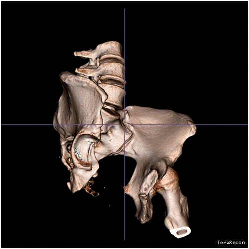
Post-operative functions were excellent because two thirds of the acetabulum could be salvaged. The current acetabulum shape can be seen clearly in a rendered image from the follow-up CT. To give a better view, the femur was removed by semi-automatic segmentation using iNtuition (TeraRecon, Inc.) software ().
Figure 4. The femur was removed using iNtuition software (TeraRecon, Inc.), applying semi-automatic, region growing-based segmentation. Subsequently, a small parts removal was performed to remove remaining bone fragments; this process also removed all the surgical clips from the image data.

Rehabilitation time was relatively short. Alternate approaches such as allograft reconstruction, (pseudo)arthrodesis, or tumor prosthesis reconstruction have been reported to result in lower post-operative function than joint salvage Citation[12]. Post-operative function with saddle prostheses is generally better than for allograft reconstruction Citation[13]; however, saddle prostheses can suffer from upward migration or dislocation, and are limited in their range of motion Citation[13], Citation[14].
Custom endoprostheses can be a good alternative to saddle prostheses in pelvic tumors, but this particular case did not warrant the use of such a prosthesis, while hip resection was not considered an option due to the limited range of motion and leg shortening. Reconstruction of the pelvic girdle is not necessary after resection.
The CAS navigation was vital in achieving the desired safe margins, providing precise, continuous 3D imaging. During the operation, the resection planes could be checked accurately for margin, greatly increasing confidence in achieving a good resection. Preplanning the operation on the CAS system has the advantage that the resection planes are already prepared and can easily be followed intra-operatively. In this case, the CAS system was used as a guide for the resection planes. In more recent operations, however, the system has also been used for guiding the instruments by attaching a tracker to, for example, the osteotome.
Surgical time for this case, based on OR data, was just over six hours. Set-up time for the CAS system was only approximately 8 minutes, and we believe this set-up time was recouped during surgery.
The musculoskeletal pathologist reported the sample as an R0, en bloc, resection with a minimal margin of 1.0 cm and adequate barriers on all sides. As stated in the Introduction, achieving wide margins, based on MSTS criteria, is important in order to minimize disease recurrence and mortality Citation[1], Citation[6], Citation[7]. However, such margins can be very hard to achieve in pelvic neoplasms due to the close proximity of other anatomical structures in the pelvis. Furthermore, the definition of margin differs from one article to the next. Pring et al. stated that if “the tumor abutted the surrounding genitourinary structures or bowel and the peritoneum or bladder could be clearly delineated and separated from the tumor, the resection was considered marginal” Citation[1], whereas Ozaki et al. defined a wide margin as “removal of the tumor plus more the 2 cm of normal tissue surrounding the tumor in all directions” Citation[15]. However, based on the overview by Lee et al. of chondrosarcoma treatment Citation[7] and the classification of Enneking et al. Citation[5], Citation[10], resection outside the reactive zone could be considered as wide. Both classifications, wide and marginal, can be applied to this patient. All the above-mentioned articles also use the same classification in grade 2 and grade 3/4 chondrosarcoma. The resection planes were achieved as planned, with, in our view, adequate barriers. “Minimal wide” would be a good classification in this case.
Computer assisted surgery can help with the resection, as the reactive zone is clearly visible on the MRI. Thus, a properly planned resection can avoid a contaminated or intra-lesional resection. Pring et al. [1] reported initially insufficiently wide margins in 17% of cases (3 intra-lesional, 5 compromised wide, and 3 compromised marginal out of 64 total).
With the progression of knowledge in local awareness, the availability of high-resolution MRI imaging, increased understanding of tumor characteristics, and advances in limb salvage surgery, there is a trend towards less destructive resections. Margins are still of primary importance, in order to avoid local recurrence, but margins that are too wide can compromise function.
Conclusion
Chondrosarcoma of the pelvis is difficult to treat due to the anatomical location, and the tumors often recur. As the surgical margins are a very important predictor of outcome, the first goal should always be to achieve adequate margins. Computer assisted surgery has already been reported to be helpful in the resection of pelvic tumors, and CAS was used in this case to safely enable a joint-salvaging approach in a modified type 2/3 resection of a grade 2 chondrosarcoma of the os ischium and os pubis. A joint-salvaging approach is preferable to allograft reconstruction or saddle prosthesis placement whenever possible. The current follow-up period is 3.5 years, and the patient is in remission. Post-operative function is excellent, with good MSTS and SF36 scores. This case serves as a good example of the additional value which CAS provides in certain situations, and we believe that a joint-sparing resection like the one reported cannot safely be performed without it.
Declaration of interest: The authors report no conflicts of interest and full compliance with ethical guidelines.
References
- Pring ME, Weber KL, Unni KK, Sim FH. Chondrosarcoma of the pelvis. A review of sixty-four cases. J Bone Joint Surg Am 2001; 83-A: 1630–1642
- Björnsson J, McLeod RA, Unni KK, Ilstrup DM, Pritchard DJ. Primary chondrosarcoma of long bones and limb girdles. Cancer 1998; 83: 2105–2119
- Gelderblom H, Hogendoorn PC, Dijkstra SD, van Rijswijk CS, Krol AD, Taminiau AH, Bovée JV. The clinical approach towards chondrosarcoma. Oncologist 2008; 13: 320–329
- Sanerkin NG, Gallagher P. A review of the behaviour of chondrosarcoma of bone. J Bone Joint Surg Br 1979; 61-B: 395–400
- Enneking WF, Dunham WK. Resection and reconstruction for primary neoplasms involving the innominate bone. J Bone Joint Surg Am 1978; 60: 731–746
- Sheth DS, Yasko AW, Johnson ME, Ayala AG, Murray JA, Romsdahl MM. Chondrosarcoma of the pelvis. Prognostic factors for 67 patients treated with definitive surgery. Cancer 1996; 78: 745–750
- Lee FY, Mankin HJ, Fondren G, Gebhardt MC, Springfield DS, Rosenberg AE, Jennings LC. Chondrosarcoma of bone: an assessment of outcome. J Bone Joint Surg Am 1996; 81: 326–338
- Reijnders K, Coppes MH, van Hulzen AL, Gravendeel JP, van Ginkel RJ, Hoekstra HJ. Image guided surgery: New technology for surgery of soft tissue and bone sarcomas. Eur J Surg Oncol 2007; 33: 390–398
- Wong K-C, Kumta SM, Chiu KH, Antonio GE, Unwin P, Leung KS. Precision tumour resection and reconstruction using image-guided computer navigation. J Bone Joint Surg Br 2007; 89: 943–947
- Enneking WF, Spanier SS, Goodman MA. A system for the surgical staging of musculoskeletal sarcoma. 1980. Clin Orthop Relat Res 2003; 415: 4–18
- Göbel V, Jürgens H, Etspüler G, Kemperdick H, Jungblut RM, Stienen U, Göbel U. Prognostic significance of tumor volume in localized Ewing's sarcoma of bone in children and adolescents. J Cancer Res Clin Oncol 1987; 113: 187–191
- Windhager R, Karner J, Kutschera HP, Polterauer P, Salzer-Kuntschik M, Kotz R. Limb salvage in periacetabular sarcomas: review of 21 consecutive cases. Clin Orthop Relat Res 1996; 331: 265–276
- Cottias P, Jeanrot C, Vinh TS, Tomeno B, Anract P. Complications and functional evaluation of 17 saddle prostheses for resection of periacetabular tumors. J Surg Oncol 2001; 78: 90–100
- Aljassir F, Beadel GP, Turcotte RE, Griffin AM, Bell RS, Wunder JS, Isler MH. Outcome after pelvic sarcoma resection reconstructed with saddle prosthesis. Clin Orthop Relat Res 2005; 438: 36–41
- Ozaki T, Hillmann A, Lindner N, Blasius S, Winkelmann W. Chondrosarcoma of the pelvis. Clin Orthop Relat Res 1997; 337: 226–239
