Abstract
Two novel, non-destructive assays were developed to evaluate contaminant-induced lipid peroxidation (thiobarbituric acid-reacting substances, TBARS, levels) and haem biosynthesis disruption (porphyrin excretion) in decapod crabs. A laboratory experiment was conducted whereby pie-crust crabs (Cancer novaezelandiae) were fed cockles (Austrovenus stutchburyi) collected from a contaminated and reference site and TBARS levels and porphyrin excretion determined using fluorometric analysis in urine samples. Pyrene metabolite levels were also measured in the same urine samples to assess polycyclic aromatic hydrocarbon (PAH) exposure. Contaminant-exposed crabs exhibited elevated urinary TBARS and porphyrin levels and a strong correlation was found between these two assays and the urinary pyrene metabolite concentrations. However, there was large within-treatment variability, which precluded a clear separation between the control and the impacted group. Nevertheless, consistency in the direction of the response shows that the biomarkers reflect pollutant levels and validates the use of these simple techniques from human medicine for environmental assessments.
Acknowledgements
The authors gratefully acknowledge the departments of Marine Science and Chemistry, University of Otago, New Zealand, for funding and use of facilities. This work was partially funded by the Ecology Research Group at the University of Otago. Moreover, we thank the University of Otago for a Postgraduate Publishing Bursary that facilitated write up of this research as a manuscript. We also thank the anonymous reviewers whose comments improved the manuscript.
Declaration of interest: The authors report no conflicts of interest. The authors alone are responsible for the content and writing of the paper.
Appendix
Figure 5. Fixed excitation fluorescence spectra (λexcitation = 530 nm) for diluted (1:47) crab urine sample, blank and 0 nmol 1−1 L MDA standards.
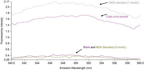
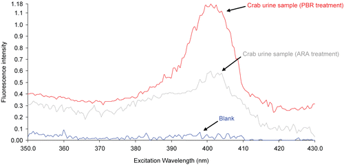
Figure 6. Fixed emission fluorescence spectra (λexcitation = 650 nm) for diluted (1:60) crab urine sample from two treatment groups (ARA and PBR) and a blank. The fluorescence spectrum of the crab urine sample reflects the typical shape of the porphyrin fluorescence signal in a human urine sample (see CitationWesterlund et al. 1988, p. 348).