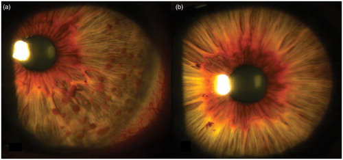
A 47-year-old woman, with no personal or familial history of Neurofibromatosis type 1 (NF1) or 2, was referred for evaluation of left unilateral Lisch nodules (LN) discovered through a routine eye care performed one month before (). Fundus oculi evaluation was normal in both eyes. The patient had occasionally attended routine eye examinations since her childhood, without discovering anomalies. Her past medical history was remarkable for migraine with aura, essential hypertension, and mild hypercholesterolemia. No other NF1 stigmata were noticed with the exception of one very pale 1.0 × 1.0 cm cafè-au-lait spot below the left leg. Magnetic resonance imaging of brain and spine, cardiac and abdominal ultrasound findings were unremarkable.
Figure 1. (a) Lisch nodules in the left eye, mainly located at the temporal portion; (b) no Lisch nodules are observed in the right iris.
The term segmental NF1 is used when the NF1 stigmata are confined to one or more body segments because of somatic mosaicism for the NF1 gene. Segmental NF1 is estimated to be rarer than the full-blown phenotype, with a prevalence of 1 in 36–40,000 subjects.Citation1 In patients with segmental form, the involved parts of the body may develop all the manifestations with the same chronological order as they appear in the generalized phenotype.Citation2 Ocular NF1 manifestations include LN, optic pathway glioma, eyelid and/or conjunctival neurofibromas, choroidal hamartomas, and orbital plexiform neurofibromas. Lisch nodules, asymptomatic melanocytic hamartomas of the iris, are the most common ocular NF1 findings and are included among diagnostic criteria.Citation1 They usually develop during childhood with their prevalence increasing with age (90–100% of adult patients with generalized NF1).Citation3
LN have been reported only occasionally in segmental NF1Citation4,Citation5 or in partial unilateral lentiginosis.Citation6 In all these cases the patients had NF1 manifestations in body areas close to the eye (ie, head, chest and face).Citation4–6 A genetic study revealed a somatic mosaicism of the NF1 gene in one case.Citation5 In the largest study on the ophthalmological manifestations in segmental NF1, no LN or other eye NF1 stigmata were observed in a cohort of 72 subjects, and no NF1 gene mutations in blood DNA were discovered. The postulated explanation for the absence of ocular involvement in these patients was that the eye was too far from tissues with NF1 mutation or that genetic anomalies involved tissues different from the eye.Citation2 Conversely, it is known that the eye and the optic pathway may be altered in segmental NF1 phenotypes primarily involving the ocular system. In fact, plexiform neurofibromas of the orbit or the eyelid can be the only manifestation of segmental NF1; furthermore, isolated optic pathway glioma, which behaved both biologically and radiographically as a typical NF1-associated tumor, has been hypothesized to represent a form of segmental NF1.Citation2,Citation7
Few reports have described unilateral LN without any other NF1 manifestations.Citation5,Citation8 There are also three families with parents having unilateral or bilateral LN and sons with full blown NF1 (Tenconi R personal communication).Citation2,Citation9,Citation10 The hypothesized genetic mechanisms existing in these families is a somatic ocular (eg, of the iris) and germinal mosaicism of the mutated NF1 gene in the parents, that results in a very limited “segmental” manifestation of NF1 (eg, LN) in the parents and in the full blown phenotype in the offspring. In contrast to the previously reported cases, our patient was older (eg, 47-years-old versus 14- and 19-year-olds), and the absence of other NF1 manifestations at this age indicates that a mosaicism for NF1 gene is limited to a very restricted area, that is the left iris, and not involving other tissues. We cannot exclude a concomitant germinal mosaicism since our patient does not have children. Isolated LN of the iris may represent a further rare form of segmental NF1 of the eye.
At present there are no means to exclude involvement of both somatic and gonadic tissue in a subject with segmental phenotype, and therefore the exact level of risk is not definable with current knowledge. Gonadic tissue may harbor the mutated NF1 gene also if the body segment affected is distant from the gonads. From this point of view, the presence – or lack – of LN in the setting of segmental NF1 should not be interpreted as a predictor for transmission of NF1 to offspring, although the effective risk cannot be precisely estimated.
Declaration of interest: The authors report no conflicts of interest. The authors alone are responsible for the content and writing the paper.
References
- Ruggieri M, Upadhyaya M, Di Rocco C, et al. Neurofibromatosis type 1 and related disorders. In: Ruggieri M, Pascual-Castroviejo I, Di Rocco C, editors. Neurocutaneous disorders: phakomatosis and hamartoneoplastic syndromes. Wien/New York: Springer-Verlag, 2008; 51–151
- Ruggieri M, Pavone P, Polizzi A, et al. Ophthalmological manifestations in segmental neurofibromatosis type 1. Br J Ophthalmol 2004;88:1429–1433
- Lubs MLE, Bauer MS, Formas ME, et al. Lisch nodules in Neurofibromatosis type 1. New Engl J Med 1991;324:1264–1266
- Weleber RG, Zonana J. Iris hamartomas (Lisch nodules) in a case of segmental neurofibromatosis. Am J Ophthalmol 1983;96:740–743
- Ceuterick SD, Van den Ende JJ, Smets RME. Clinical and genetic significance of unilateral Lisch nodules. Bull Soc Belge Ophtalmol 2005;295:49–53
- Rao GS. Partial unilateral lentiginosis with Lisch nodules: a forme frusta of segmental neurofibromatosis? Indian J Dermatol Venereol Leprol 2004;70:114–115.
- Listernick R, Mancici AJ, Charrow J. Segmental neurofibromatosis in childhood. Am J Med Genet 2003;121A:132–135
- Lal G, Leavitt JA, Lindor NM, et al. Unilateral Lisch nodules in the absence of other features of Neurofibromatosis 1. Am J Ophthalmol 2003;135:567–578
- Toonstra J, Dandrieu MR, Ippel PF, et al. Are Lisch nodules an ocular marker of neurofibromatosis gene in otherwise unaffected family members? Dermatologica 1987;174:232–235.
- Riccardi VM, Riccardi AL. Penetrance of von Recklinghausen neurofibromatosis: a distinction between predecessors and descendant. Am J Hum Genet 1988;42:284–289