Abstract
Context: The plant kingdom has become a target in the search for new drugs and biologically active lead compounds. The common Jrani Tunisian caprifig Ficus carica L. (Moraceae) is one of the large number of plant species that are used in folklore medicine yet to be investigated for the treatment of many diseases, including those of infectious nature.
Objective: Hexane extract of the Tunisian common Jrani caprifig latex was assayed for antibacterial activity against several Gram-positive and Gram-negative bacteria. Chemical composition of the extract was also investigated.
Materials and methods: The hexane extract was obtained from Tunisian Jrani caprifig latex by maceration, and then analyzed by gas chromatography–mass spectrometry. The extract was tested in vitro for antibacterial activity by the disc diffusion method and minimal inhibitory concentration (MIC) was also determined for all the test cultures.
Results: Thirty-six compounds of the extract were identified, 90.56% of the total area of peaks were coumarins. A strong bactericidal effect was demonstrated. The most sensitive bacteria were Staphylococcus saprophyticus clinical isolate, and Staphylococcus aureus ATCC 25923, with a MIC of 19 µg/mL.
Discussion and conclusion: These findings demonstrate an effective in vitro antibacterial activity of the hexane extract of caprifig latex.
Introduction
Renewed attention in recent decades to alternative medicines and natural therapies has stimulated a new wave of research interest in traditional practices (CitationBraun & Cohen, 2007). The increased prevalence of antibiotic-resistant bacteria emerging from the extensive use of antibiotics may render the current antimicrobial agents insufficient to control at least some bacterial infections. Antimicrobial resistance among key microbial pathogens continues to grow at an alarming rate worldwide. Resistance among strains of Staphylococcus aureus, Pseudomaunas ssp., Streptococcus ssp., and Enterobacteriaceae has been described (CitationBhavnani & Ballow, 2000). The screening of plant extracts and plant products for antimicrobial activity has shown that higher plants represent a potential source of new anti-infective agents (CitationArias et al., 2004), and only 5–15% have been studied for a potential therapeutic value (CitationKinghorn, 1992). Caprifig Ficus carica L. (Moraceae) a fig tree which produces both male and female flowers and is used to fertilize the female trees of the species. Unlike common figs, the caprifig produces three crops of synconia. These are known by their Italian terms, profichi, mammoni, and mamme. Fresh leaves and syconia of caprifigs in Tunisian folk medicine are used by agriculturalists as an additive in chicken alimentation for prevention against bacteria and virus infections; their high amount of latex should be an effective source of protection.
CitationLazreg Aref et al. (2010) have investigated the antimicrobial activity of F. carica latex extracts of Bidhi Bither variety (Saint-Pedro type) against some resistant bacteria. This study investigated the antibacterial activity of the hexane extract of caprifig latex and defined its chemical composition using gas chromatography–mass spectrometry (GC-MS) technique.
Materials and methods
Plant material
The caprifig Jrani latex was collected from unripe inedible fig fruit from Mesjed Aissa agricultural field located in the central cost of Tunisia in June 2010. The latex was held in ice during the period of collection. The identification of this variety was established by Prof. Massoud Mars, professor of arboriculture at the high school of horticulture of Chott Meriam Sousse, Tunisia, Department of Agriculture and Arboriculture, and the code collection of the tree is JR1.
Plant extract
The fresh latex was macerated in 1 L of hexane (Merck, Germany) for 10 days then filtered and evaporated under reduced pressure to give a gummy extract.
GC-MS analysis
The analysis of Jrani caprifig latex hexane extract was performed on a GC-MS HP model 1909S-433 inert MSD (Agilent Technologies, J&W Scientific Products, Palo Alto, CA, USA), equipped with an Agilent Technologies capillary DB-5MS column (30 m in length; 0.25 mm i.d.; 0.25 mm film thickness), and coupled to a mass selective detector (MSD1909S-433, ionization voltage 70 eV; all Agilent, Santa Clara, CA, USA). The carrier gas was He and was used at 1 mL/min flow rate. The oven temperature program was as follows: 2 min at 150°C ramped from 150°C to 240°C at 10°C/min and 1 min at 240°C then ramped from 240°C to 280°C at 5°C/min and 15 min at 280°C. The chromatograph was equipped with a split/splitless injector used in the split mode. The split ratio was 1:100. Bis-(trimethylsilyl)-acetamide (BSTFA) (100 mL) was added to 100 mL of extract. The control of the GC-MS system and the data peak processing were carried out by means of MSDCHEM software. Identification of components was assigned by matching their mass spectra with Wiley and NIST library data and standards of the main components.
Microorganisms
A collection of 10 test microorganisms including Gram-positive and Gram-negative bacterial strains was used. The groups included five organisms of American Type Culture Collection (ATCC): Pseudomonas aeruginosa ATCC 27950, Pseudomonas aeruginosa ATCC 2783, Staphylococcus aureus ATCC 25923, Escherichia coli ATCC 25922, Enterococcus faecalis ATCC 29212, and five clinical organisms were obtained from the laboratory of microbiology, Faculty of Pharmacy, Monastir, Tunisia: Staphylococcus epidermidis, Staphylococcus saprophyticus, Staphylococcus aureus, Enterobacter cloacae, and Escherichia coli. All the test cultures were stored at +4°C on Muller-Hinton Agar (MH) (Biorad), subcultured every 2 weeks, and checked for purity.
Antimicrobial activity
Disc diffusion assay
The conventional disc diffusion method (CitationMurray et al., 1995) was employed for the initial assessment of antibacterial potential of the extract. The dried Jrani (caprifig) extract was dissolved in dimethyl-sulfoxide (DMSO; Merck) and then in sterile water, to reach a final concentration of 20 mg/mL, and sterilized by filtration by 0.45 µm Millipore filters. The media used was Muller-Hinton Agar (Biorad). The discs (6 mm in diameter) were impregnated with 10 µL of the extract (200 µg/disc) at a concentration of 20 mg/mL and placed on the inoculated Agar (for the preparation of the inocula, colonies of bacteria were suspended in Mueller-Hinton (MH) Broth (Biorad); the suspensions contained 108 CFU/mL of bacteria). Negative controls were prepared using the DMSO solvent employed to dissolve the latex extract. Oxacillin (5 µg/disc; Gibco), tetracyclin (30 µg/disc; Gibco), and erythromycin (15 µg/disc; Gibco) served as positive reference standards to determine the sensitivity of bacterial stains tested. The inoculated plates were incubated at 37°C for 24 h. The growth inhibition was assessed as the diameter (in mm) of the zone of inhibited microbial growth. Each assay in this experiment was performed in triplicate.
Micro-dilution assay
The minimal inhibitory concentration (MIC) values were also studied for the microorganisms that were determined as sensitive to the extract in disc diffusion assay. The inocula were prepared in broth cultures, and suspensions were adjusted to 0.5 Mc Farland standard turbidity. Extract dissolved in 10% DMSO was first diluted to the highest concentration (2.5 mg/mL) to be tested, and then serial two-fold dilutions were made in a concentration range from 2.5 to 0.0195 mg/mL in 10 mL sterile test tubes containing sterile water. The final concentration of DMSO in tubes was 0.78%. The MIC values of the extract against bacterial strains were determined based on a micro-well dilution method according to NCCLS [National Committee for Clinical Laboratory Standards (NCCLS), 2008] as described with slight modifications as follows.
The 96-well plates were prepared by dispensing into each well 95 µL of MH Broth and 5 µL of the inocula. Extract (100 µL) initially prepared at a concentration of 2.5 mg/mL was added into the first well. Then, 100 µL from its serial dilution was transferred into six consecutive wells. The last well, containing 195 µL of MH Broth without compound and 5 µL of the inoculum on each strip, was used as negative control. The final volume in each well was 200 µL. Tetracycline (Gibco®) at the concentration range of 400–0.39 µg/mL was prepared in sterile water and used as standard drug for positive control, and the DMSO was maintained as negative control. The plates were incubated at 37°C for 24 h, and the MIC was determined from the lowest concentration of the compound to inhibit the growth of microorganisms. Inhibition of proliferation was assessed by optical density measurements (625 nm). These experiments were replicated three times.
Results
The average percentage of individual compounds of Jrani caprifig (Ficus carica) latex extract is presented in . GC-MS analysis resulted in the identification of 36 compounds representing 96.12% of the total extract. Among the identified compounds, three were sesquiterpens (2.83%), two were triterpens (0.46%), two were oxygenated triterpens (0.74%), only one monoterpene [bornanone-3. (1.04)], 14 were coumarins (90.56%) and six were alcans (0.34%). Furthermore, the most abundant compounds (>8%) of extract were lanosta-8 (13.17%), urs-12-en-24-oic acid (21.52%), aristolone (15.63%), olean-12-en-3-ol, acetate (23.47%), and maragenin I acetate (8.78%). represents the chemical formula and molecular weight of some major components of the extract.
Table 1. Chemical composition of hexane extract obtained by GC-MS.
Table 2. Chemical formula and molecular weight of major component of Jrani hexane latex extract.
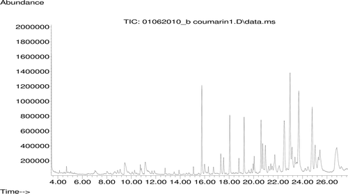
shows the GS-MS chromatogram of analyzed hexane extract of latex. Antimicrobial activity of extract was tested against five ATCC cultures and five clinical bacterial isolates. shows diameters of inhibition zones of latex extracts and shows the MIC values.
Table 3. Antimicrobial activity of the extract in Agar diffusion assay.
Table 4. Minimum inhibitory concentration (µg/mL) of the extract.
The highest inhibition zones recorded were 28, 28, and 26 mm, against S. saprophyticus, S. aureus ATCC25923, and S. epidermidis, respectively, whereas the lowest zones observed were 14, 14, and 15 mm against P. aeruginosa ATCC27950, P. aeruginosa ATCC2783, and E. faecalis ATCC29212, respectively.
The MIC values ranged from 19 to 312 µg/mL indicated that the extract was an effective antimicrobial agent. The antibacterial activity of bioactive compound produced by hexane extract is comparable with tetracycline as standard antibiotic. Interestingly, the hexane extract from caprifig latex had MIC values of 19 µg/mL against S. aureus ATCC 25923 and 39 µg/mL against S. epidermidis, which was approximately two and three times lower than that of tetracycline (MIC 25 and 100 µg/mL), respectively (). This extract could be good candidates for further studies of its antibacterial bioactive compounds.
Discussion
The phytochemical analysis reveals that the aqueous extract of ripe dried fruit of F. carica contains alkaloids, flavonoids, coumarins, saponins, and terpenes (CitationTeixeira et al., 2006; CitationVaya & Mahmood, 2006). Some phenolic compounds with reported pharmacological properties have already been isolated from fig leaves, namely, furanocoumarins like psoralen and bergapten; flavoloids like rutin, quercetin, and luteolin; phenolic acids like ferrulic acid; and also phytosterols like toraxasterol (CitationRoss & Kasum, 2002; CitationVaya & Mahmood, 2006). Coumarins constitute an important class of phytochemicals. Antimicrobial activities of the hexane extract of latex containing mostly coumarins were evaluated. There are reports on efficacies of pure coumarins against Gram-positive and Gram-negative bacteria as well as fungi (CitationKayser & Kolodziej, 1997; CitationBisignano et al., 2000). Free 6-OH in coumarin nucleus has been found to be important for antifungal activity, the free hydroxyl group at position 7 is important for antibacterial activity (CitationSardari et al., 1999). Interestingly, coumarins have also inhibitory effect on DNA gyrase which may be linked to the anti-HIV (human immunodeficiency virus) activity (CitationMatern et al., 1999).
In the present study, we describe the antibacterial activity for the first time of caprifig (F. carica) latex extract which showed a strong activity against S. saprophyticus, S. aureus, E. coli, S. epidermidis, E. cloacae, E. faecalis, and P. aeruginosa (MIC, 19 to 312 µg/mL) that can be due to the presence of coumarins with 90.56% of the total area.
Conclusion
Based on the results of this study, it can be concluded that caprifig latex is an agent which is used as folklore medicine and could be a source for finding new antibacterial agents in order to treat and control infections.
Acknowledgments
The authors are grateful to Prof. Ben Ouada Hafed (Directeur de l’Institut Supérieur des Sciences Appliquées et de Technologie de Mahdia) and Prof. Rachid Chamli (Laboratoire de Pharmacognosie et phytothérapie, Faculté de pharmacie 5000 Monastir Tunisie) for providing facilities for this work.
Declaration of interest
The authors report no conflicts of interest. The authors alone are responsible for the content and writing of the paper.
References
- Arias ME, Gomez JD, Cudmani NM, Vattuone MA, Isla MI. (2004). Antibacterial activity of ethanolic and aqueous extracts of Acacia aroma Gill. ex Hook et Arn. Life Sci, 75, 191–202.
- Bhavnani SM, Ballow CH. (2000). New agents for Gram-positive bacteria. Curr Opin Microbiol, 3, 528–534.
- Bisignano G, Sanogo R, Marino A, Aquino R, D’Angelo V, Germanò MP, De Pasquale R, Pizza C. (2000). Antimicrobial activity of Mitracarpus scaber extract and isolated constituents. Lett Appl Microbiol, 30, 105–108.
- Braun L, Cohen M. (2007). Herbs and Natural Supplements: An Evidence-based Guide, second ed. Elsevier, Australia, ISBN: 072953796X, p. 791.
- Kayser O, Kolodziej H. (1997). Antibacterial activity of extracts and constituents of Pelargonium sidoides and Pelargonium reniforme. Planta Med, 63, 508–510.
- Kinghorn AD. (1992). Plants as sources of medicinally and pharmaceutically important compounds. In: Nigg HN, Seigler D, (eds.), Phytochemical Resources for Medicine and Agriculture. New York. Plenum Press, pp. 75–95.
- Lazreg Aref H, Bel Hadj Salah K, Chaumont JP, Fekih A, Aouni M, Said K. (2010). In vitro antimicrobial activity of four Ficus carica latex fractions against resistant human pathogens. Pak J Pharm Sci, 23, 53–58.
- Matern U, Lüer P, Kreusch D. (1999). Biosynthesis of coumarins. In: Barton D, Nakanishi K, Meth-Cohn O, Sankawa U (eds.): Comprehensive Natural Products Chemistry, Vol. 1, Polyketides and Other Secondary Metabolites Including Fatty Acids and Their Derivatives. Elsevier Science Ltd., Oxford, UK, pp. 623–637.
- Murray PR, Baron EJ, Pfaller MA, Tenover FC, Yolke RH. (1995). Manuel of Clinical Microbiology. 6th Edn. Washington. DC, ASM, p. 300.
- NCCLS. (2008). Performance Standards for Antimicrobial Susceptibility Testing; Ninth Informational Supplement. NCCLS document M100-S9. National Committee for Clinical Laboratory Standards, Wayne, PA, pp. 120–126.
- Ross JA, Kasum CM. (2002). Dietary flavonoids: Bioavailability, metabolic effects, and safety. Annu Rev Nutr, 22, 19–34.
- Sardari S, Mori Y, Horita K, Micetich RG, Nishibe S, Daneshtalab M. (1999). Synthesis and antifungal activity of coumarins and angular furanocoumarins. Bioorg Med Chem, 7, 1933–1940.
- Teixeira DM, Patão RF, Coelho AV, da Costa CT. (2006). Comparison between sample disruption methods and solid-liquid extraction (SLE) to extract phenolic compounds from Ficus carica leaves. J Chromatogr a, 1103, 22–28.
- Vaya J, Mahmood S. (2006). Flavonoid content in leaf extracts of the fig (Ficus carica L.), carob (Ceratonia siliqua L.) and pistachio (Pistacia lentiscus L.). Biofactors, 28, 169–175.
