
Abstract
Context Eucommiae Cortex and Radix Dipsaci, occurring in a ratio of 1:1 in Du-Zhong-Wan (DZW), a Chinese herbal medicine, is available as a water extract followed by ethanol precipitation for the treatment of osteoporosis, fractures and menopausal syndrome.
Objective This study investigates the protective effects of DZW in ovariectomy (OVX)-induced bone loss in a rat osteopenia model.
Materials and methods Sixty Sprague–Dawley rats were randomly divided into the sham-operated group (SHAM) and five OVX subgroups: OVX with vehicle (OVX), 17β-estradiol (E2) and with three graded doses of DZW. Daily oral administration of the different samples started on the fifth week and lasted for 12 weeks, respectively. The body weight, uterus wet weight, serum biochemical parameters, bone mineral density (BMD), bone biomechanical properties, bone microarchitecture and immunohistochemistry were examined.
Results Compared with the SHAM group, the DZW treatment significantly reversed the osteoporotic changes in OVX rats. The DZW-H group showed that serum tartrate-resistant acid phosphatase 5b (TRACP-5b) levels reduced by 152.25% (p < 0.01) and osteocalein (OCN) levels dose dependently increased by 118.43% (p < 0.01) as compared with the OVX group. Compared with the OVX group, the DZW at different three dosages of DZW evidently increased the right femur BMD by 112.43, 114.56 and 116.45%, and dramatically promoted bone quality and bone strength (p < 0.05). Further, immunohistochemical evaluation also showed that DZW administration increased ER expression in uteri (p < 0.01).
Conclusions DZW exhibits an anti-osteoporotic effect, probably mediated via phyto-estrogenic effects. It might be a potential herbal alternative for the management of postmenopausal osteoporosis.
Introduction
Osteoporosis, characterized by low bone mass and structural deterioration of bone tissue, is one of the most significant health challenges, especially for post-menopausal women regardless of racial or ethnic differences (Ko et al. Citation2012). The population afflicted with osteoporosis was estimated to be approximately 200 million worldwide (Reginster & Burlet Citation2006). Currently, hormone replacement therapy (HRT) and bisphosphonate administration are the two therapeutic approaches for the prevention and treatment of postmenopausal osteoporosis (Li et al. Citation2012). However, long-term HRT has been found to increase the risk of cardiovascular diseases, biliary disease, breast and endometrial cancers (Persson et al. Citation1999; Davison & Davis Citation2003), and bisphosphonates cause bone atraumatic fracture. Therefore, plant-derived oestrogens with minimal adverse effects are desirable.
Traditional Chinese medicines, derived from natural medicinal plants with fewer side effects, have been historically used in the prevention and treatment of postmenopausal osteoporosis, and will undoubtedly continue to be used as a cost-effective alternative to commercial pharmaceutical products. Du-Zhong-Wan (DZW) is a traditional Chinese formulation, first recorded in Collation and Annotation of Well-tried Recipes for Women (Jiaozhu Furen Liangfang, volume 12) in 1237 AD by Song dynasty. It contains Eucommiae Cortex (Du Zhong in Chinese), dried and salted bark of Eucommia ulmoides Oliv. (Eucommiaceae), and Radix Dipsaci (Xu Duan in Chinese), dried and salted root of Dipsacus asperoides Wall. ex Henry (Caprifoliaceae), and was developed hundreds of years ago as a restorative formula. An equal weight ratio (1:1) of Eucommiae Cortex and Radix Dipsaci is widely used to prevent and treat various kidney and aging diseases by “improving the tone of the liver and kidneys, reinforcing the muscles and bones” (Hou et al. Citation2010). Additionally, these herbal medicines could be used for the treatment of bone-related diseases such as osteoporosis and bone fractures. Eucommiae Cortex and Radix Dipsaci need to be salted to improve efficacy according to the traditional Chinese medicine theory. Therefore, DZW is considered effective in the treatment of osteoporosis. However, evidence supporting the water extract using alcohol precipitation which is a method used to remove the impurity of water extract is limited. No data are available supporting DZW in osteoporosis induced by oestrogen deficiency and its potential mechanism(s) in OVX rats.
Therefore, this study aimed to systematically investigate whether DZW fraction may have a role in the treatment of osteoporosis induced by OVX, and its potential mechanism(s) in rats.
Materials and methods
Preparation of DZW water extract by alcohol precipitation
Dried and salted Eucommiae Cortex and Radix Dipsaci were purchased from Simcere Drugstore (Nanjing, China) in July 2013. The materials were identified by Professor Ping Li, School of Traditional Chinese Medicine of China Pharmaceutical University. The voucher specimens of Eucommiae Cortex and Radix Dipsaci were deposited under Nos. 10644289-DZ and 10644289-XD, respectively, in the herbarium of China Pharmaceutical University.
DZW was prepared according to the National Pharmacopoeia Commission of P. R. China (Board Citation2010), and extracted as follows: 3000 g of Eucommiae Cortex and Radix Dipsaci (weight ratio 1:1) was triturated, extracted with water (3 × 30 L, 1.5 h each time), combined and concentrated to 1:1.2 (g/mL) to obtain the water extract. The water extract was subjected to ethanol precipitation with 60% alcohol to remove the impurity. The liquid supernatant was filtered and lyophilized, with a yield of 30.5%. The DZW fraction was analysed and quantified using high-performance liquid chromatography ( and ). The powder was stored in desiccators at room temperature before use.
Figure 1. HPLC analysis of the plant extract reveals a complex composition of DZW water extract (Table 1).
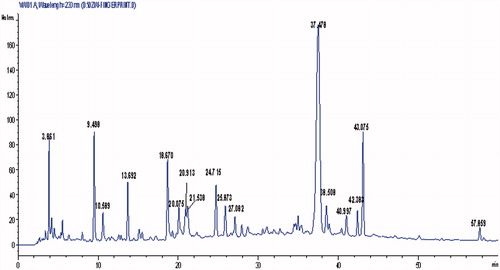
Table 1. Characterization of DZW water extract compounds from HPLC chromatogram.
Animals and treatments
Sixty female Sprague–Dawley-specific pathogen-free (SPF) rats aged 6 months and weighting 280 ± 20 g were caged in a group of five for 7 d prior to experiment. Animals were fed with a standard rat chow and water ad libitum under climate-controlled conditions (25 °C, 55% humidity, a 12 h light/dark cycle). Experimental Animal Facility and protocols were approved by the Animals Ethics Committee of the University, and all protocols were performed according to the Guide for Humane Use and Care of Laboratory Animals in China.
Rats underwent either bilateral sham operation (SHAM, n = 10) or bilateral ovariectomy (OVX, n = 50). The surgical procedure was performed under aseptic conditions following the China Pharmaceutical University Animal Care protocol. Rats were housed for 4 weeks to allow rats to develop osteopenia and rehabilitate. OVX rats were randomly assigned to five groups (10 rats each) as follows: OVX with normal saline as vehicle (OVX); OVX with 17β-estradiol (E2, 50 μg/kg body weight/d) as positive control; and OVX with DZW in three doses (DZW-L, 2.0 g/kg/d; DZW-M, 4.0 g/kg/d; DZW-H, 6.0 g/kg/d). Subsequently, all the rats were treated for 12 weeks. The DZW or E2 doses were adjusted according to body weights which were measured every week.
Specimen collection
At the end of treatment, whole blood samples were collected by carotid artery under general anaesthesia using 300 mg/kg chloral hydrate (i.p.). Blood was allowed to clot for 30 min, and serum was separated by centrifugation at 3000 × g for 10 min and stored at −80 °C for biochemical assays. After sacrifice, femora were cleaned and stored in tubes with physiological saline and stored at −20 °C for measurement of bone quality parameters including trabecular microarchitecture, bone mineral density (BMD) and bone mechanics.
Immunochemistry
The serum osteocalein levels (OCN) were measured using enzyme-linked immunosorbent assay (ELISA) kits (R&D Systems Inc., Minneapolis, MN). Tartrate-resistant acid phosphatase 5b (TRACP-5b), secreted by osteoclasts, correlates with bone resorption activity in abnormal bone metabolism. For markers of bone resorption, TRACP-5b levels were also measured with ELISA kits (R&D Systems Inc., Minneapolis, MN) in serum. All ELISA procedures were performed in accordance with the protocols of the manufacturers.
Dual-energy X-ray absorptiometry (DEXA)
The right total femur BMD was measured using Discovery W dual energy X-ray absorptiometry (DEXA, Hologic Inc., Boston MA) equipped with suitable software (edition 13.1.2) for bone density assessment in small animal scan mode. The operator was unaware of the rat's group. The BMD and the bone mineral content in total femur were determined for statistical analysis.
Biomechanical parameters
The mechanical properties of isolated right femurs were evaluated by three-point bending test (Zhang et al. Citation2012) using CSS-4420 material testing machine (Changchun Research Institute for Testing Machines Co. Ltd., Jilin, China). The load–deformation curve was drawn by a computer which was linked to the material-testing machine and recorded force and displacement data simultaneously. The biomechanical parameters were directly obtained from the load–deformation curve as follows: (a) ultimate load (Newtons, N), which showed the maximum bone load, (b) extrinsic stiffness (Newtons per millimeter, N/mm), calculated as the slope in the linear region between 40% and 80% of the ultimate load and (c) energy to ultimate load (millijoules, mJ), which is indicated the area under the curve until ultimate load.
Micro-CT analysis
To evaluate structural loss in distal femur, which is rich in trabecular bone, morphometric parameters of right distal femur from each group were scanned using Micro-CT (μCT-Sharp, ZKKS-MCT, Guangzhou, China). The scanning conditions were set to 60 kV, 40 W, with an isotropic voxel size of 22 μm. The pictures were reconstructed using ZKKS Micro-CT 3D analysis software version 3.0 (ZKKS-MCT, Guangzhou, China). Bone volume over total volume (BV/TV), trabecular separation (Tb.Sp), trabecular number (Tb.N), trabecular thickness (Tb.Th) and structure model index (SMI) were obtained by counting volume of interest (VOI).
Uterus immunohistochemistry
Uteri (n = 6/group) were trimmed, and fixed in 10% neutral buffered formalin at 4 °C for 2 d for assessment of E2 expression. The processed tissues were washed, dehydrated in gradient alcohol concentrations, embedded in paraffin wax and cut into 4 μm thick serial sagittal sections using a microtome (Leica RM2235, Nussloch, Germany). E2 immunohistochemical localization was performed using commercially available antibodies according to the recommended protocol of the manufacturer (Bioss, Beijing, China). Negative controls omitted the primary antibody. The operator conducting analysis was blinded to the divided groups, and stained sections were examined qualitatively under light microscopy (Leica DM 100, Nussloch, Germany) with Mini See 1.0.9.37 image analyzing system.
Statistical analysis
Data are presented as mean ± standard deviation (SD). Group differences were analyzed by one-way analysis of variance (ANOVA) followed by a post hoc multiple comparison with Fisher's least significant difference (LSD) t-test using SPSS Version 14.0 for Windows (SPSS, Chicago, IL). A p value of < 0.05 was defined as statistically significant.
Results
Body weight and uteri wet weight
The body weight changes in each group throughout the experimental period are shown in . No significant baseline differences in the average body weight were seen among all groups. After 12 weeks of treatment, the body weight of OVX group was obviously higher than that of the SHAM group (p < 0.01). All the DZW administration groups showed markedly inhibited OVX-induced weight increase (p < 0.01), similar to the E2 group. However, there were no significant differences in the decreased body weight among all DZW groups (). OVX caused significant atrophy of uterine tissue compared with that of the SHAM group (p < 0.01), indicating the success of the surgical procedure. DZW or E2 significantly increased the uterine/body weight ratio compared with OVX group (p < 0.01).
Table 2. Body weight and uterus wet weight, serum parameters after 12 weeks administration of DZW (n = 10, mean ± SD).
Biochemical assay
Serum bone turnover markers OCN and TRACP-5b, and serum E2 levels were determined. In , OVX resulted in a significant increase in serum TRACP-5b, and decrease in OCN and E2 levels compared with the SHAM group (p < 0.01). After 12-week DZW administration, serum TRACP-5b levels were apparently reduced in all DZW groups. In addition, compared with the OVX group, DZW-H group showed the lowest levels of 152.25% (p < 0.01). Further, DZW treatment increased OCN levels dose dependently, to 118.43% especially in the DZW-H group, compared with the OVX group (p < 0.01). Furthermore, three doses of DZW administration groups increased serum E2 levels to 120.98, 124.25 and 129.88%, compared with the OVX group (p < 0.01). All doses of DZW treatments exerted a similar effect as E2 in changing bone turnover markers and serum E2 level (p < 0.01) in this study.
Bone mineral density
In order to investigate the anti-osteoporotic activity of DZW different dosages on bone mass of OVX animals, the BMD of the right total femur was measured by DXA. The results were statistically significant (p < 0.01) with the total femur BMD in the OVX group compared with the SHAM group (). Compared with the OVX group, a 12-week administration of DZW evidently increased the BMD of right femur by 112.43, 114.56 and 116.45%, which was similar to E2. Additionally, a significant difference was observed among DZW groups.
Table 3. BMD, Micro-CT properties of femoral trabeculae and biomechanical test of femur after 12 weeks administration of DZW (n = 10, mean ± SD).
Micro CT evaluation
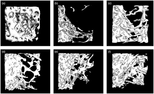
The quantitative results of distal femur Micro-CT evaluation were expressed as BV/TV, Tb.N, Tb.Sp, Tb.Th and SMI in and . The distal femur morphometric parameters analysis indicated that OVX significantly decreased trabecular BV/TV, Tb.N and Tb.Th (p < 0.01), compared with the SHAM group. In contrast, Tb.Sp and SMI (p < 0.01) in the distal femur were significantly increased in response to OVX compared with the SHAM group. Treatment with DZW or E2 reversed these indices significantly compared with the OVX group. The 3D Micro-CT images () also demonstrated the preventive effects of DZW on trabecular bone mass and microarchitecture deterioration. Compared with the SHAM group, the trabecular number and the area in the OVX group showed obvious reduction. The 12-week treatment of DZW and E2 partially prevented OVX-induced bone loss, and conspicuously promoted trabecular bone mass and prevented microarchitecture damage (p < 0.05).
Figure 2. Representative Micro-CT images of trabecular bone microarchitecture in the distal femurs. (a) SHAM group, (b) OVX group, (c) E2 group, (d) DZW-L group, (e) DZW-M group and (f) DZW-H group. The OVX rats presented notable reduction in the trabecular number, trabecular area compared with the SHAM rats. DZW and E2 partially prevented OVX-induced trabecular bone loss and significantly improved trabecular bone mass and microarchitecture.
Biomechanics
The biomechanical three-point bending experiment results are shown in . Twelve weeks of OVX resulted in oestrogen deficiency in OVX rats. Therefore, biomechanical parameters such as stiffness, ultimate load and energy absorption showed a significant decline compared with the SHAM group (p < 0.01). DZW treatment for 12 weeks improved the levels of stiffness, ultimate load and energy absorption (p < 0.01), which represented parameters of bone mechanical strength, similar to the E2 group. However, there was no significant difference among DZW groups.
Immunohistochemistry of ER
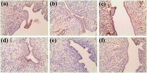
We examined the ER levels in the endometrium after the 12-week treatment using a fixed amount of E2 and three doses of DZW. The nuclei of all the intact endometrial epithelial cells were labelled. Compared with the SHAM group, an obvious ER labelling reduction was observed in the OVX group. After 12 weeks of treatment with E2 and DZW, all the treated groups showed enhanced ER labelling (). The three DZW-treated groups evidently improved ER expression by 65.83, 107.35 and 132.91%, compared with the OVX group, which was similar to the E2 group that increased by 328.79%.
Figure 3. Representative ER expression of uterus in the endometrium. (a) SHAM group, (b) OVX group, (c) E2 group, (d) DZW-L group, (e) DZW-M group and (f) DZW-H group. The OVX rats presented notable reduction of ER labelling compared with the SHAM rats. DZW and E2 partially prevented OVX-induced ER expression reduction.
Discussion
Osteoporosis is a disorder in which loss of bone mass and strength leads to fragility fractures. Oestrogen deficiency is a causative factor in the pathogenesis of osteoporosis (Dalle et al. Citation2001; Deal Citation2009). The ovariectomized (OVX) rat is a validated and widely used experimental model of post-menopausal osteoporosis, mirroring bone loss in humans. Ovariectomy resulted in increased bone turnover and marrow cavity of tibial diaphysis, and decreased cortical area within several weeks (Ishihara et al. Citation1999). These features are attributed to bone resorption on the endocortical surface, leading to bone loss (Turner et al. Citation1987; Ke et al. Citation1993). In the present study, we assessed the effect of DZW in preventing bone loss in OVX rats and used E2, with a higher biological efficacy, as a positive control for representing oestrogenic activity on bone modelling and remodelling. We demonstrated that DZW prevented bone loss in ovariectomized rats by increasing bone mineral density, mechanical properties and attenuated the microarchitectural deterioration of femora.
OCN are widely accepted phenotype markers for bone formation, while TRACP-5b serves a similar role in monitoring bone resorption. The present study shows that 12 weeks of treatment with DZW induces significant increase in bone turnover markers OCN and decrease in TRACP-5b level. These results may indicate the potential protective role of DZW by increasing bone formation and reducing bone resorption in OVX rats. After ovariectomy, serum E2 levels are markedly decreased in the OVX rats compared with SHAM group rats. In our study, DZW-treated groups and the E2 group improved serum E2 levels in OVX rats compared with the SHAM group.
BMD represents bone quality and strength (Bouxsein Citation2003). Compared with the SHAM group, BMD is notably decreased due to increased bone turnover in the OVX rats. In this study, the results showed that ovariectomy resulted in significant BMD reduction in femur after 12 weeks. Orally administrated doses of DZW increased femur BMD in a dose-dependent manner, compared with the OVX group. Additionally, bone mechanical tests are also necessary to evaluate the efficacy of treatment on quality of trabecular bone, which is more readily lost in this animal model. The three-point bending test results further support the BMD findings, evidenced by dose-dependent improvement in bone mechanical properties such as ultimate load (N), stiffness (N/mm) and energy absorption (mJ).
BMD is a vital indicator of bone strength and quality. However, the architectural changes occurring in trabecular bone were overlooked (Kleerekoper et al. Citation1985; Snyder et al. Citation1993). Clinically, the trabecular bone microarchitecture is considered as an acceptable predictor of bone loss and structural deterioration in osteoporosis (Chappard et al. Citation2008). Micro-CT, as a new high resolution digital imaging technique equipped with statistical software, has recently been extensively used to provide detailed quantitative non-destructive analysis of 3D microscopic bone architecture in non-clinical studies (Sran et al. Citation2007). Our data show that ovariectomy resulted in decreased distal femoral trabecular BV/TV, Tb.N and increased Tb.Th decreasing and SMI and Tb.Sp, which confirms an earlier study (Qi et al. Citation2012). Daily administration of DZW for 12 weeks reversed the effects of OVX and prevented further deterioration of bone structure, but the effects were not significant enough to restore the trabecular bone, consistent with previous studies (Laib et al. Citation2001).
The protective mechanism(s) of DZW on inhibition of bone loss may be mediated by the oestrogen receptor (ER)-dependent activation of oestrogen. Consequently, we determined the ER levels in the rat endometrium after administration of fixed doses of E2 and three different doses of DZW for 12 weeks. The result showed that DZW treatment obviously increased ER expression in the endometrium similar to the E2-treated group demonstrating that DZW prevented OVX induced rats bone mass loss, at least partially. Our previous HPLC results suggested that the DZW fraction contented mostly flavonoids, lignans and iridoid glycosides, with oestrogen-like effects distinct from the classical actions of oestrogen (Liu et al. Citation2013; Resende et al. Citation2013; Xiao et al. Citation2014). Therefore, daily administration of DZW for 12 weeks also improved serum E2 levels in this study. However, the molecular mechanism(s) underlying prevention of bone mass and preservation of bone quality by DZW following ovariectomy requires further investigation.
In conclusion, our data show that daily oral administration of DZW for 12 weeks in the adult female OVX rats prevented oestrogen deficiency-induced bone loss, and also increased OCN and E2 levels, decreased bone turnover markers TRACP-5b levels and preserved bone strength and quality. DZW treatment also resulted in the prevention of trabecular microarchitecture deterioration and maintenance of bone biomechanical properties. Further, we found that treatment with 6.0 g/kg/d of DZW was more effective in increasing the BMD of the femur than the other two doses of DZW. DZW can be prepared easily, conveniently and cost-effectively, suggesting a clinical advantage as an alternative to oestrogen therapy for preventing postmenopausal osteoporosis. Studies are required to (1) identify the bioactive compounds associated with the DZW bone-protective effects in vivo; and (2) elucidate the molecular mechanism(s) of action underlying the therapeutic activity of DZW.
Declaration of interest
This research was supported by the Natural Science Foundation of Jiangsu province (No. BK20140674), the National Natural Science Fund (No. 81403080) and the Fundamental Research Funds for the Central Universities. The author have no conflict of interest in this research.
References
- Board P-E, editor. 2010. Pharmacopoeia of the People's Republic of China. Beijing, China: Chemical Industry Press.
- Bouxsein M-L. 2003. Mechanisms of osteoporosis therapy: a bone strength perspective. Clin Cornerstone. Suppl 2:S13–S21.
- Chappard D, Basle M-F, Legrand E, Audran M. 2008. Trabecular bone microarchitecture: a review. Morphologie 92:162–170.
- Dalle C-L, Arlot M-E, Chavassieux P-M, Roux J-P, Portero N-R, Meunier P-J. 2001. Comparison of trabecular bone microarchitecture and remodeling in glucocorticoid-induced and postmenopausal osteoporosis. J Bone Miner Res. 16:97–103.
- Davison S, Davis S-R. 2003. Hormone replacement therapy: current controversies. Clin Endocrinol (Oxford). 58:249–261.
- Deal C. 2009. Potential new drug targets for osteoporosis. Nat Clin Pract Rheumatol. 5:20–27.
- Hou X, Lv Z-Q, Zhao C-Z, Lv W-H. 2010. The analysis of three fractions between Du-Zhong-Wan stir-fried with saltwater and compatibility. Chinese Tradit Patent Med. 32:1442–1444.
- Ishihara A, Sasaki T, Debari K, Furuya R, Kawawa T, Ramamurthy N-S, Golub L-M. 1999. Effects of ovariectomy on bone morphology in maxillae of mature rats. J Electron Microsc (Tokyo) 48:465–469.
- Ke H-Z, Jee W-S, Zeng Q-Q, Li M, Lin B-Y. 1993. Prostaglandin E2 increased rat cortical bone mass when administered immediately following ovariectomy. Bone Miner 21:189–201.
- Kleerekoper M, Villanueva A-R, Stanciu J, Rao D-S, Parfitt A-M. 1985. The role of three-dimensional trabecular microstructure in the pathogenesis of vertebral compression fractures. Calcif Tissue Int. 37:594–597.
- Ko Y-J, Wu J-B, Ho H-Y, Lin W-C. 2012. Antiosteoporotic activity of Davallia formosana. J Ethnopharmacol. 139:558–565.
- Laib A, Kumer J-L, Majumdar S, Lane N-E. 2001. The temporal changes of trabecular architecture in ovariectomized rats assessed by Micro-CT. Osteoporos Int. 12:936–941.
- Li F, Yang Y-N, Zhu P-P, Chen W-N, Qi D-L, Shi X-P, Zhang C-F, Yang Z-L, Li P. 2012. Echinacoside promotes bone regeneration by increasing OPG/RANKL ratio in MC3T3-E1 cells. Fitoterapia 83:1443–1450.
- Liu H-W, Yu X-Z, Padula D, Pescitelli G, Lin Z-W, Wang F, Ding K, Lei M, Gao J-M. 2013. Lignans from Schisandra sphenathera Rehd. et Wils. and semisynthetic schisantherin A analogues: absolute configuration, and their estrogenic and anti-proliferative activity. Eur J Med Chem. 59:265–273.
- Persson I, Weiderpass E, Bergkvist L, Bergstrom R, Schairer C. 1999. Risks of breast and endometrial cancer after estrogen and estrogen-progestin replacement. Cancer Causes Control 10:253–260.
- Qi W, Yan Y-B, Lei W, Wu Z-X, Zhang Y, Liu D, Shi L, Cao P-C, Liu N. 2012. Prevention of disuse osteoporosis in rats by Cordyceps sinensis extract. Osteoporos Int. 23:2347–2357.
- Resende F-A, de Oliveira A-P, de Camargo M-S, Vilegas W, Varanda E-A. 2006. Osteoporosis: a still increasing prevalence. Bone 38:S4–S9.
- Resende F-A, de Oliveira A-P, de Camargo M-S, et al. 2013. Evaluation of estrogenic potential of flavonoids using a recombinant yeast strain and MCF7/BUS cell proliferation assay. PLoS One 8:e74881.
- Snyder B-D, Piazza S, Edwards W-T, Hayes W-C. 1993. Role of trabecular morphology in the etiology of age-related vertebral fractures. Calcif Tissue Int. 53 Suppl 1:S14–S22.
- Sran M-M, Boyd S-K, Cooper D-M, Khan K-M, Zernicke R-F, Oxland T-R. 2007. Regional trabecular morphology assessed by micro-CT is correlated with failure of aged thoracic vertebrae under a posteroanterior load and may determine the site of fracture. Bone 40:751–757.
- Turner R-T, Vandersteenhoven J-J, Bell N-H. 1987. The effects of ovariectomy and 17 beta-estradiol on cortical bone histomorphometry in growing rats. J Bone Miner Res. 2:115–122.
- Xiao H-H, Fung C-Y, Mok S-K, Wong K-C, Ho M-X, Wang X-L, Yao X-S, Wong M-S. 2014. Flavonoids from Herba epimedii selectively activate estrogen receptor alpha (ER-alpha) and stimulate ER-dependent osteoblastic functions in UMR-106 cells. J Steroid Biochem Mol Biol. 143:141–151.
- Zhang R, Hu S-J, Li C, Zhang F, Gan H-Q, Mei Q-B. 2012. Achyranthes bidentata root extract prevent OVX-induced osteoporosis in rats. J Ethnopharmacol. 139:12–18.