
Abstract
Context Despite the reported anticarcinogenic activity of lophirones B and C, no scientific information exists for its activity in rat hepatocytes.
Objective Effect of lophirones B and C on aflatoxin B1 (AFB1)-induced oxidative stress, and DNA fragmentation in rat hepatocytes was investigated.
Materials and methods Wistar rat hepatocytes were incubated with lophirones B and C (1 mg/mL) or sylimarin (1 mg/mL) in the presence or absence of AFB1. For an in vivo study, rats were orally administered with lophirones B and C, and/or AFB1 (20 μg/d) for 9 weeks.
Results Lophirones B and C lowered AFB1-mediated increase in nitric oxide, superoxide anion radicals, caspase-3 and fragmented DNA. Lophirones B and C attenuated AFB1-mediated decrease in superoxide dismutase, catalase, glutathione peroxidase, glutathione reductase, and reduced glutathione. Also, lophirones B and C attenuated AFB1-mediated increase in conjugated dienes, lipid hydroperoxides and malondialdehyde in rat hepatocytes. Furthermore, AFB1-mediated alterations in alkaline phosphatase, alanine aminotransferase, aspartate aminotransferase, albumin, total bilirubin and globulin in rat serum were significantly annulled in lophirones B and C-treated rats.
Conclusion This study revealed that lophirones B and C prevented AFB1-induced oxidative damage in rat hepatocytes.
Introduction
Aflatoxin B1 (AFB1), a mutagenic food contaminant, is widely recognized as one of the most potent hepatocarcinogens in humans and experimental animals (Josse et al. Citation2012). It is responsible for the increased liver cancer, growth retardation, decreased immunity and vaccination failure in most African countries (Bankole et al. Citation2006). AFB1 is activated in the liver by cytochrome P450 2E1 to AFB1-8,9-epoxide, which forms adducts with both DNA and protein (Scholl et al. Citation1997). The genetic toxicity of AFB1 is partly due to the accumulation of reactive oxygen species (ROS) such as superoxide anion radical, hydrogen peroxide and hydroxyl radical (Shen & Ong Citation1996). Studies have shown that AFB1 alters cell cycle and apoptosis-signalling pathways in liver cell models (Ellinger-Ziegelbauer et al. Citation2004; Li et al. Citation2004; Tilton et al. Citation2005; Josse et al. Citation2012). In particular, the p53-related genes are upregulated, leading to the induction of target genes involved in cell-cycle arrest, apoptosis and stimulation of DNA repair activity (Jennen et al. Citation2010; Josse et al. Citation2012).
Several studies have shown that dietary antioxidants, widely distributed in cereals, fruits, vegetables and medicinal plants, halt oxidative stress and apoptosis in AFB1-induced hepatocarcinogenesis (Rastogi et al. Citation2000, Citation2001). Thus, the wise use of fruits, medicinal plants and vegetables requires investigations into the phytochemicals and antioxidants as well as possible medicinal properties and prospective products, such as nutraceuticals and phytomedicines (Oloyede et al. Citation2013).
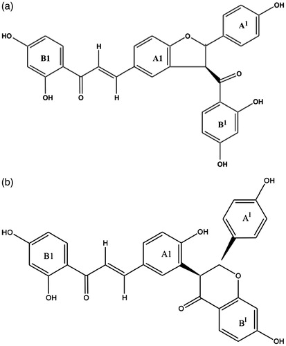
Lophirones B and C () are a member of numerous chalcone dimers found in Lophira alata (Ochnaceae) Van Tiegh. Ex Keay stem bark. Lophirones B and C inhibit 4-nitro-o-phenylenediamine and benzo[a]pyrene-induced frame shift mutation in TA98 and AFB1-induced base pair substitution in TA100 (Ajiboye et al. Citation2014a). It also inhibits proliferation of Ehrlich ascite carcinoma cell (Ajiboye et al. Citation2014b). The presence of α,β-conjugated double bonds, hydroxyl groups at C2′ and C4′ in ring A and at C4 of ring B, in lophirones B and C, is responsible for the inhibiting proliferation of Ehrlich ascite carcinoma cells (Ajiboye et al. Citation2014a). In addition, the chalcone dimers possess free radical and ROS (hydroxyl radical, hydrogen peroxide and superoxide anion radical) scavenging properties (Ajiboye et al. Citation2014a). Also, lophirones B and C enhance the expressions and activities of ROS and electrophilic species through the nuclear erythroid-related factor-2 (Nrf-2) (Ajiboye et al. Citation2014b). These enhanced expressions and activities prevent oxidative stress, lipid peroxidation and DNA fragmentation in AFB1-induced hepatocarcinogenesis (Ajiboye et al. Citation2014c).
Further to understanding the mechanism by which lophirones B and C mitigate against AFB1 carcinogenesis, we investigated the effects of lophirones B and C in AFB1-induced apoptosis, oxidative stress and DNA fragmentation in rat hepatocytes.
Figure 1. (a) Lophirone B. (b) Lophirone C.
Materials and methods
Chemicals
Glutathione disulphide, ephinephrine, glucose 6-phosphate, glutathione (reduced form), 2,6-dichlorophenol-indophenol, reduced β-nitocinamide adenide dinucleotide (β-NADH), oxidized β-nicotineamide adednide dinucleotide (β-NAD+) and uridine 5′-diphosphoglucuronic acid were purchased from Sigma Chemical Co., St. Loius, MO. Glutathione peroxidase (GPx) assay kit was procured from Randox Laboratories Co-Artrim, Crumlin, UK. Caspase-3 kit was purchased from Sigma Chemical Co., St. Loius, MO. All other reagents were of analytical grade.
Plant materials
Lophira alata stem bark was collected in Chaza village, Suleja, Niger State. The plant sample was authenticated and deposited in the Herbarium unit of Medicinal Plant Research Department, National Institute of Pharmaceutical Research and Development, Idu-Abuja, Nigeria, with voucher no. 4534
Plant processing, extraction and isolation of lophirones B and C
The extraction and the isolation of lophirones B and C from Lophira alata stem bark were done according to the procedure reported by Ajiboye et al. (Citation2014a,Citationb). Briefly, plant materials were chopped and dried under shade. The dried plant materials were thereafter weighed, extracted in methanol for 96 h and concentrated to give 50.03 g, which was re-extracted in ethyl acetate to yield 24.22 g. The ethyl acetate extract was subjected to thin-layer chromatography (TLC) using dichloromethane:methanol (10:1) (a solvent system that produced a distinct separation of lophirones B, C, D and E). The ethylacetate extract was thereafter subjected to dry column chromatography using the same solvent system for the TLC, with the adsorbent as silica gel. The separated bands were sliced after developing the dry column chromatography and eluted with acetone. The structures of isolated compounds were elucidated with 1H and 13C NMR spectra. Nuclear magnetic resonance (1H NMR, 13C NMR and spin echo Fourier transform) spectra were recorded on a Bruker-AMX 400 instrument (Bruker Daltonics Inc., Billerica, MA) using TMS as a solvent and an internal standard, respectively.
Isolation of rat hepatocytes
Rat hepatocytes were isolated following the procedures described by Fry (Citation1981). Briefly, male albino rat (210 g) was sacrificed by cervical dislocation and liver excised. A portion of excised liver tissue (2 g) was minced with clean razor blades to thin slices. The minced tissue was mixed and shaken with Dulbecco’s phosphate buffer saline, containing glucose (1 mg/mL), 37 °C for 10 min (90 oscillations per min). The solution was removed with Pasteur pipette. This washing stage is repeated twice and serves to remove debris, RBCs and some connective tissues from the slices. Calcium ions were removed from the minced tissue by exposing the washed slices to 0.5 mM ethylene glycol-bis(β-aminoethyl ether) N,N′-tetraacetic acid (EGTA) in two 10 min cycles in order to aid cell dissociation. The solution present after the second cycle is carefully removed and replaced with 10 mL of the enzyme solution (Hanks buffer containing 5 mg collagenase, 10 mg hyaluronidase, 250 mM CaCl2, 1 M NaHCO3 and 100 mg/mL glucose). Then the solution was shaken at 37 °C for up to 1 h. When the slices appeared well digested (usually after 45–60 min of incubation), the resulting cell suspension was filtered through a muslin cloth. Then the filtered cell solution was centrifuged at 1000 g for 5 min, the supernatant fluid was removed and the remaining cell pellet washed twice with Dulbecco’s phosphate buffer saline, containing glucose (1 mg/mL).
In vitro experimental treatment
The rat hepatocytes were incubated in a HEPES-buffered salt solution, pH 7.45 (NaC1, 8.00 g; KC1, 0.40 g; KH2PO4, 0.06 g; Na2HPO4ċ12H2O, 0.125 g; CaCI2ċ2H2O, 0.185 g; MgSO4ċ7H2O, 0.246 g; HEPES (free acid), 4.76 g; glucose, 1.0 g; 5 N NaOH, 2 ml; all dissolved in 1 L of distilled water). The hepatocytes were incubated with or without AFB1 and chalcone dimers (lophirones B and C) as follows:
Treatment 1: 0.25% DMSO
Treatment 2: 9 nM AFB1
Treatment 3: 1 mg/mL lophirone B
Treatment 4: 1 mg/mL lophirone C
Treatment 5: 9 nM AFB1 + 1 mg/mL lophirone B
Treatment 6: 9 nM AFB1 + 1 mg/mL lophirone C
Treatment 7: 9 nM AFB1 + 1 mg/mL sylimarin
Treatments were done in triplicate and the incubation was done after 4 h incubation period. The concentration (1 mg/mL) was chosen based on the previous report by Ajiboye et al. (Citation2014a), while the cell viability of AFB1 (9 nM) was reported by Ribeiro et al. (Citation2010).
Animal treatment
Thirty male rats were randomly grouped into six (A–F) of five rats each. The rats in group A served as the control and received the vehicle, 0.5% dimethyl sulphoxide (DMSO). Animals in groups B, E and F received aflatoxin B1 (20 μg/d) in 0.5% DMSO through gavage starting from the third week until the end of the experimental period (6 weeks). Rats in groups C and E received 20 mg/kg body weight of lophirone B for 6 weeks, while those in groups D and F were treated with 20 mg/kg bodyweight of lophirone C for the same periods. The rats were sacrificed 24 h after the last dose, and liver from individual animals was used for isolation of subcellular fractions. The animals were used according to the NIH Guide for the Care and Use of Laboratory Animals (National Research Council (NRC) Citation2010) following approval by Al-Hikmah University Ethical Committee on the Use of Laboratory Animals (HUI/ECULA/013/17/003).
Biochemical assays
Proapoptotic indexes
The level of nitric oxide (NO) in rat hepatocytes was determined using the colorimetric method based on the Griess reaction (Green et al. Citation1982). The concentration of intracellular superoxide radical anion in rat hepatocytes was measured using the procedure described by Ajiboye et al. (Citation2016). Briefly, hepatocytes (0.5 mL) were incubated with 2.5 mL nitroblue tetrazolium (1 mg/mL) for 30 min at 37 °C. Then 0.5 mL of 0.1 M HCl was added. The blue colour was read at 575 nm. The amount of superoxide radical anion generated was calculated using the molar extinction coefficient of 3-(4,5-dimethylthiazol-2-yl)-2,5-diphenyltetrazolium bromide formazan (17 000 M − 1 cm − 1 at pH 7.4–8.0). The activity of caspase-3 was determined based on the procedure outlined in Sigma assay kit (Sigma Chemical Co., St. Loius, MO) for caspase-3, colorimetrically using Molecular Devices Vmax kinetic microplate reader (Molecular Devices Inc., Sunnyvale, CA).
Fragmented DNA
The quantity of fragmented DNA in the hepatocytes was determined using the procedure described by Burton (Citation1956). Briefly, hepatocytes were centrifuged at 15 000 g, for 15 min at 4 °C. The supernatant was separated from the pellet and treated with trichloroacetic acid (1.50 mL, 10%). The pellet was also treated with trichloroacetic acid (0.65 mL, 5%). The reaction mixtures were allowed to precipitate overnight (≥ 4 h) in a refrigerator (4 °C), centrifuged at 2500 g for 10 min. The reaction mixtures were boiled at 100 °C for 15 min, cooled to room temperature and centrifuged at 2500 g for 5 min. The supernatants (0.5 mL) were treated with diphenylamine reagent (1 mL) and incubated at 37 °C for 4 h. The absorbance was read at 600 nm using spectrophotometer. The fragmented DNA was calculated using the following expression:
(1)
Superoxide dismutase (SOD)
The activity of SOD was determined as described by Misra and Fridovich (Citation1972). Briefly, 0.2 mL of hepatocyte cells was added to 2.5 mL of 0.05 M carbonate buffer (pH 10.2) to equilibrate and the reaction was started by addition of 0.3 mL of freshly prepared 0.3 mM epinephrine. An increase in the absorbance was recorded at 480 nm every 30 s for 150 s. One unit of enzyme activity is 50% inhibition of the rate of auto-oxidation of pyrogallol as determined by the change in absorbance/min at 480 nm.
Catalase
Catalase activity was determined using the method described by Aebis (Citation1984). Hepatocytes suspension (50 μL) was added to a cuvette containing 2 mL of phosphate buffer (pH 7.0) and 1 mL of 30 mM H2O2. Catalase activity is measured at 240 nm for 1 min using a spectrophotometer. The molar extinction coefficient of H2O2, 43.6 M cm − 1, was used to determine the catalase activity.
Glutathione peroxidase and glutathione reductase
The activities of glutathione peroxidase and glutathione reductase were determined using the procedure outlined in commercial kits (Randox Laboratories Ltd., Antrim, UK).
Reduced glutathione (GSH) and glutathione disulphide (GSSG)
The level of GSH in the hepatocytes suspension was determined using the procedure described by Ellman (Citation1959). Briefly, 1.0 mL of hepatocytes was added to 0.1 mL of 25% trichloroacetic acid (TCA) and the precipitate was removed by centrifuge at 5000 g for 10 min. Supernatant (0.1 mL) was added to 2 mL of 0.6 mM DTNB prepared in 0.2M sodium phosphate buffer (pH 8.0). The absorbance was read at 412 nm.
GSSG level was determined using the procedure described by Hissin and Hilf (Citation1976). Hepatocytes (50 μL) were mixed with 20 μL of 0.04 M N-ethylmaleimide (NEM) to prevent the oxidation of GSH to GSSG. It was incubated at room temperature for 30 min and 1.68 mL of 0.3 M Na2HPO4 solution was added to it followed by 250 μl of DTNB reagent. The absorbance of the sample was measured at 412 nm.
Lipid peroxidation products and protein carbonyl
The levels of conjugated dienes, lipid hydroperoxides and malondialdehyde were assayed according to Reilly and Aust (Citation2001).
Conjugated dienes
Briefly, hepatocytes suspension was mixed with 5 mL, 2:1 (v/v) chloroform/methanol, and centrifuged at 1000 g for 10 min at 4 °C, to separate the aqueous layer from the organic layer. The chloroform layer containing the lipids was separated from the aqueous layer and evaporated to dryness at 45 °C. The lipid residue was re-suspended in 1.5 mL cyclohexane and the absorbance read at 233 nm against a blank containing only cyclohexane using Simtronic microprocessor UV/VIS spectrophotometer, model 807 (Simmtronics Infotech Pvt. Ltd., Delhi, India). Conjugated dienes was estimated using 2.52 × 10⁴ extinction coefficient.
Lipid hydroperoxides
Hepatocytes suspension was mixed with an equal volume of EDTA (1 mM). The resulting mixture was added to 5 mL of chloroform/methanol (2:1; v/v) and centrifuged at 1000 g for 10 min at 4 °C, to separate the aqueous layer from the organic layer. The chloroform layer containing the lipids was evaporated to dryness at 45 °C. The lipid residue was re-suspended in 1 mL of acetic acid/chloroform (3:2), mixed with 0.05 mL of 1.2 g/mL potassium iodide and incubated for 5 min in the dark at room temperature. Cadmium acetate (0.5%; 3 mL) was added, mixed and centrifuged at 1000 g for 10 min at room temperature. The absorbance of the upper aqueous phase was read at 353 nm versus that of the extracted blank (containing all components except liver homogenate) using Simtronic microprocessor UV/VIS spectrophotometer, MODEL 807 (Simmtronics Infotech Pvt. Ltd., Delhi, India). Lipid hydroperoxides were quantified using the extinction coefficient of 1.73 × 10⁶.
Malondialdehyde
Briefly, hepatocytes suspension was mixed with TBA/TCA/HCl (15%, 0.2 N, 0.37%) at a reagent/sample ratio of 2:1 (v/v), placed in a boiling water bath for 15 min, cooled to room temperature and centrifuged at 1000 g for 10 min at room temperature. The absorbance of the solution was read at 535 nm against the blank (containing all reagents except hepatocytes suspension) using Simtronic microprocessor UV/VIS spectrophotometer, MODEL 807 (Simmtronics Infotech Pvt. Ltd., Delhi, India). MDA content was determined using the extinction coefficient of 1.56 × 10⁶.
Protein carbonyl
Protein carbonyl concentration was determined according to the procedure described by Levine et al. (Citation1990). Briefly, hepatocytes suspension (100 μL) was mixed with an equal volume of 10 mM 2,4-dinitrophenylhydrazine (DNPH) prepared in 2.5 M HCl, and incubated in the dark for 1 h at room temperature, with intermittent shaking at every 15 min. After 1 h of incubation, TCA (10% w/v) was added and centrifuged at 3500 g for 20 min. The pellet was washed three times with ethanol/ethyl acetate (1:1 v/v) to remove unreacted DNPH and lipid remnants. The pellet was dissolved in 2 mL of 6 M guanidine hydrochloride and incubated at 37 °C for 10 min, centrifuged at 1000 g for 10 min and the carbonyl content was determined by reading the absorbance of the samples at 370 nm using Simtronic microprocessor UV/VIS spectrophotometer, MODEL 807 (Simmtronics Infotech Pvt. Ltd., Delhi, India). The carbonyl content (μmol/mg protein) was estimated using an absorption coefficient of 22 000 M − 1 cm − 1. Blank was treated the same way except that DNPH was replaced with 2.5 M HCl.
Liver function indices
The activities of alkaline phosphatase (ALP), alanine aminotransferase (ALT) and aspartate aminotransferase were determined spectrophotometrically using the procedures described by Wright et al. (Citation1972) and Bergmeyer et al. (Citation1986a,Citationb). The serum concentrations of albumin, total bilirubin and globulin were estimated using procedure outlined in commercial assay kits (Randox Laboratories Ltd., Antrim, UK).
Statistical analysis
Analysis of variance (ANOVA) followed by the Tukey–Kramer test was used to detect any significant differences among different means as well as the interactions between the variables used in this study using StatPlus, 2011 (SAS Inc., Cary, NC). Differences were considered statistically significant at p < 0.05.
Results
Proapoptotic factors and fragmented DNA
The levels of superoxide anion radical and NO significantly increased (p < 0.05) in AFB1-treated hepatocytes when compared with the control. Lophirones B and C completely attenuated AFB1-mediated increase in superoxide anion radical by 0.65- and 0.75-fold and nitric oxide by 0.80 and 0.84-fold, respectively (). This was compared significantly with the control and the sylimarin-treated group (). Conversely, there was 0.96- and 0.97-fold, and 0.80- and 0.81-fold attenuation of AFB1-mediated increase in caspase-3 and fragmented DNA (%) by lophirones B and C, respectively ().
Table 1. Levels of apoptotic index in rat hepatocytes incubated with aflatoxin B1 and lophirones B and C.
Enzymatic and non-enzymatic antioxidants
Treatment of hepatocytes with AFB1 resulted to 74.10, 82.89, 69.74 and 62.16% decrease in the activities of superoxide dismutase, catalase, glutathione peroxidase and glutathione reductase, respectively. Lophirones B and C significantly reversed AFB1-mediated decrease in the activities of these enzymes by 90.43 and 106.84, 82.54 and 95.29, 96.27 and 102.46, and 75.57 and 100%. These values compared significantly with the control and the sylimarin-treated group (). In addition, AFB1 significantly lowered the level of reduced glutathione with corresponding increase in glutathione disulphide in the hepatocytes (). Lophirones B and C significantly annulled AFB1-mediated alterations in glutathione (reduced and disulphide) compared significantly (p > 0.05) with control and sylimarin treatments ().
Table 2. Activities of antioxidant enzymes in rat hepatocytes incubated with aflatoxin B1 and lophirones B and C.
Table 3. Levels of non-enzymatic antioxidants and lipid peroxidation products in rat hepatocytes incubated with aflatoxin B1 and lophirones B and C.
Lipid peroxidation products
Levels of lipid peroxidation products, i.e., conjugated dienes, lipid hydroperoxides and malondialdehyde in AFB1-treated hepatocytes increased significantly by 56.56, 72.45 and 89.63%, respectively, when compared with the control treatment (). AFB1-mediated increase in these products was significantly (p < 0.05) reversed by lophirones B and C and compared significantly with the sylimarin treatment group ().
Liver function indices
The serum activities of ALP, ALT and AST increased significantly (p < 0.05) in AFB1-treated rats by 85.37, 85.01 and 77.86%, respectively, when compared with the control. Although lophirones B and C produced no significant change in the activities of these enzymes, a significant attenuation of AFB1-mediated increase in these enzymes was produced following the administration of lophirones B and C to AFB1-treated rats (). The attenuation of AFB1-mediated increase in these enzymes compared significantly (p < 0.05) with the control and the sylimarin-treated group ().
Table 4. Liver function indices in the serum of aflatoxin B1-treated rats following oral administration of lophirones B and C for 1 week.
Albumin and globulin levels in the serum of AFB1-treated rats decreased significantly (p < 0.05) by 2.94- and 1.82-fold, while there was 9.24-fold increase in total bilirubin (). Lophirones B and C significantly (p < 0.05) attenuated AFB1-mediated alterations (). This attenuation was compared significantly (p < 0.05) with the control and the sylimarin-treated group ().
Discussion
Apoptosis
Uncontrolled ROS production has been implicated in AFB1-mediated genetic damage and cell death (Eaton & Gallagher Citation1994; Janicke Citation1998). Peroxynitrite anion, product of superoxide anion and nitric oxide, is proapototic, damage mitochondria, depletes GSH and damages DNA (Pryor & Squadrito Citation1995; Meki et al. Citation2001). The elevated levels of these reactive species in AFB1-treated hepatocytes could enhance the expression of enzymes and transcription factors involved in apoptosis leading to uncontrolled oxidative attack and cell death (Kolb Citation2000). The reversal of AFB1-mediated increase in these reactive species may be attributed to the antioxidant properties of lophirones B and C (Ajiboye et al. Citation2014a, Citation2014b), affirming the protective importance of these chalcones.
Caspase-3 activation in AFB1 intoxication has been linked to ROS production leading to apoptosis and cell death (Duan et al. Citation2005; Van Vleet et al. Citation2006). The increase in caspase-3 activity as observed in this study could be due to the elevated levels of NO and superoxide anion radical, suggesting that apoptosis is one of the mechanisms by which AFB1 mediates cell death. The reversal in caspase-3 activity shows the capability of lophirones B and C to prevent cellular apoptosis (Reddy et al. Citation2006). Similarly, the reversal of increased fragmented DNA indicates stalled apoptosis, possibly due to the antioxidant properties of the chalcone dimers (Ajiboye et al. Citation2014a). Although how it regulates apoptosis is yet to be known, lophirones B and C evidently regulate apoptosis, possibly by modulating ROS generation.
Oxidative stress
The activities of antioxidant enzymes provide valuable insight to the biochemical mechanism of toxicity of drugs and chemical substances (Ajiboye et al. Citation2010). Decrease in the activities of antioxidants enzymes as observed in this study is prominent in AFB1 carcinogenesis/toxicity (Souza et al. Citation1999; Meki et al. Citation2001, Citation2004; Ajiboye et al. Citation2013; Adeleye et al. Citation2014). The AFB1-mediated decrease in the activities of these enzymes in rat hepatocytes, attributed to overwhelmed antioxidant enzymes, indicates oxidative stress arising from ROS (superoxide anion and nitric oxide) generated. The attenuation of AFB1-mediated decrease in the activities of these enzymes (SOD, CAT, GSH-Px and GSH-red) in rat hepatocytes shows the capability of lophirones B and C to prevent oxidative stress possibly by enhancing Nrf-2 expression (Ajiboye et al. Citation2014c). Meki et al. (Citation2004) reported similar reversal of AFB1-mediated decrease in antioxidant enzymes following treatment of rats with melatonin. Reduction in the hepatocytes GSH level may be linked to overwhelmed antioxidant enzymes. Accumulation of GSSG in rat hepatocytes indicates cytotoxic effect of AFB1. This could lead to oxidative damage in the hepatocytes as well as initiates apoptosis (Sanchez et al. Citation1997). The reversal of alterations in hepatocytes levels of GSH and GSSG by lophirones B and C shows cytoprotection.
Elevated levels of lipid peroxidation products such as conjugated dienes, lipid hydroperoxides and malondialdehyde have been established in both in vitro and in vivo models of AFB1 toxicity (Shen et al. Citation1994; Towner et al. Citation2002; Lee et al. Citation2005; Theumer et al. Citation2010). This elevation signals perturbed membrane organization and functional loss (Niki Citation2009), which might have resulted from uncompromised free radicals and ROS productions. The reversal in the levels of conjugated dienes, lipid hydroperoxides and malondialdehyde by lophirones B and C may be attributed to antioxidants activities and capability to enhance ROS-detoxifying enzymes (Ajiboye et al. Citation2014b).
Liver function indices
ALP, ALT and AST are useful biomarkers of hepatic injury shown to be elevated in AFB1-mediated hepatotoxicity (Ajiboye et al. Citation2010; Ajiboye Citation2011; Oloyede et al. Citation2013; Adeleye et al. Citation2014; Ajiboye Citation2015). The AFB1-mediated increase in serum ALP, ALT and AST might have resulted from disruption of plasma membrane by ROS (e.g. O, O2− OH and H2O2) generated during AFB1 metabolism (Shen et al. Citation1994, Citation1995, Citation1996). The attenuation of these alterations by lophirones B and C may be due its capability to upregulate cytoprotective enzymes (Ajiboye et al. Citation2014b,Citationc), indicating the protection.
Serum levels of albumin, globulins and total bilirubin are indicators of the functional capacity of liver (Dufour et al. Citation2000). Thus, AFB1-mediated reduction in these parameters indicates hepatocellular dysfunction (Dufour et al. Citation2000), which may be due to protein loss (nephrotic syndrome, burns and protein-losing enteropathy), and liver disease (Doumas et al. Citation1997). The reversal of AFB1-mediated alterations in these liver function indices shows the protection against nephrotic syndrome, burns, protein-losing enteropathy and liver disease.
Conclusion
It is evident from this study that lophirones B and C attenuated AFB1-induced oxidative stress and DNA fragmentation in rat hepatocytes possibly by enhancing the activities of antioxidant enzymes. This was corroborated by the attenuation of alterations in makers of hepatic damage in AFB1-treated rats.
Funding information
The authors received no financial support for the research, authorship, and/or publication of this article.
Disclosure statement
The authors declared no potential conflicts of interest with respect to the research, authorship, and/or publication of this article.
References
- Adeleye AO, Ajiboye TO, Iliasu GA, Abdussalam FA, Balogun A, Ojewuyi OB, Yakubu MT. 2014. Phenolic extract of Dialium guineense pulp enhances reactive oxygen species detoxification in aflatoxin B1 hepatocarcinogenesis. J Med Food. 17:875–885.
- Aebi H. 1984. Catalase in vitro. Methods Enzymol. 105:121–126.
- Ajiboye TO, Yakubu MT, Salau AK, Oladiji AT, Akanji MA, Okogun JI. 2010. Antioxidant and drug detoxification potential of aqueous extract of Annona senegalensis leaves in carbon tetrachloride-induced hepatocellular damage. Pharm Biol. 48:1361–1370.
- Ajiboye TO. 2011. In vivo antioxidant potentials of Piliostigma thonningii (Schum) leaves: studies on hepatic marker enzyme, antioxidant system, drug detoxifying enzyme and lipid peroxidation. Human Exp Toxicol. 30:55–62.
- Ajiboye TO. 2015. Standardized extract of Vitex doniana Sweet stalls protein oxidation, lipid peroxidation and DNA fragmentation in acetaminophen-induced hepatotoxicity. J Ethnopharmacol. 164:273–282.
- Ajiboye TO, Salau AK, Yakubu MT, Oladiji AT, Akanji MA, Okogun JI. 2010. Aqueous extract of Securidaca longepedunculata root induce redox imbalance in male rat liver and kidney. Hum Exp Toxicol. 29:679–688.
- Ajiboye TO, Raji HO, Muritala HF, Ojewuyi OB, Yakubu MT. 2013. Anthocyanin extract of Lannea microcarpa fruits stall Aflatoxin B1-mediated oxidative rout on cellular proteins, lipids and DNA. Food Bioscience. 4:58–67.
- Ajiboye TO, Naibi AM, Abdulazeez IO, Alege IO, Mohammed AO, Bello SA, Yusuf II, Ibitoye OB, Muritala HF. 2016. Involvement of oxidative stress in bactericidal activity of 2-(2-nitrovinyl) furan against Escherichia coli, Pseudomonas aeruginosa and Staphylococcus aureus. Microbial Pathogen. 91:107–114.
- Ajiboye TO, Yakubu MT, Oladiji AT. 2014a. Cytotoxic, antimutagenic, and antioxidant activities of methanolic extract and chalcone dimers (lophirones B and C) derived from Lophira alata (Van Tiegh. Ex Keay) stem bark. J Evid Based Complement Altern Med. 19:20–30.
- Ajiboye TO, Yakubu MT, Oladiji AT. 2014b. Electrophilic and reactive oxygen species detoxification potentials of chalcone dimers is mediated by redox transcription factor Nrf-2. J Biochem Mol Toxicol. 28:11–22.
- Ajiboye TO, Yakubu MT, Oladiji AT. 2014c. Lophirones B and C extenuate AFB1-mediated oxidative onslaught on cellular proteins, lipids, and DNA through Nrf-2 expression. J Biochem Mol Toxicol. 28:558–567.
- Bankole S, Schollenberger M, Drochner W. 2006. Mycotoxins in food systems in sub-Saharan Africa: a review. Mycotoxin Res. 22:163–169.
- Bergmeyer HU, Hørder M, Rej R. 1986a. International Federation of Clinical Chemistry (IFCC) Scientific Committee, Analytical Section: approved recommendation (1985) on IFCC methods for the measurement of catalytic concentration of enzymes. Part 2. IFCC method for aspartate aminotransferase. J Clin Chem Clin Biochem. 24:497–510.
- Bergmeyer HU, Hørder M, Rej R. 1986b. International Federation of Clinical Chemistry (IFCC) Scientific Committee, Analytical Section: approved recommendation (1985) on IFCC methods for the measurement of catalytic concentration of enzymes. Part 3. IFCC method for alanine aminotransferase. J Clin Chem Clin Biochem. 24:481–495.
- Burton K. 1956. A study of the conditions and mechanism of the diphenylamine reaction for the colorimetric estimation of deoxyribonucleic acid. Biochem J. 62:315–323.
- Doumas BT, Watson WA, Biggs HG. 1997. Albumin standards and the measurement of serum albumin with bromcresol green. Clin Chim Acta. 258:21–30.
- Duan XX, Ou JS, Li Y, Su JJ, Ou C, Yang C, Yue HF, Ban KC. 2005. Dynamic expression of apoptosis-related genes during development of laboratory hepatocellular carcinoma and its relation to apoptosis. World J Gastroenterol. 11:4740–4744.
- Dufour DR, Lott JA, Nolte FS, Gretch DR, Koff RS, Seeff LB. 2000. Diagnosis and monitoring of hepatic injury I. Perform Charact Lab Tests 2068:2050–68.
- Eaton DL, Gallagher EP. 1994. Mechanisms of aflatoxin carcinogenesis. Annu Rev Pharmacol Toxicol. 34:135–172.
- Ellinger-Ziegelbauer H, Stuart B, Wahle B, Bomann W, Ahr HJ. 2004. Characteristic expression profiles induced by genotoxic carcinogens in rat liver. Toxicol Sci. 77:19–34.
- Ellman GL. 1959. Tissue sulfhydryl groups. Arch Biochem Biophys. 82:70–77.
- Fry JR. 1981. Preparation of mammalian hepatocytes. Methods Enzymol. 77:130–137.
- Green LC, Wagner DA, Glogowski J, Skipper PL, Wishnok JS, Tannenbaum SR. 1982. Analysis of nitrate, nitrite, and [15N]nitrate in biological fluids. Anal Biochem. 126:131–138.
- Hissin PJ, Hilf R. 1976. A fluorometric method for determination of oxidized and reduced glutathione in tissues. Anal Biochem. 74:214–226.
- Janicke RU. 1998. Caspase-3 is required for DNA fragmentation and morphological changes associated with apoptosis. J Biol Chem. 73:9357–9360.
- Jennen DGJ, Magkoufopoulou C, Ketelslegers HB, van Herwijnen MHM, Kleinjans JCS, van Delft JHM. 2010. Comparison of HepG2 and HepaRG by whole-genome gene expression analysis for the purpose of chemical hazard identification. Toxicol Sci. 115:66–79.
- Josse R, Dumont J, Fautrel A, Robin MA, Guillouzo A. 2012. Identification of early target genes of aflatoxin B1 in human hepatocytes, inter-individual variability and comparison with other genotoxic compounds. Toxicol Appl Pharmacol. 258:176–187.
- Kolb JP. 2000. Mechanisms involved in the pro- and anti-apoptotic role of NO in human leukemia. Leukemia. 14:1685–1694.
- Lee J, Hye E, Lee K, Sook H. 2005. Alleviation of aflatoxin B1-induced oxidative stress in HepG2 cells by volatile extract from Allii fistulosi bulbus. Life Sci. 77:2896–2910.
- Levine RL, Garland D, Oliver CN, Amici A, Clement I. 1990. Determination of carbonyl content in oxidatively modified proteins. Methods Enzymol. 186:464–478.
- Li Y, Wan DF, Su JJ, Cao J, Ou C, Qiu XK, Ban KC, Yang C, Qin LL, Luo D, et al. 2004. Differential expression of genes during aflatoxin B(1)-induced hepatocarcinogenesis in tree shrews. World J Gastroenterol. 10:497–504.
- Meki AR, Abdel-Ghaffar SK, El-Gibaly I. 2001. Aflatoxin B1 induces apoptosis in rat liver: protective effect of melatonin. Neuro Endocrinol Lett. 22:417–426.
- Meki ARMA, Esmail EEDF, Hussein AA, Hassanein HM. 2004. Caspase-3 and heat shock protein-70 in rat liver treated with aflatoxin B1: effect of melatonin. Toxicon. 43:93–100.
- Misra HP, Fridovich I. 1972. The role of superoxide anion in the autoxidation of epinephrine and a simple assay for superoxide dismutase. J Biol Chem. 247:3170–3175.
- National Research Council (NRC). 2010. Guide for the care and use of laboratory animals. Laboratory animals. Available from: http://grants.nih.gov/grants/olaw/Guide-for-the-care-and-use-of-Laboratory-animals.pdf.
- Niki E. 2009. Lipid peroxidation: physiological levels and dual biological effects. Free Radic Biol Med. 47:469–484.
- Oloyede HOB, Ajiboye TO, Komolafe YO, Salau AK. 2013. Polyphenolic extract of Blighia sapida arilli prevents N-nitrosodiethylamine-mediated oxidative onslaught on microsomal protein, lipid and DNA. Food Biosci. 1:48–56.
- Pryor WA, Squadrito GL. 1995. The chemistry of peroxynitrite: a product from the reaction of nitric oxide with superoxide. Am J Physiol. 268:L699–L722.
- Rastogi R, Srivastava AK, Srivastava M, Rastogi AK. 2000. Hepatocurative effect of picroliv and silymarin against aflatoxin B1 induced hepatotoxicity in rats. Planta Med. 66:709–713.
- Rastogi R, Srivastava AK, Rastogi AK. 2001. Long term effect of aflatoxin B(1) on lipid peroxidation in rat liver and kidney: effect of picroliv and silymarin. Phytother Res. 15:307–310.
- Reddy L, Odhav B, Bhoola K. 2006. Aflatoxin B1-induced toxicity in HepG2 cells inhibited by carotenoids: morphology, apoptosis and DNA damage. Biol Chem. 387:87–93.
- Reilly CA, Aust SD. 2001. Measurement of lipid peroxidation. Curr Protoc Toxicol. Chapter 2, Unit 2.4.
- Ribeiro DHB, Ferreira FL, da Silva VN, Aquino S, Correa B. 2010. Effects of aflatoxin B1 and fumonisin B1 on the viability and induction of apoptosis in rat promary hepatocytes. Int J Mol Sci. 11:1944–1955.
- Sanchez A, Alvarez AM, Benito M, Fabregat I. 1997. Cycloheximide prevents apoptosis, reactive oxygen species production, and glutathione depletion induced by transforming growth factor beta in fetal rat hepatocytes in primary culture. Hepatology. 26:935–943.
- Scholl PF, Musser SM, Groopman JD. 1997. Synthesis and characterization of aflatoxin B1 mercapturic acids and their identification in rat urine. Chem Res Toxicol. 10:1144–1151.
- Shen HM, Ong CN. 1996. Mutations of the p53 tumor suppressor gene and ras oncogenes in aflatoxin hepatocarcinogenesis. Mutat Res. 366:23–44.
- Shen HM, Shi CY, Lee HP, Ong CN. 1994. Aflatoxin B1-induced lipid peroxidation in rat liver. Toxicol Appl Pharmacol. 127:145–150.
- Shen HM, Ong CN, Shi CY. 1995. Involvement of reactive oxygen species in aflatoxin B1-induced cell injury in cultured rat hepatocytes. Toxicology. 99:115–123.
- Shen HM, Shi CY, Shen Y, Ong CN. 1996. Detection of elevated reactive oxygen species level in cultured rat hepatocytes treated with aflatoxin B1. Free Radic Biol Med. 21:139–146.
- Souza MF, Tomé AR, Rao VS. 1999. Inhibition by the bioflavonoid ternatin of aflatoxin B1-induced lipid peroxidation in rat liver. J Pharm Pharmacol. 51:125–129.
- Theumer MG, Cánepa MC, López AG, Mary VS, Dambolena JS, Rubinstein HR. 2010. Subchronic mycotoxicoses in Wistar rats: assessment of the in vivo and in vitro genotoxicity induced by fumonisins and aflatoxin B(1), and oxidative stress biomarkers status. Toxicology. 268:104–110.
- Tilton SC, Gerwick LG, Hendricks JD, Rosato CS, Corley-Smith G, Givan SA, Bailey GS, Bayne CJ, Williams DE. 2005. Use of a rainbow trout oligonucleotide microarray to determine transcriptional patterns in aflatoxin B1-induced hepatocellular carcinoma compared to adjacent liver. Toxicol Sci. 88:319–330.
- Towner RA, Mason RP, Reinke LA. 2002. In vivo detection of aflatoxin-induced lipid free radicals in rat bile. Biochim Biophys Acta. 1573:55–62.
- Van Vleet TR, Watterson TL, Klein PJ, Coulombe RA. 2006. Aflatoxin B1 alters the expression of p53 in cytochrome P450-expressing human lung cells. Toxicol Sci. 89:399–407.
- Wright PJ, Leathwood PD, Plummer DT. 1972. Enzymes in rat urine: alkaline phosphatase. Enzymologia. 42:317–327.