Sirs,
In this report, we demonstrate two cases of acquired bilateral nevus of Ota-like macules (ABNOM) located on the lower eyelids mimicking periorbital hyperpigmentation, so-called dark circles under the eyes. The patients were treated with the 1064-nm Q-switched (QS) Nd:YAG laser with low fluence and presented clinical improvements.
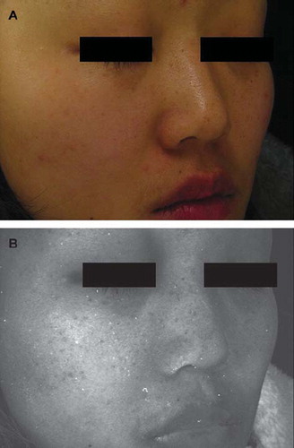
A 27-year-old Korean female with Fitzpatrick skin type IV visited our clinic complaining of dark circles under the eyes, which had persisted for 8 years. The patient had no treatment history for the pigmented lesions and no pertinent family or past medical history. Photographs were taken using the Canfield Visia CR System (Canfield, NJ, USA) with standard light () and ultraviolet light (). The periorbital hyperpigmented lesions were mainly localized to the lower eyelids and composed of several discrete brownish macules. The light brownish lesions, which were slightly accentuated with the ultraviolet light, were also noted on the nose and malar regions, but not on the temples and upper eyelids. The patient was clinically diagnosed with ABNOM.
Figure 1. The ABNOM lesions in the first patient on the lower eyelids, nose, and malar regions taken under (A) standard light and (B) ultraviolet light.
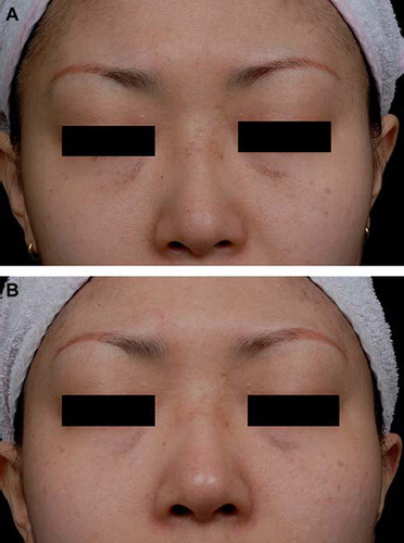
We also observed a 30-year-old Korean female with Fitzpatrick skin type IV presenting the pigmented lesions on the infraorbital regions, which had been developed for 7 years. The patient had neither treatment history of the lesions nor pertinent family or past medical history. The several discrete brownish lesions were concentrated on the lower eyelids as well as on the root of the nose (). The lesions on the temples were brownish blue. A clinical diagnosis of ABNOM was made.
Figure 2. The ABNOM lesions in the second patient are on the lower eyelids as well as on the root of the nose and temples before (A) and 2 months after (B) two successive 1064-nm QS Nd:YAG treatments.
Patients were treated with the 1064-nm QS Nd:YAG laser using MedLite C6™ (HoyaConBio Inc., Fremont, CA, USA) at 2-week intervals. For local anesthesia, the face was cleansed and topical EMLA cream (eutectic mixture of 2.5% lidocaine HCl and 2.5% prilocaine; AstraZeneca AB, Södertälje, Sweden) was applied to the entire face or both cheeks under occlusion an hour prior to the laser therapy. The entire face or both cheeks, including the ABNOM, were treated with the settings of 2.2–2.6 J/cm2, 6-mm spot size, and two to three passes with appropriate overlapping as described in the previous report (Citation1). Additional treatment on the ABNOM was directly followed with the settings of 4–6 J/cm2, 4-mm spot size, and two to three passes or until the appearance of fine petechiae, with appropriate overlapping. The patients presented clinical improvements with QS Nd:YAG treatments ().
Dark circles under the eyes are referred to as round and homogeneous pigmented lesions on both infraorbital areas (Citation2). Post-inflammatory hyperpigmentation, superficial vasculature, periorbital edema, shadowing by skin laxity, and dermal melanin deposition may cause the formation of dark rings (Citation2). Histologic studies on periorbital lesions have suggested that the dermal melanosis could be defined as dermal melanocytosis (Citation3). However, the relationship between the dermal melanocytosis in dark circles and ABNOM has not been evaluated.
ABNOM or Hori's nevus is dermal melanocytic hyperpigmentation, which usually present discrete brown and blue macules depending on the location of the melanocytes (Citation4,Citation5). Mostly, ABNOM appears on malar regions, temples, upper eyelids, and the root and alae of the nose. However, the infraorbital regions have not been sufficiently emphasized as one of the characteristic locations of the ABNOM. We believe that to differentiate the patients complaining of dark circles under the eyes with ABNOM is a significantly effective way not only to choose the proper treatment modalities, but also to discover and to treat the lesions distributed in areas other than the eyelids, such as the malar regions, upper eyelids, temples, and nose. The additional ultraviolet light examination will be useful to find unnoticeable and light-colored lesions for solid diagnosis.
In this study, we treated our patients by using QS Nd:YAG with low-fluence therapy, as described in a recent report (Citation1). Treatment of the entire face or both cheeks prior to the delivery of additional laser treatment on the ABNOM lesions was performed with a low fluence of QS Nd:YAG because most Korean patients with pigmentary disorders expect overall improvement in skin tone in addition to treatment of the pigmentary lesions. Moreover, effective obscuring of the borderline between the ABNOM lesions and the non-lesional skin can be expected by using this method.
Acknowledgement
The authors have declared no conflict of interest.
Declaration of interest: The authors report no conflicts of interest. The authors alone are responsible for the content and writing of the paper.
References
- Cho SB, Park SJ, Kim MJ, Bu TS. Treatment of acquired bilateral nevus of Ota-like macules (Hori's nevus) using 1064-nm Q-switched Nd:YAG laser with low fluence. Int J Dermatol. 2009;48:1308–12.
- Freitag FM, Cestari TF. What causes dark circles under the eyes? J Cosmet Dermatol. 2007;6:211–15.
- Watanabe S, Nakai K, Ohnishi T. Condition known as “dark rings under the eyes” in the Japanese population is a kind of dermal melanocytosis which can be successfully treated by Q-switched ruby laser. Dermatol Surg. 2006;32:785–9.
- Hori Y, Kawashima M, Oohara K, Kukita A. Acquired bilateral nevus of Ota-like macules. J Am Acad Dermatol. 1984;10:961–4.
- Ee HL, Wong HC, Goh CL, Ang P. Characteristics of Hori naevus: A prospective analysis. Br J Dermatol. 2006; 154:50–3.