Abstract
We report on a couple with a five-year history of idiopathic primary infertility. Two early miscarriages had followed intrauterine insemination (IUI). The man's fertility was then re-evaluated, in order to establish whether or not IUI was the best treatment option. Although the semen parameters were normal (sperm concentration: 89 million/ml; progressive motility: 40%; percentage of typical forms: 20%), a computer-assisted sperm morphology analysis with strict criteria found that 12% of the spermatozoa had enlarged heads. All of the latter had a normal form and none had multiple flagella. Using fluorescence in situ hybridization (FISH) analysis, we found that the proportion of aneuploid and diploid spermatozoa was 78% for the sample as a whole and 68% for normally-shaped spermatozoa with a normal-sized head. Although treatment options are well documented for men with macrocephalic sperm head syndrom, there is no consensus on individuals with a low but non-negligible proportion of spermatozoa with enlarged heads. Here, our FISH results contraindicated the use of assisted reproductive technology with the man's sperm. The couple decided to resort to donor sperm.
Introduction
In the literature, the terms ‘macrocephalic head’ and ‘enlarged head’ are both used to define spermatozoa for which the head length and width measurements are outside the normal range. In fact, the term ‘macrocephalic head’ is not used in the fifth edition of the World Health Organization (WHO) laboratory manual for the examination of human semen [WHO 2010] but stems from Nistal and colleagues’ description of macrocephalic sperm head syndrome 25 years ago [Nistal et al. 1977]. The presence of a high proportion (> 90%, according to most authorities) of spermatozoa with a large, amorphous head (some of which may have several tails) generally constitutes a counter-indication to the use of assisted reproductive technology (ART) even though some spermatozoa with a normal-sized head can be selected for intracytoplasmic sperm injection (ICSI) [Guthauser et al. 2006; Chelli et al. 2009]. The term ‘enlarged head’ is found in the literature in general and in the WHO Laboratory Manual in particular. It defines a spermatozoon with a smooth, normally-shape head that is nevertheless abnormally long and wide. Spermatozoa with enlarged heads can be present to a variable extent in semen samples with otherwise normal characteristics. At present, there is no consensus on whether the use of ART with partner sperm is indicated when some spermatozoa with enlarged heads are present. Spermatozoa with enlarged heads are uncommon (1.6%) in the semen of fertile men [Schwartz et al. 1984]. Hence, the presence of this type of spermatozoon in semen raises a number of questions and may be relevant when evaluating fertility.
Case Report
This case study reports on a couple who were a 35-year-old man and a 36-year-old woman, with a five-year history of idiopathic primary infertility. After the third and fourth attempts at intrauterine insemination (IUI), early miscarriages occurred after 4 and 5 weeks of amenorrhea, respectively (i.e., biochemical pregnancies). Neither the man nor the women had a family history of infertility or miscarriage.
Day-3 serum hormone concentrations of the woman were within the normal range: FSH: 7.8 IU/l; estriol: 33 pg/mL; AMH: 2.1 ng/mL. Neither thrombophilia, infections, nor miscarriage-associated endocrine, anatomical, and immune factors were observed.
The man did not have a history of cryptorchidism, orchitis, or testicular torsion. He had not been exposed to toxics (pesticides, tobacco, alcohol, etc.) and had not undergone chemo- or radiotherapy. The results of a physical examination were unremarkable. No varicocele was found and the testicular volumes were symmetrical.
Results
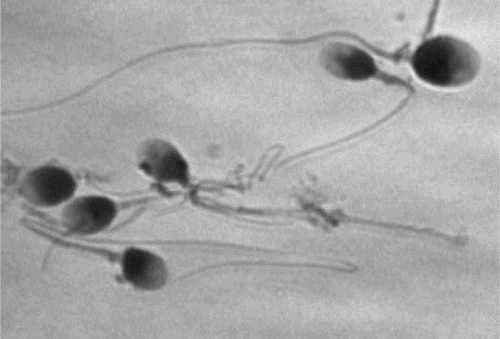
The sperm count (89 x 106/ml) and progressive motility (40%) were normal. According to strict criteria [Menkveld et al. 1990], 20% of the spermatozoa had a typical form (i.e., above the normal cut-off value of 15%). When spermatozoa were observed solely by eye under the microscope, 7% were judged to have enlarged heads. However, the use of image-processing software (as recommended by the WHO Laboratory Manual [2010]) revealed that 12% of the spermatozoa had enlarged heads (i.e., head length > 4.7 µm and head width > 3.2 µm). For the sample as whole, the mean sperm head surface area was 9.3 ± 2.8 µm2 (range: 5.1-18.0), the mean sperm head length was 4.1 ± 0.6 µm (range: 3.1-5.6), and the mean sperm head width was 2.8 ± 0.6 µm (range: 2.0-4.1). All the enlarged heads were oval and normally shaped. No spermatozoa with multiple flagella were found. A typical image of the sample is shown in . For spermatozoa with enlarged heads, the mean head surface was 14.4 ± 1.6 µm (range: 12.5-18.0) with a mean length of 5.0 ± 0.3 µm, and a mean width of 3.7 ± 0.3 µm. We found that 11% of the spermatozoa had not undergone chromatin condensation; this is below the threshold (20%) for abnormal samples.
Figure 1. Spermatozoa after Papanicolaou staining.
The FISH results for three chromosomes are given in . In the original semen sample, 22% of the spermatozoa were haploid-euploid, 25% were diploid, and 53% were haploid-aneuploid. For selected, normal spermatozoa, 22% were diploid and 46% were haploid-aneuploid. We sought to determine whether the non-disjunctions had occurred at meiosis I or meiosis II by analyzing the percentage of aneuploid or diploid spermatozoa. Twenty-nine percent and 1% of the non-disjunctions occurred at meiosis I or meiosis II, respectively. We were unable to draw firm conclusions in the remaining 70% of cases.
Table 1. Results of sperm chromosome analysis using fluorescence in situ hybridization.
The karyotypes were normal (46, XY and 46, XX).
Discussion
For spermatozoa with enlarged heads, measurement of the head size is a recommended way of distinguishing between normal and abnormal spermatozoa [WHO 2010]. The presence of an abnormally high proportion of spermatozoa with enlarged heads is associated with abnormal chromosomal content [Achard et al. 2007] and chromatin condensation dysfunction [Guthauser et al. 2011].
In the case presented here, all the spermatozoa had a regular-shaped head (including those with enlarged heads) and none had multiple flagella. The overall sperm parameters were normal. However, computer-assisted measurements revealed that 12% of the spermatozoa had enlarged heads. For the sample as a whole, the mean sperm head length and width were normal and the mean sperm head surface area was similar to that published (9.6 µm± 2.2 µm2) for normocephalic spermatozoa [Guichaoua et al. 2009]. In 2007, Achard et al. reported on a chromosome content analysis (with three chromosomes analyzed) of four sperm samples with high proportions of spermatozoa with enlarged heads (19%, 22%, 29%, and 49%, respectively). The researchers found an abnormal chromosome number in 25.6%, 43.6%, 51.4%, and 71.7% of the spermatozoa, respectively. Even though only 12% of our patient's spermatozoa had enlarged heads, 78% of his spermatozoa displayed an abnormal chromosome number. These values are higher than the proportions observed for infertile patients with a normal karyotype [Shi and Martin 2001; Tempest and Martin 2009], i.e., not more than 2% (when three chromosomes are analyzed).
Most of the spermatozoa were haploid-aneuploid. Since few supernumerary chromosomes were involved in the aneuploidy and those involved were small chromosomes, the spermatozoon's nucleus was not significantly larger. The fact that the chromatin was well condensed may also explain the lack of an abnormal increase in the mean sperm head surface area.
We did not screen for a homozygous truncating mutation in the aurora kinase C gene, since this was not a homogeneous, macrocephalic phenotype (as previously discussed [Dieterich et al. 2007]). Here, we considered that the FISH results for whole semen contraindicated a fifth IUI attempt. Intracytoplasmic sperm injection was not indicated, since most of the normal potentially selectable spermatozoa (68% of the total) displayed an abnormal number of chromosomes. Even though it may have been possible to obtain pregnancies, the risk of obtaining aneuploid embryos would be very high. We considered that the 5-year history of infertility and the two early miscarriages after IUI were causally linked to the high aneuploidy rate. Hence, the couple was invited to participate in an ART program with donor sperm.
As discussed recently [Menkveld 2010], software-based sperm measurement appears to be a relevant tool for further improving the morphological assessment of semen in which a certain proportion of spermatozoa display enlarged heads. To the best of our knowledge, the present report is the first to highlight such a high abnormal chromosome content rate in a sample with a relatively low percentage of spermatozoa displaying enlarged heads.
In conclusion, the case reported here indicates that the data generated by FISH studies can usefully inform accurate genetic counseling and the choice of the best type of ART for patients with a low but non-negligible proportion of spermatozoa with enlarged heads. However, further studies are needed to determine the ‘enlarged head’ threshold above which sperm chromosome content analysis should be performed.
Materials and Methods
Semen analysis parameters
After informed consent, a semen sample was obtained after three days of sexual abstinence. Semen parameters were evaluated according to the WHO [2010] guidelines. Concentration and motility were evaluated by light microscopy (400x magnification; Leica, France) and sperm motility was evaluated at 37°C (for 200 spermatozoa). Using an air-dried, Papanicolaou-stained sperm sample, the morphology of 200 spermatozoa was assessed (1,000x magnification, Leica, France) according to strict criteria [Menkveld et al. 1990; WHO 2010]. Sperm migration was studied on a two-layer (45%-90%) density gradient (Puresperm, Nidacon, Sweden), with centrifugation at 300 x g for 20 min. The sperm pellet was washed with Universal IVF Medium (Medicult, Origio, France), as described previously [Frainais et al. 2010]. A 10 µL droplet was spread on a glass slide and air-dried to enable further sperm analysis.
Sperm head length, width, and surface area (n = 100 spermatozoa) were measured on the original semen sample under a light microscope (100x magnification; Leica) equipped with a camera (Sony, Tokyo, Japan) and imaging software (Histolab, Microvision Instrument, France). According to the fifth edition of the WHO Laboratory Manual [2010] and following Papanicolaou staining, a normal spermatozoon head was defined as being oval and regular-shaped, with a length of between 3.7 and 4.7 µm (median: 4.1 µm) and a width of between 2.5 and 3.2 µm (median: 2.8 µm). Heads measuring > 4.7 µm in length and > 3.2 µm in width were considered to be enlarged.
Sperm nucleus assessment
The sperm chromatin condensation rate was evaluated on the original semen sample by aniline blue staining. Slides were stained for 5 min with 5% acetified aniline blue (Farmitalia Carlo Erba, Italy), washed with distilled water, and dehydrated with 95% and 100% ethanol. Sperm exhibiting heads with more than 50% staining were considered to have non-condensed chromatin. In normal semen samples, less than 20% of spermatozoa have non-condensed chromatin [Auger et al. 1990].
FISH analysis
After the removal of seminal liquid, the sperm sample was washed twice with sterile water and fixed with Carnoy's solution. The spermatozoa were spread on a slide, air dried, and fixed with methanol. Sperm DNA was decondensed with 1 N NaOH. After dehydration, FISH was performed with centromeric probes for chromosomes X, Y, and 18 (Abbott Laboratories, Chicago, IL). The slides were washed and then counterstained with 6-diamino-2-phenylindole solution. The spermatozoa were analyzed using an Olympus BX60 microscope (Olympus, Hamburg, Germany) with Pathavision software (Digital Scientific, Cambridge, UK). The normative reference value for the proportion of aneuploid spermatozoa was < 2%, according to Shi and Martin [2001] and Tempest and Martin [2009]. Furthermore, after migration on density gradient, normal spermatozoa were selected individually under an inverted microscope (400x magnification), placed in a 2 µl drop of distilled water, and fixed with 95% ethanol for 5 min, as previously described [Guthauser et al. 2006].
Karyotypes
Both male and female karyotypes were determined from a sample of peripheral blood lymphocytes. Our protocols for sperm assays and genetic analyses were approved by an institutional review board and registered with the French health authorities.
Statistical analysis
Descriptive statistics were applied. Variables were quoted as the mean ± the standard error of the mean (SEM).
Declaration of interests: The authors report no conflicts of interest.
Author contributions: Conceived and designed the experiments, performed part of the analysis (sperm measurements), analyzed the data, and wrote the manuscript: BG; Analyzed the results and revised the manuscript: FB; Designed the experiments: MA; Attending embryologist: FK; Woman's gynecologist: CM; Performed some of the analyses (the FISH analysis): FF; Director of the IVF unit: JS; Conceived and designed the experiment, analyzed the data, and revised the manuscript: FV.
Related Research Data
References
- Achard, V., Paulmyer-Lacroix, O., Mercier, G., Porcu, G., Saias-Magnan, J., Metzler-Guillemain, C., (2007) Reproductive failure in patients with various percentages of macronuclear spermatozoa: high level of aneuploid and polyploid spermatozoa. J Androl 28:600–606.
- Auger, J., Mesbah, M., Huber, C. and Dadoune, J.P. (1990) Aniline blue staining as a marker of sperm chromatin defects associated with different semen characteristics discriminates between proven fertile and suspected infertile men. Int J Androl 13:452–462.
- Chelli, M.H., Albert, M., Ray, P.F., Guthauser, B., Izard, V., Hammoud, I., (2009) Can intracytoplasmic morphologically selected sperm injection be used to select normal-sized sperm heads in infertile patients with macrocephalic sperm head syndrome? Fertil Steril 93:1347.e1–5.
- Dieterich, K., Soto Rifo, R., Faure, A.K., Hennebicq, S., Ben Amar, B., Zahi, M., (2007) Homozygous mutation of AURKC yields large-headed polyploidy spermatozoa and causes male infertility. Nat Genet 39:661–665.
- Frainais, C., Vialard, F., Rougier, N., Aegerther, P., Damon, F., Ayel, J.P. (2010) Impact of freezing/thawing technique on sperm DNA integrity in HIV-I patient. J Assist Reprod. Genet 27:415–421.
- Guichaoua, M.R., Mercier, G., Goeffroy-Siraudin, C., Paulmyer-Lacroix, A., Lanteaume, A., Metzler-Guillemin, C., (2009) Macrocephalic spermatozoa. What would be the impact on reproduction? Gynecol Obstet Fertil 37:703–711. French.
- Guthauser, B., Vialard, F., Dakouane, M., Izard, V., Albert, M. and Selva, J. (2006) Chromosomal analysis of spermatozoa with normal-sized heads in two infertile patients with macrocephalic sperm head syndrome. Fertil Steril 85(3):750.e5–750.e7.
- Guthauser, B., Albert, M., Ferfouri, F., Ray, P.F., Rabiey, G., Selva, J., (2011) Inverse correlation between chromatin condensation and sperm head size in case of enlarged sperm heads. RBM Online 23:711–716.
- Menkveld, R., Stander, F.S., Kotze, T.J., Kruger, T.F. and Van Zyl, J.A. (1990) The evaluation of morphological characteristics of human spermatozoa according to stricter criteria. Hum Reprod 5:586–592.
- Menkveld, R. (2010) Clinical significance of the low normal sperm morphology value in the fifth edition of the WHO Laboratory Manual for the examination and Processing of Human Sperm. Asian J Androl 12:47–58.
- Nistal, M., Paniagua, R. and Herruzo, A. (1977) Multi-tailed spermatozoa in a case with asthenospermia and teratospermia. Virchows Arch B Cell Pathol 26:111–118.
- Schwartz, D., Mayaux, M.J., Guihard-Moscato, M.L., Spira, A., Jouannet, P., Czyglik, F., (1984) Study of sperm morphologic characteristics in a group of 838 fertile men. Andrologia 16:423–428.
- Shi, Q. and Martin, R.H. (2001) Aneuploidy in human spermatozoa: FISH analysis in men with constitutional chromosomal abnormalities, and in infertile men. Reproduction 121:655–666.
- Tempest, H.G. and Martin, R.H. (2009) Cytogenetic risks in chromosomally normal infertile men. Curr Opin Obstet Gynecol 21:223–227.
- WHO (2010) World Health Organization laboratory manual for the examination of human semen, fifth edition. WHO Press, Geneva.