Abstract
Schizophrenia is a heterogeneous psychiatric disorder of unknown cause or characteristic pathology. Clinical neuroscientists increasingly postulate that schizophrenia is a disorder of brain network organization. In this article we discuss the conceptual framework of this dysconnection hypothesis, describe the predominant methodological paradigm for testing this hypothesis, and review recent evidence for disruption of central/hub brain regions, as a promising example of this hypothesis. We summarize studies of brain hubs in large-scale structural and functional brain networks and find strong evidence for network abnormalities of prefrontal hubs, and moderate evidence for network abnormalities of limbic, temporal, and parietal hubs. Future studies are needed to differentiate network dysfunction from previously observed gray- and white-matter abnormalities of these hubs, and to link endogenous network dysfunction phenotypes with perceptual, behavioral, and cognitive clinical phenotypes of schizophrenia.
La esquizofrenia es un trastorno psiquiátrico heterogéneo de causa o patología específica desconocidas. Los neurocientíficos clínicos postulan cada vez más que la esquizofrenia es un trastorno de la organización de la red cerebral. En este artículo se presenta el marco conceptual de la hipótesis de la desconexión, se describe el paradigma metodológico predominante para probar esta hipótesis y se revisa la evidencia reciente de una alteración de las regiones cerebrales centrales/concentradoras, como un ejemplo prometedor de esta hipótesis. Se resumen los estudios de los concentradores cerebrales en las redes cerebrales estructurales y funcionales a gran escala y se encuentra una fuerte evidencia para las alteraciones de la red de concentradores prefrontales y evidencias menores para las alteraciones de la red de concentradores límbicos, temporales y parietales. Se requiere de futuros estudios para diferenciar la disfunción de la red de las alteraciones observadas previamente en la sustancia gris y blanca de estos concentradores y relacionar fenotipos endógenos de disfunción de la red con fenotipos clínicos de perception, comportamiento y cognición en la esquizofrenia.
La schizophrénie est un trouble psychiatrique hétérogène sans cause connue ni anatomo-pathologie caractéristique. Les neurosciences cliniques suggèrent de plus en plus que la schizophrénie est un trouble de l'organisation des réseaux cérébraux. Nous analysons dans cet article le cadre conceptuel de I'hypothèse de cette déconnexion, nous décrivons le modèle méthodologique prédominant pour vérifier cette hypothèse et nous étudions les preuves récentes d'une perturbation des régions centrales du cerveau comme exemple prometteur de cette hypothèse. Nous résumons les études sur les centres cérébraux des réseaux cérébraux anatomiques et fonctionnels à grande échelle. Les preuves d'anomalies de réseaux des centres préfrontaux sont solides ; elles le sont modérément pour les anomalies des réseaux des centres limbiques, temporaux et pariétaux. D'autres études seront nécessaires pour différencier les troubles des réseaux des anomalies déjà connues des substances grise ou blanche de ces centres, et pour établir des correspondances entre les dysfonctions endogènes des réseaux et les phénotypes cliniques cognitifs, comportementaux et perceptuels de la schizophrénie.
Schizophrenia and integration
Schizophrenia is a psychiatric disorder with an onset in early adulthood, a chronic course, and serious morbidity only modestly controlled by currently available treatments.Citation1,Citation2 The cause and characteristic abnormalities of schizophrenia are unknown; the disorder is thought to be underpinned by neurodevelopmental abnormalities of brain structure and function, but is onlydiagnosed using subjective criteria of psychiatric diagnostic manuals.Citation3,Citation4 These definitions emphasise the co-occurrence of positive/psychotic symptoms (eg, hallucinations and delusions), negative/deficit symptoms (eg, poverty of thought and loss of motivation), and cognitive symptoms (eg, impairment of memory and attention); the definitions encompass a wide range of heterogeneous presentations such that two patients with schizophrenia may share no common symptoms.Citation5 These definitions may plausibly represent several distinct diseases and/or a spectrum of diseaseCitation6; we return to this important point in the discussion, but for now we assume — as do most current investigators — that schizophrenia is a single entity.
Our current understanding of the causes of schizophrenia emphasizes interactions between diverse genetic and environmental factors.Citation2,Citation7 Conceptually, these diverse causes should converge on a small set of brain abnormalities pathognomonic of the disorder. Modern neuroimaging methods reveal a wide range of brain abnormalities in schizophrenia, including reductions in whole brain volume, increases in ventricular volume, reductions in frontal, temporal, limbic, and thalamic grey matter, and abnormalities in frontal and temporal white matter.Citation8-Citation10
Despite these promising findings, the abnormalities are insufficiently sensitive or specific to be individually or collectively diagnostic or prognostic of the disease in the clinical setting. In addition, the abnormalities have yet to be integrated into a clinically validated model of schizophrenia; such a model would for instance allow a rational approach to the search for treatment and prevention of the disorder.
Clinical neuroscientists increasingly postulate that schizophrenia is a disorder of integration of information between specialized brain regions. The emergence of complex perceptual, behavioral, and cognitive functions — the functions predominantly affected in schizophrenia — is contingent on such integrationCitation11,Citation12; the binding of visual and other sensory stimuli into a unified perceptual whole is a well-studied instance of this phenomenon.Citation13 Abnormality of integration hence represents an intuitive unifying hypothesis of schizophrenia. An early version of this hypothesis was posited by psychiatrists in the 19th century; modern versions of this hypothesis — including the constructs of dysconnection and cognitive dysmetria — have emerged in the last 20 years,Citation14-Citation19 driven by advances in neuroimaging and the consequent possibility of the study of integration in living humans. As part of the same broader trend, investigators recently proposed a roadmap towards reclassification of schizophrenia and other psychiatric disorders from entities based on subjective clinical diagnoses towards entities based on objective abnormalities of integration or brain networks.Citation20
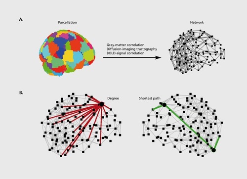
The study of integration is the study of brain networks.Citation21 While the precise nature of integration remains an important question, neuroscientists increasingly emphasize the central role of reciprocal, distributed, and parallel interactions between brain regions, over serial and feedforward interactions in this process.Citation22,Citation23 Recent characterizations of large-scale structural and functional networks of the human brain broadly reveal several organizational principles supporting these properties; for instance, organization of brain networks is conducive to reciprocal interactions through a preponderance of symmetric connections and the presence of clusters; at the same time organization of brain networks is conducive to distributed and parallel interactions through the presence of high interconnectedness between most brain regions ().Citation24,Citation25
An additional important property of large-scale brain network organization is the presence of central regions, or hubs. Hubs are brain regions which, by virtue of their many, diverse, strategic, or long-range connections are important in facilitating integration.Citation26,Citation27 Prominent hubs have been identified in prefrontal, temporal, and parietal multimodal association areas, and in limbic and subcortical areas.Citation28-Citation32 Abnormalities of brain hubs are increasingly implicated in brain diseaseCitation32,Citation33 and have potentially powerful explanatory capacity for a wide range of symptoms of schizophrenia. In this article we review methods used to describe hubs in large-scale brain networks and summarize recent studies which have begun to test abnormalities of these hubs in schizophrenia. Fornito et alCitation34 comprehensively review more general properties of large-scale brain networks in schizophrenia.
Brain networks and hubs
Brain networks are maps of structural or functional interactions (termed links) between brain regions (termed nodes). The studied regions and interactions may span multiple spatial or temporal scales, although in practice the nature of these elements is limited by the spatiotemporal resolution of imaging methods. The present spatiotemporal resolution of magnetic resonance imaging (MRI) makes it the dominant method for imaging whole-brain networks; for instance, functional MRI is the only current method which allows noninvasive visualization of whole-brain networks of functional interactions, due to a reasonable trade-off between millimetre-scale spatial (node) resolution and second-scale temporal (link) resolution.Citation35 However, it remains unclear if this resolution is sufficient for a fundamental understanding of integration, and alternative future approaches may define individual neurons as nodes,Citation36 study structural and functional synaptic interactions in post-mortem brainsCitation37 or stem-cell-derived neuronal cultures,Citation38 or improve spatial resolution in neurophysiological recordingsCitation39 to examine neuronal oscillations at the millisecond scale.Citation40,Citation41
The definition of brain networks involves the definition of nodes and links (Figure 1a). Nodes in brain networks represent structurally and functionally homogeneous brain regions. Parcellation of whole-brain MRI scans into a collection of such nodes is an active and important area of research.Citation42 Links in brain networks represent anatomical or functional interactions. In MRI datasets, anatomical links are defined using two main methods. The method of structural correlation is based on the principle that anatomically connected regions share common trophic factors and correlate in size. This method defines links as correlations in interregional gray-matter volume or thickness inferred from a group of subjects, such that one network is constructed for the whole group.Citation43 The method of diffusion-imaging tractography is based on the principle of anisotropic water diffusion along white-matter tracts and detects large-scale interregional anatomical links more directly such that one network is constructed for each subject.Citation44 Functional links are defined as correlations of interregional low-frequency fluctuations in blood-oxygen-level-dependent (BOLD) signal, an indirect measure of neural activity based on concentration of oxygenated hemoglobin in brain tissueCitation45; with this method one network is likewise constructed for each subject. These different types of connectivity are complementary and each offers distinct insights into interactions between brain regions.
The empirical study of brain networks has broadly and somewhat arbitrarily proceeded along two methodologically distinct lines of work. One line of work studies small networks of several brain regionsCitation46; the other line of work studies large networks of the whole brain.Citation25 Investigators define small networks with methods such as seed correlation analysisCitation47 (the detection of structurally or functionally similar neighbors for an a priori defined “seed” region) and independent component analysisCitation48 (the delineation of the brain into a set of maximally independent small networks), and with modeling approaches.Citation49,Citation50 Small networks are usually associated with specific functional tasks: a classic example is of the language network associated with the comprehension and production of languageCitation51; a prominent recent example is the default network associated with internallyfocused cognition.Citation52 Associations with functional tasks make small networks comparatively easy to interpret and the study of such networks has a long tradition in neurology.Citation53 Nonetheless small networks may provide limited insight into characteristic abnormalities of schizophrenia if such abnormalities involve widespread disturbances of integration, as is likely to be the case. Calhoun et alCitation54 review abnormalities of small functional brain networks in schizophrenia.
In this article we focus on large networks involving many nodes and describing the complete structural or functional maps of interregional interactions of the brain, the human connectome (Figure 1a).Citation55,Citation56 Large networks offer a powerful and general framework for the study of brain function in health and disease but — as a consequence of their size, nontrivial organization, and absence of associations with specific functional tasks — are more difficult to interpret. These networks are characterized with concepts from graph theory (the mathematical study of networks) and statistical physics.Citation57 Early characterizations of these networks included computation of statistics for the propensity of networks to segregate into clusters (termed “the clustering coefficient”), the propensity of networks to be globally interconnected (termed “the characteristic path length”), and the simultaneous combination of these two properties (termed “small-worldness”), as recently reviewed.Citation58,Citation59
The analysis of whole-brain networks is however arguably at its most powerful when it localizes functionally distinct or functionally important brain regions solely on the basis of connection patterns associated with these regions.Citation27 The concept of brain hubs is an example of this analysis, and is defined with measures of network centrality (Figure 1b). The archetypal measure of centrality is the degree which equals the total number of connections associated with a node. Other common measures are the closeness centrality and regional efficiency, both based on the average length of shortest paths from a node to all other nodes, the betweenness centrality, based on the fraction of all shortest paths traversing a node, and the eigenvector centrality, based on the extent with which a node is connected to important nodes in the network. Individual measures of centrality are often highly correlated, and hub nodes should score highly on several distinct measures.Citation60
Brain hubs in schizophrenia
In this section we summarize all recent studies of abnormal hub organization in schizophrenia. These nine patient-control MRI studies have all been published in the last 5 years, and are evenly divided between structural correlation studies, diffusion-imaging tractography studies, and functional correlation studies. We summarize the main findings of these studies below and in Table I.
TABLE I. A summary of studies of hub abnormalities in schizophrenia. N, number of subjects; A, age of subjects; M, proportion of male subjects; PSS, positive-and-negative-symptom-scale positive symptoms; NSS, positive-and-negative-symptom-scale negative symptoms; AAL atlas, anatomical-automatic-labelling atias; BOLD signal, blood-oxygen-level-dependent signal
Three studies examine hubs in structural correlation networks of patients with schizophrenia.Citation61-Citation63 Two of these studiesCitation61,Citation62 construct networks from structural images of large cohorts of middle-aged subjects with schizophrenia and detect less central hubs in frontal and limbic association areas in schizophrenia; one studyCitation62 additionally detects hubs in paralimbic areas in healthy controls, and an increased number of these hubs in schizophrenia. The third studyCitation63 constructs correlation networks from structural images of a smaller cohort of neonates born to mothers with schizophrenia, and hence at high risk of developing the disorder. This study finds hubs in frontal, temporal, parietal, and subcortical areas in healthy control neonates, and a reduction in the total number of hubs with an absence of parietal and subcortical hubs in high-risk neonates.
Three studies examine hubs in diffusion-imaging tractography networks of patients with schizophrenia.Citation64-Citation66 These studies find hubs in frontal and parietal association areas, as well as in limbic, paralimbic, and subcortical areas. All three studies find less central hubs in frontal association areas. Two studiesCitation64,Citation65 additionally find less central hubs in limbic areas (), while the third studyCitation66 extends the simple description of hubs and describes the pattern of interconnections between individual hubs as part of a so-called “rich club,” a small group of high-degree nodes which are additionally highly connected to each other. The study finds a weaker rich club in schizophrenia, reflecting a lower level of connectivity between hubs of schizophrenia subjects.
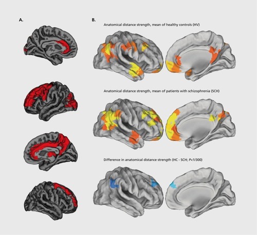
Three studies examine hubs in functional correlation networks of patients with schizophrenia.Citation67-Citation69 Two of these studies examine cohorts of middle-aged subjects and find less central hubs in temporal and limbic association areasCitation67 or in frontal, temporal, limbic, and occipital association areas.Citation68 The third studyCitation69 examines a group of adolescent subjects with childhood-onset schizophrenia, a rare and severe form of schizophrenia. This studyfocuses on the relationship between connection weight and anatomical distance, and finds hubs in frontal, temporal, and parietal association areas, and a greater proportion of long distance connections between hubs in the schizophrenia group (Figure 2b).
Discussion and conceptual issues
The above body of work broadly implicates abnormalities of association hubs and limbic hubs in schizophrenia. More specifically, the studies strongly implicate abnormalities of prefrontal hubs (9/9 studies) and moderately implicate abnormalities of limbic (6/9 studies), temporal (6/9 studies) and parietal (5/9 studies) hubs. The evidence points to a decreased centrality of these hubs in schizophrenia, at least in studies with adult populations.
The involvement of multimodal and limbic association areas has a relatively straightforward neurobiological interpretation in schizophrenia; these transmodal areas of the cerebral cortex are important in facilitating brain functions most visibly impaired in the disorder. Such functions include the integration or binding of distinct perceptual stimuli into complex unified representations, the cognitive and emotional contextualization of these representations, and higher cognitive functions such as executive control and working memory.Citation11 In addition, the relatively late white-matter myelination and neuronal pruning of areas in multimodal cortexCitation71 suggests that inherent neurodevelopmental abnormalities of these areas may only symptomatically manifest in adolescence or early adulthood, a common age of onset for schizophreniaCitation70.
Despite this seemingly straightforward interpretation, there inevitably remain empirical and conceptual questions. Empirically, previous studies have already reported schizophrenia-associated reductions in gray matter in all presently implicated hubs, as well as abnormalities in white-matter tracts connecting most of these hubsCitation8-Citation10; indeed two of the present studies directly examine the relationship between regional gray-matter volumes and regional network centrality, and report substantial associations between these two properties.Citation62,Citation68 Hence, while the present body of work builds on previous studies to present a more global-network view of hub disorganization, the pathogenetic precedence of gray- or white-matter abnormalities and global network disorganization remains undetermined. On the one hand, it is simpler to consider the emergence of global network dysfunction following local abnormalities of gray and white matter. On the other hand, it is simpler to formulate a unitary model of schizophrenia based on the notion of abnormal disruption of integration and hubs, rather than on the notion of multiple focal lesions. For instance, one studyCitation69 reports that brain hubs have longer-distance and more metabolically costly functional connections; this arrangement implies that hubs are likely to be more susceptible to metabolic insult, and provides a conceptually straightforward potential pathogenetic mechanism.
An additional empirical question concerns the specificity of hub disruption as a characteristic phenotype of schizophrenia. It is possible that the same hubs are implicated in many other psychiatric and neurological disorders,Citation32,Citation33 making it difficult to associate endogenous phenotypes of hub dysfunction with perceptual, behavioural and cognitive clinical phenotypes of schizophrenia. Other empirical concerns include the absence of a standard methodological framework for the construction and characterization of brain networks, and low statistical power associated with some studies, resulting in potential for bias and inconsistent findings.Citation72-Citation74
Conceptually, the schizophrenia dysconnection hypothesis is hampered by the imprecision of both the notion of schizophrenia, and the notion of dysconnection. Neuroscientists commonly motivate the dysconnection hypothesis by invoking its long history dating back to psychiatrists in the 19th century. Yet there is no clear link between the work of early psychiatrists and present researchCitation75 nor would the presence of such a link imply conceptual validity. Schizophrenia remains a heterogeneous and poorly defined disorder, to the extent that a search for a single characteristic abnormality may be an a priori futile task. Brain networks are likewise heterogeneous, and the spatiotemporal resolution on which characteristic abnormalities of schizophrenia optimally manifest remains undetermined. The sceptically inclined may hence describe the dysconnection hypothesis of schizophrenia as an attempt at explanation of one imprecise concept with another.
In this context there is possibility for systematic progress if the constructs of schizophrenia and brain networks are both sufficiently close approximations to real and coherent entities. Progress may occur for instance through a series of iterative and mutual conceptual modifications of both constructs. In the case of schizophrenia, increased coherence may be achieved through a focus on more specific forms of the disorder, such as paranoid (primarily psychotic-symptom) and disorganized (primarily deficit-symptom) subtypes,Citation3,Citation4 or the focus on forms of the disorder with a seemingly higher genetic component, such as childhood-onset schizophreniaCitation76 or 22qll.2 deletion syndrome, a genetic syndrome associated with a high occurrence of schizophrenia.Citation77 In the case of brain networks, increased coherence is likely to follow from increasing spatial and temporal resolution associated with future methodological innovations. We hope that these developments will eventually lead to a substantial clarification in our understanding of schizophrenia.
Conclusion
There is now considerable conceptual and empirical evidence for the importance of network integration in healthy brain function, for the importance of topologically central nodes or hubs in brain network integration, and for abnormalities of both integration and hubs in schizophrenia. Despite this we will not be able to claim conclusively that schizophrenia is a disease of brain hubs, a hubopathy, until future studies have consolidated the preliminary findings based mainly on small- to medium-sized samples; resolved some of the discrepancies between functional and structural network phenotypes of schizophrenia; clarified how abnormal network hubs might emerge developmentally and in the context of growing awareness of the role of synaptic and postsynaptic risk alleles in the genetic predisposition to schizophrenia; and established the specificity of hub abnormalities in schizophrenia compared with other brain disorders. There is much still to do to substantiate and contextualize the new results arising from complex network analysis of the schizophrenia connectome. However, we suggest that the basic insight that brain network hubs may be central to the systems-level pathophysiology of schizophrenia is at least likely to prove heuristically valuable as we continue to make progress in understanding the neurobiological basis of psychotic disorders.
MR is supported by the NARSAD Young Investigator Grant and the Isaac Newton Trust. ETB is employed part-time by GlaxoSmithKline and part-time by the University of Cambridge. The Behavioural and Clinical Neuroscience Institute is supported by the Wellcome Trust and the Medical Research Council.
REFERENCES
- van OsJ.KapurS.Schizophrenia.Lancet.200937463564519700006
- InselTR.Rethinking schizophrenia.Nature.201046818719321068826
- American Psychiatric Association.Diagnostic and Statistical Manual of Mental Disorders.4th ed. Text Revision. Washington, DC: American Psychiatric Association;2000
- World Health Organization.The ICD-10 Classification of Mental and Behavioral Disorders. Clinical descriptions and diagnostic guidelines. Geneva, Switzerland: World Health Organization;1992
- AndreasenNC.A unitary model of schizophrenia: Bleuler's “fragmented phrene” as schizencephaly.Arch Gen Psychiatry.19995678178712884883
- JablenskyA.The diagnostic concept of schizophrenia: its history, evolution, and future prospects.Dialogues Clin Neurosci.20101227128720954425
- van OsJ.KenisG.RuttenBP.The environment and schizophrenia.Nature.201046820321221068828
- Ellison-WrightI.BullmoreE.Anatomy of bipolar disorder and schizophrenia: a meta-analysis.Schizophr Res.201011711220071149
- Ellison-WrightI.BullmoreE.Meta-analysis of diffusion tensor imaging studies in schizophrenia.Schizophr Res.200910831019128945
- ShentonME.WhitfordTJ.KubickiM.Structural neuroimaging in schizophrenia: from methods to insights to treatments.Dialogues Clin Neurosci.20101231733220954428
- MesulamMM.From sensation to cognition.Brain.1998121(Pt 6)101310529648540
- VarelaF.LachauxJP.RodriguezE.MartinerieJ.The brainweb: phase synchronization and large-scale integration.Nat Rev Neurosci.2001222923911283746
- RobertsonLC.Binding, spatial attention and perceptual awareness. WatRev Neurosci.2003493102
- FristonKJ.FrithCD.Schizophrenia: a disconnection syndrome?Clin Neurosci.1995389977583624
- BullmoreET.FrangouS.MurrayRM.The dysplastic net hypothesis: an integration of developmental and dysconnectivity theories of schizophrenia.Schizophr Res.1997281431569468349
- AndreasenNC.ParadisoS.O'LearyDS.“Cognitive dysmetria” as an integrative theory of schizophrenia: a dysfunction in cortical-subcorticalcerebellar circuitry?Schizophr Bull.1998242032189613621
- PeledA.Multiple constraint organization in the brain: a theory for schizophrenia.Brain Res Bull.19994924525010424844
- TononiG.EdelmanGM.Schizophrenia and the mechanisms of conscious integration.Brain Res Brain Res Rev.20003139140010719167
- StephanKE.BaldewegT.FristonKJ.Synaptic plasticity and dysconnection in schizophrenia.Biol Psychiatry.20065992993916427028
- InselT.CuthbertB.GarveyM.et alResearch domain criteria (RDoC): toward a new classification framework for research on mental disorders.Am J Psychiatry.201016774875120595427
- SpornsO.Networks of the Brain.Cambridge, Mass: MIT Press;2011
- CalvertGA.ThesenT.Multisensory integration: methodological approaches and emerging principles in the human brain.J Physiol Paris.20049819120515477032
- GhazanfarAA.SchroederCE.Is neocortex essentially multisensory?Trends Cogn Sci.20061027828516713325
- SpornsO.ChialvoDR.KaiserM.HilgetagCC.Organization, development and function of complex brain networks.Trends Cogn Sci.2004841842515350243
- BullmoreE.SpornsO.Complex brain networks: graph theoretical analysis of structural and functional systems.Nat Rev Neurosci.20091018619819190637
- BullmoreE.SpornsO.The economy of brain network organization.Nat Rev Neurosci.20121333634922498897
- SpornsO.Network attributes for segregation and integration in the human brain.Curr Opin Neurobiol.20132316217123294553
- AchardS.SalvadorR.WhitcherB.SucklingJ.BullmoreE.A resilient, low-frequency, small-world human brain functional network with highly connected association cortical hubs.J Neurosci.200626637216399673
- HagmannP.CammounL.GigandetX.etal.Mapping the structural core of human cerebral cortex.PLoS Biol.20086e15918597554
- ColeMW.PathakS.SchneiderW.Identifying the brain's most globally connected regions.Neuroimage.2010493132314819909818
- TomasiD.VolkowND.Functional connectivity density mapping. ProcNatl Acad Sci USA.201010798859890
- BucknerRL.SepulcreJ.TalukdarT.et alCortical hubs revealed by intrinsic functional connectivity: mapping, assessment of stability, and relation to Alzheimer's disease.J Neurosci.2009291860187319211893
- AchardS.Delon-MartinC.VertesPE.etal.Hubs of brain functional networks are radically reorganized in comatose patients.Proc Natl Acad Sci USA.2012109206082061323185007
- FornitoA.ZaleskyA.PantelisC.BullmoreET.Schizophrenia, neuroimaging and connectomics.Neuroimage.2012622296231422387165
- Meyer-LindenbergA.From maps to mechanisms through neuroimaging of schizophrenia.Wature.2010468194202
- LewisDA.SweetRA.Schizophrenia from a neural circuitry perspective: advancing toward rational pharmacological therapies.J Clin Invest.200911970671619339762
- ChungK.WallaceJ.KimSY.etal.Structural and molecular interrogation of intact biological systems.Nature.201349733233723575631
- BrennandKJ.SimoneA.JouJ.etal.Modelling schizophrenia using human induced pluripotent stem cells.Nature.201147322122521490598
- BassettDS.BullmoreET.Meyer-LindenbergA.ApudJA.WeinbergerDR.CoppolaR.Cognitive fitness of cost-efficient brain functional networks.Proc Natl Acad Sci USA.2009106117471175219564605
- UhlhaasPJ.SingerW.Abnormal neural oscillations and synchrony in schizophrenia.Nat Rev Neurosci.20101110011320087360
- UhlhaasPJ.Dysconnectivity, large-scale networks and neuronal dynamics in schizophrenia.Curr Opin Neurobiol.20132328329023228430
- BehrensTE.SpornsO.Human connectomics.Curr Opin Neurobiol.20122214415321908183
- Alexander-BlochA.GieddJN.BullmoreE.Imaging structural co-variance between human brain regions.Nat Rev Neurosci.20131432233623531697
- Le BihanD.Looking into the functional architecture of the brain with diffusion MRI.Nat Rev Neurosci.2003446948012778119
- FornitoA.BullmoreET.What can spontaneous fluctuations of the blood oxygenation-level-dependent signal tell us about psychiatric disorders?Curr Opin Psychiatry.20102323924920216219
- FoxMD.RaichleME.Spontaneous fluctuations in brain activity observed with functional magnetic resonance imaging.Nat Rev Neurosci.2007870071117704812
- FoxMD.SnyderAZ.VincentJL.CorbettaM.Van EssenDC.RaichleME.The human brain is intrinsically organized into dynamic, anticorrelated functional networks.Proc Natl Acad Sci USA.20051029673967815976020
- DamoiseauxJS.RomboutsSA.BarkhofF.etal.Consistent resting-state networks across healthy subjects.Proc Natl Acad Sci USA.2006103138481385316945915
- McintoshAR.Gonzalez-LimaF.Structural equation modeling and its application to network analysis in functional brain imaging.Hum Brain Mapp.19942222
- FristonKJ.HarrisonL.PennyW.Dynamic causal modelling.Neuroimage.2003191273130212948688
- BookheimerS.Functional MRI of language: new approaches to understanding the cortical organization of semantic processing.Annu Rev Neurosci.20022515118812052907
- BucknerRL.Andrews-HannaJR.SchacterDL.The brain's default network: anatomy, function, and relevance to disease.Ann N Y Acad Sci.2008112413818400922
- CataniM.ffytcheDH.The rises and falls of disconnection syndromes.Brain.2005128(Pt 10)2224223916141282
- CalhounVD.EicheleT.PearlsonG.Functional brain networks in schizophrenia: a review.Front Hum Neurosci.200931719738925
- SpornsO.TononiG.KotterR.The human connectome: a structural description of the human brain.PLoS Comput Biol.20051e4216201007
- BiswalBB.MennesM.ZuoXN.etal.Toward discovery science of human brain function.Proc Natl Acad Sci U S A.20101074734473920176931
- NewmanMEJ.xx.Networks: an Introduction. Oxford, UK: New York, NY: Oxford University Press;2010
- RubinovM.SpornsO.Complex network measures of brain connectivity: uses and interpretations.Neuroimage.2010521059106919819337
- BullmoreET.BassettDS.Brain graphs: graphical models of the human brain connectome.Annu Rev Clin Psychol.2011711314021128784
- SpornsO.HoneyCJ.KotterR.Identification and classification of hubs in brain networks.PLoS One.20072e104917940613
- BassettDS.BullmoreE.VerchinskiBA.MattayVS.WeinbergerDR.Meyer-LindenbergA.Hierarchical organization of human cortical networks in health and schizophrenia.J Neurosci.2008289239924818784304
- ZhangY.LinL.LinCP.etal.Abnormal topological organization of structural brain networks in schizophrenia.Schizophr Res.201214110911822981811
- ShiF.YapPT.GaoW.LinW.GilmoreJH.ShenD.Altered structural connectivity in neonates at genetic risk for schizophrenia: a combined study using morphological and white matter networks.Neuroimage.2012621622163322613620
- van den HeuvelMP.MandlRC.StamCJ.KahnRS.Hulshoff PolHE.Aberrant frontal and temporal complex network structure in schizophrenia: a graph theoretical analysis.J Neurosci.201030159151592621106830
- WangQ.SuTP.ZhouY.etal.Anatomical insights into disrupted smallworld networks in schizophrenia.Neuroimage.2012591085109321963918
- vandenHeuvelMP.SpornsO.CollinG.etal.Abnormal rich club organization and functional brain dynamics in schizophrenia.JAMA Psychiatry.20137078379223739835
- BassettDS.NelsonBG.MuellerBA.CamchongJ.LimKO.Altered resting state complexity in schizophrenia.Neuroimage.2012592196220722008374
- WangL.MetzakPD.HonerWG.WoodwardTS.Impaired efficiency of functional networks underlying episodic memory-for-context in schizophrenia.J Neurosci.201030131711317920881136
- Alexander-BlochAF.VertesPE.StiddR.et alThe anatomical distance of functional connections predicts brain network topology in health and schizophrenia.Cereb Cortex.20132312713822275481
- LynallME.BassettDS.KerwinR.etal.Functional connectivity and brain networks in schizophrenia.J Neurosci.2010309477948720631176
- RossCA.PearlsonGD.Schizophrenia, the heteromodal association neocortex and development: potential for a neurogenetic approach.Trends Neurosci.1996191711768723199
- loannidisJP.Why most published research findings are false.PLoS Med.20052e12416060722
- CarpJ.The secret lives of experiments: methods reporting in the fMRI literature.Neuroimage.20126328930022796459
- ButtonKS.loannidisJP.MokryszC.etal.Power failure: why small sample size underminesthe reliability of neuroscience. WatRev Neurosci.201314365376
- BerriosGE.LuqueR.VillagranJM.Schizophrenia: a conceptual history.Int J Psychol Psychol Ther.20033111140
- AsarnowJR.Annotation: childhood-onset schizophrenia.J Child Psychol Psychiatry.199435134513717868633
- BassettAS.ChowEW.22q11 deletion syndrome: a genetic subtype of schizophrenia.Biol Psychiatry.19994688289110509171