Nano-sized lipid vectors for the pulmonary co-delivery of anticancer drugs and siRNA in targeted lung cancer treatment
Novel inhaled lung cancer therapy may address the adverse side effects of conventional chemotherapy and achieve a greater level of efficacy
Researchers from The Cancer Institute of New Jersey (NJ, USA), in collaboration with Oregon State University (OR, USA) and Ernest Mario School of Pharmacy (NJ, USA), have developed and tested a multifunctional nanostructured lipid nanocarrier-based system (NLCS) for the efficient delivery of an anticancer drug and siRNA directly into the lungs by inhalation for the treatment of lung cancer.
The delivery system consists of nanostructured lipid carriers, anticancer drug (doxorubicin or paclitaxel), siRNA – targeted to suppress pump drug resistance – and to stop non-pump cellular resistance, and a synthetic analog of LHRH, which acts as a targeting moiety directing the delivery system to specific receptors that are overexpressed in the plasma membrane of lung cancer cells.
Research leader, Tamara Minko, a Professor and the Chair at the Department of Pharmaceutics at Rutgers University (NJ, USA) and a member of the Cancer Institute of New Jersey, commented: “The proposed novel treatment much more effectively kills resistant cancer cells when compared with conventional anticancer drugs and shows superior efficiency over the traditional chemotherapy.”
The team tested the NLCS in vitro using human lung cancer cells and in vivo employing a mouse orthotopic model of human lung cancer. Minko and co-workers reported that data demonstrated that the NLCS effectively delivered its payload to the targeted lung cancer cells, efficiently suppressing tumor growth.
Minko, who has been leading the project since 2006, commented: “Up until now, limited clinical efficiency and significant toxicity have represented two critical barriers restricting progress in the therapy of advanced lung cancer.” Speaking to Therapeutic Delivery, Minko explained that the proposed approach begins to address this issue as it “delivers the drug directly to lung cancer cells, thereby enhancing the efficiency of therapy of resistant lung cancer and limiting the adverse side effects of chemotherapy on healthy organs and tissues.”
Whereas conventional chemotherapy treatment for lung cancer results in accumulation of the drugs in the liver, kidney and spleen, reducing the amount of drugs that reach the lungs, the results of the study indicate that 83% of the drugs are delivered directly to the lungs by inhalation therapy, relative to the 23% that is achieved by intravenous injection.
The nanoparticle system is currently delivered using a dosage form suitable to nebulizers, though the team plans to develop the therapy for delivery by dry powder inhaler. Speaking to Therapeutic Delivery, Minko commented that, in terms of future work, the team now intends to move from preclinical studies to clinical trials.
– Written by Hannah Coaker
Sources: Taratula O, Kuzmov A, Shah M, Garbuzenko OB, Minko T. Nanostructured lipid carriers as multifunctional nanomedicine platform for pulmonary co-delivery of anticancer drugs and siRNA. J. Control. Release (2013) (In Press). Inhalation Therapy for Lung Cancer Shows Promise in Rutgers Study: http://news.rutgers.edu/news/inhalation-therapy-lung-cancer-shows-promise-rutgers-study/20130625#.Uc1Iu9xwbSA
Peptide-coated gold nanoparticles capable of controlling in vitro angiogenesis
Researchers from the University of Southampton (Southampton, UK) have reported on the manipulation of in vitro angiogenesis using functional peptide-coated gold nanoparticles, which may be significant in preventing the spread of cancer.
The team, which was led by Antonios Kanaras, anchored peptides to oligo-ethylene glycol capped gold nanospheres, enabling selective interaction with cell receptors responsible for activation or inhibition of angiogenesis. According to the report, functional particles demonstrated the ability to significantly influence the extent and morphology of vascular structures, without causing toxicity. Mechanistic studies also revealed that the nanoparticles are capable of altering the balance between naturally secreted pro- and anti-angiogenic factors.
Speaking to Therapeutic Delivery regarding the potential of this system in inhibiting tumor growth, Timothy Miller, who collaborated on the study, commented: “Being able to regulate new vessel growth by manipulating angiogenesis starves the tumor of nutrients and also reduces its ability to break out to other sites that are more difficult to treat.”
Using these techniques, control of angiogenesis could also be exploited to induce blood vessels to switch on their recruitment of immune cells, which have an important biological role in detecting and removing cancers. “This ability is often lost in tumors so being able to manipulate this activity should lead to greater targeting and immune-system-dependent removal,” explained Miller.
Kanaras added that ultimately, “any biological system that involves angiogenesis, for example, wound healing, may be a potential domain for employing advanced nanoparticles for its manipulation.”
– Written by Hannah Coaker
Source: Bartczak D, Muskens OL, Sanchez-Elsner T, Kanaras AG, Millar TM. Manipulation of in vitro angiogenesis using peptide-coated gold nanoparticles. ACS Nano 7(6), 5628–5636 (2013).
Illuminated microbubbles could advance medical applications in targeted drug delivery
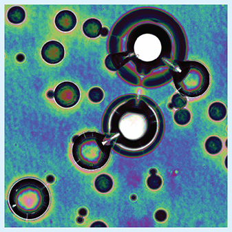
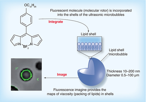
Marina Kuimova, from Imperial College London (London, UK) explained the objectives behind the research. “Microbubbles are widely used to enhance contrast in ultrasound imaging; however, no reliable method exists to characterize their ‘coats‘, which is in fact what is responsible for their function. We wanted to develop a method to characterize the viscosity of microbubble coats; that is, to be able to say how tightly the lipids are packed on the outside of each bubble.”
The scientists used a small fluorophore, termed a ‘molecular rotor‘, which was embedded within the surface of the bubbles. The fluorescence lifetime directly correlated with the viscosity of the surroundings, and imaging demonstrated that shell viscosities vary widely across the population of the microbubbles and are influenced both by shell composition and the manufacturing process. Eleanor Stride (University of Oxford, UK), one of the authors of the study, commented, “Our results demonstrate that the surface properties of microbubbles, which are crucial to their use in both imaging and drug delivery, are extremely sensitive to the bubble coating composition, environment and manufacturing technique used. This will have a significant effect on how we, as engineers, design our fabrication processes and future experiments and ultimately how we apply microbubbles clinically.”
The researchers now believe that this work will bring forward further applications of microbubbles in the drug-delivery field, demonstrating the need for careful design and also maximizing the potential of microbubble-mediated therapies by being able to characterize the bubbles individually.
Kuimova stated the groups future directions following the results of the study, “We are now planning to look at different ways to prepare microbubbles and correlate the resulting ultrasound properties with the manufacturing methods. Another option is to look at bubble interaction with tissues and look at the shell properties under the ultrasound. The latter two are particularly relevant for drug-delivery applications.”
– Written by James Potticary
Source: Hosny NA, Mohamedi G, Rademeyer P et al. Mapping microbubble viscosity using fluorescence lifetime imaging of molecular rotors. Proc. Natl Acad. Sci. USA 110(23) 9225–9230 (2013).
With special thanks to Marina Kuimova (Imperial College London) and Eleanor Stride (University of Oxford). Images kindly provided by Maria Kuimova.
Novel topical nanoparticle treatment for Candida wound infection
Researchers at Albert Einstein College of Medicine (NY, USA) have recently developed a nanoparticle-based treatment for severe thermal injury, wound infection and sepsis. A great risk to wound patients due to its antibiotic resistance is the yeast species Candida. One of the most effective antifungal agents is Amphotericin B (AmB), although until now, this could only be administered intravenously and has been clinically limited by its adverse-advent profile.
The team, led by Adam Friedman, Professor at the Albert Einstein College of Medicine, has developed a therapy involving the encapsulation of AmB into nanoparticles, resulting in enhanced efficacy and greater payloads. Friedman discussed his findings, “We demonstrated that AmB could be effectively encapsulated into a nanovehicle that allowed for controlled and sustained release of drug over time. Given the known occlusive effect of nanoparticles on the skin, the controlled release nature in combination with the resulting increased residence time on the epidermis due to their size will allow for enhanced cutaneous drug penetration, which AmB alone can not accomplish.” He continued, “The AmB nanoparticles were found to be more effective in vitro in both killing candidal species at analogous concentrations to solubilized AmB, and importantly, more effective at inhibiting biofilm formation and disrupting mature biofilms.”
The researchers expect their discovery to have broader implications beyond fungal infections as AmB has activity against parasitic infections such as cutaneous leishmaniasis. The results are expected to lead to further research in higher animals in order to enter the clinical space as a topical antifungal agent.
Friedman highlighted that, “The future of nanotechnology in the antimicrobial arena is not only bright, but a requisite to successfully combat the medical crisis that has resulted from the continued emergence of resistant infectious organisms.”
– Written by Phoebe Heseltine
Source: Sanchez DA, Schairer D, Tuckman-Vernon C et al. Amphotericin B releasing nanoparticle topical treatment of Candida spp. in the setting of a burn wound. Nanomedicine doi:10.1016/j.nano.2013.06.002 (2013) (Epub ahead of print).