Abstract
With the rapidly growing availability of the entire genome sequences of microbial pathogens, there is unmet need for increasingly sensitive systems to monitor the gene-specific markers for diagnosis of bacteremia that enables an earlier detection of causative agent and determination of drug resistance. To address these challenges, a novel FISH-type genomic sequence-based molecular technique is proposed that can identify bacteria and simultaneously detect antibiotic resistance markers for rapid and accurate testing of pathogens. The approach is based on a synergistic combination of advanced Peptide Nucleic Acid (PNA)-based technology and signal-enhancing Rolling Circle Amplification (RCA) reaction to achieve a highly specific and sensitive assay. A specific PNA-DNA construct serves as an exceedingly selective and very effective biomarker, while RCA enhances detection sensitivity and provide with a highly multiplexed assay system. Distinct-color fluorescent decorator probes are used to identify about 20-nucleotide-long signature sequences in bacterial genomic DNA and/or key genetic markers of drug resistance in order to identify and characterize various pathogens. The technique's potential and its utility for clinical diagnostics are illustrated by identification of S. aureus with simultaneous discrimination of methicillin-sensitive (MSSA) versus methicillin-resistant (MRSA) strains. Overall these promising results hint to the adoption of PNA-based rapid sensitive detection for diagnosis of other clinically relevant organisms. Thereby, new assay enables significantly earlier administration of appropriate antimicrobial therapy and may, thus have a positive impact on the outcome of the patient.
Acknowledgements
We thank Dr. Peter Nielsen for providing us with PNA oligomers, Mark Fiandaca for S. aureus bacterial strains and Dr. Karen Quillen for expired packed human red blood cells.
This work was supported by the Wallace C. Coulter Foundation to Maxim D. Frank-Kamenetskii and Nancy S. Miller and a NIH research grant (1R21RR025371-01) to Irina Smolina.
Figures and Tables
Figure 1 PNA-based diagnostic scheme. Major steps of the proposed assay for in situ detection of S. aureus and its methicillin-resistant strains.
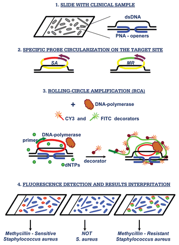
Figure 2 Detection discrimination of methicillin-sensitive (MSSA) versus methicillin-resistant (MRSA) strains. Images of S. aureus bacterial cells observed by fluorescent microscopy in experiments performed with simulated matrix (packed red cells and blood culture media) spiked with S. aureus bacterial cells. A combination of two probes (SA-3 and MR-1) was applied to simulated matrix specimens spiked with: the regular MSS A strain (A1 and B1); the Mu3 MRSA strain (A2 and B2) and their mixture (A3–C3). The fluorescent signals were acquired separately using three filter sets: A1–A3 present a superposition of two separate images, with DAPI and CY3; B1–B3 present a superposition of two separate images, with DAPI and FITC; C3 presents a superposition of three separate images, with DAPI, CY3 and FITC.
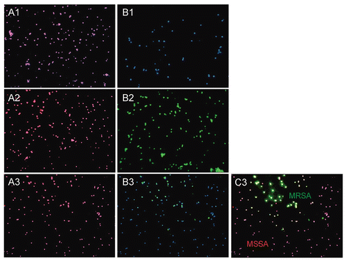
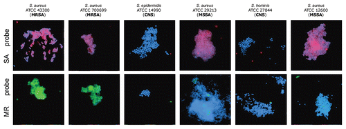
Figure 3 Experimenter-blinded tests with de-identified, codified bacteria contained S. aureus (both MSSA and MRSA) and coagulase-negative staphylococcal species (CNS). Images of methicillin-sensitive S. aureus (MSSA), coagulase-negative staphylococcus (CNS) and methicillin-resistant S. aureus (MRSA) cells observed by fluorescent microscopy when probes specific to SA (MSSA/MRSA) or the MR (to mec cassette—MRSA only) were applied. The fluorescent signals were acquired separately using three filter sets (DAPI for DNA and Cy3 or FITC for the RCA product). Images are superposition of two images: (SA-probe) DAPI and CY3; (MR-probe) DAPI and FITC. Signals were pseudocolored in blue for DAPI, red for CY3 and green for FITC. SA-probes (PNA binding sites are underlined): SA-1: AAA GAA AAA GCA ACA AGA GGA A, SA-2: AGA GGA AGC AGA GCG CAA GGG AAA, SA-3: AAA AGA AGA AAG ATT CAG AGG AAG; MR-probes (within mecA gene for penicillin-binding protein): MR-1: AAG GAG GAT ATT GAT GAA AAA GA, MR-2: GGA AGA AAA ATA TTA TTT CCA AAG AAA A.